. 2018 May 18:9:565.
doi: 10.3389/fphys.2018.00565. eCollection 2018.

Epidural Spinal Cord Stimulation of Lumbosacral Networks Modulates Arterial Blood Pressure in Individuals With Spinal Cord Injury-Induced Cardiovascular Deficits

Affiliations

Epidural Spinal Cord Stimulation of Lumbosacral Networks Modulates Arterial Blood Pressure in Individuals With Spinal Cord Injury-Induced Cardiovascular Deficits

Sevda C Aslan et al. Front Physiol. .

Abstract

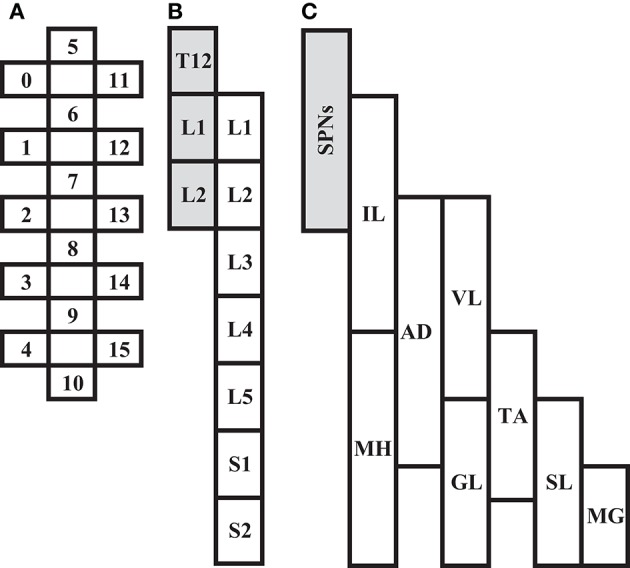
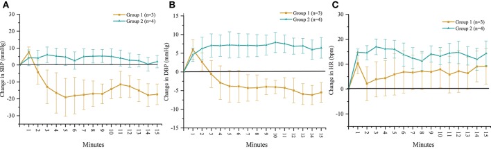
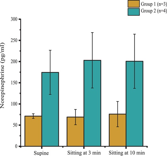
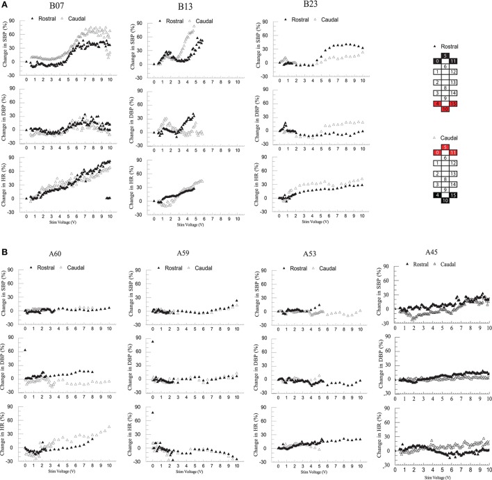
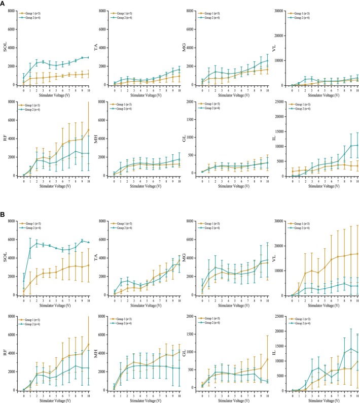
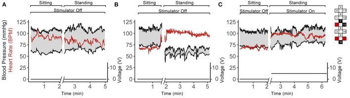
Disruption of motor and autonomic pathways induced by spinal cord injury (SCI) often leads to persistent low arterial blood pressure and orthostatic intolerance. Spinal cord epidural stimulation (scES) has been shown to enable independent standing and voluntary movement in individuals with clinically motor complete SCI. In this study, we addressed whether scES configured to activate motor lumbosacral networks can also modulate arterial blood pressure by assessing continuous, beat-by-beat blood pressure and lower extremity electromyography during supine and standing in seven individuals with C5-T4 SCI. In three research participants with arterial hypotension, orthostatic intolerance, and low levels of circulating catecholamines (group 1), scES applied while supine and standing resulted in increased arterial blood pressure. In four research participants without evidence of arterial hypotension or orthostatic intolerance and normative circulating catecholamines (group 2), scES did not induce significant increases in arterial blood pressure. During scES, there were no significant differences in electromyographic (EMG) activity between group 1 and group 2. In group 1, during standing assisted by scES, blood pressure was maintained at 119/72 ± 7/14 mmHg (mean ± SD) compared with 70/45 ± 5/7 mmHg without scES. In group 2 there were no arterial blood pressure changes during standing with or without scES. These findings demonstrate that scES configured to facilitate motor function can acutely increase arterial blood pressure in individuals with SCI-induced cardiovascular deficits.

Keywords: blood pressure; epidural stimulation; human spinal cord injury; orthostatic hypotension; systemic hypotension.


Figures

Figure 1
Figure 1
Depiction of scES 16-electrode array (A) relative to spinal cord segments L1 to S2 (B), and corresponding muscles (C; IL, Iliopsoas; MH, Medial Hamstrings, AD, Adductor Magnus; VL, Vastus Lateralis; GL, Gluteus Maximus; TA, Tibialis Anterior; SL, Soleus; MG, Medial Gastrocnemius). Shaded areas (B,C) represent localization of spinal sympathetic preganglionic neurons (SPNs) at T12-L2 levels.
Figure 2
Figure 2
Time course of change in (A) systolic blood pressure (SBP), (B) diastolic blood pressure (DBP) and (C) heart rate (HR) in response to orthostatic stress test performed without scES. Group 1 (n = 3) SBP (p < 0.001), DBP (p < 0.001), and HR (p < 0.01) changed significantly compared with baseline; group 2 (n = 4) demonstrated no significant changes to SBP, DBP, or HR from baseline. Data are represented as mean ± SD.
Figure 3
Figure 3
Plasma norepinephrine levels in supine position, and during minutes 3 and 10 of orthostatic stress. Norepinephrine levels were significantly lower (p = 0.02) in Group 1 (n = 3) compared with Group 2 (n = 4) throughout the orthostatic stress test. Data are represented as mean ± SD.
Figure 4
Figure 4
Effect of rostral (0–5–11–/4+10+15+) and caudal (4–10–15–/0+5+11+) scES at 2 Hz on supine blood pressure and heart rate. Illustrated from group 1 (A; n = 3) and group 2 (B; n = 4) are mean percent-change in systolic blood pressure (SBP), diastolic blood pressure (DBP), and heart rate (HR) from baseline (open triangles: rostral; black triangles: caudal stimulations) concurrent with increases in stimulator voltage. Supine voltage increased from 0 V to 10 V by 0.1 V and 0.5 V intervals. Electrode configuration and color map are presented on the right side of the figure; black boxes are cathode, red boxes are anode, and white boxes are inactive electrodes.
Figure 5
Figure 5
Effect of scES at 2 Hz with (A) rostral (0–5–11–/4+10+15+) and (B) caudal (4–10–15–/0+5+11+), stimulation configurations on muscle activity. Illustrated is mean electromyography (EMG) of leg muscles (SOL, Soleus; TA, Tibialis Anterior; MG, Medial Gastrocnemius; VL, Vastus Lateralis; RF, Rectus Femoris; MH, Medial Hamstrings; GL, Gluteus Maximus; IL, Iliopsoas) from group 1 (gold circle; n = 3) and group 2 (green triangle, n = 4) as simulator voltage increased from 0 V to 10 V by 0.1 V and 0.5 V intervals. There were no significant differences in EMG activity between groups during rostral and caudal scES configurations. Data are represented as mean ± SD.
Figure 6
Figure 6
Continuous blood pressure and heart rate recordings from A59 (A) and B23 (B,C) in sitting and standing positions while participant was sitting and standing without scES (A,B) and using scES (C). Top and bottom black lines indicate systolic and diastolic blood pressure; red line indicates heart rate. The stimulator intensity and electrode configuration are given on the right side; black boxes are cathode, red boxes are anode, and white boxes are inactive electrodes. The stimulation frequency was 15 Hz. Note that: subject A59 did not experience a drop in blood pressure upon standing (A), while subject B23 experienced such drop (B), and this decrease in blood pressure was abolished in the presence of the stimulation.

References

    1. Angeli C. A., Edgerton V. R., Gerasimenko Y. P., Harkema S. J. (2014). Altering spinal cord excitability enables voluntary movements after chronic complete paralysis in humans. Brain 137, 1394–1409. 10.1093/brain/awu038 - DOI - PMC - PubMed
    1. Blackmer J. (1997). Orthostatic hypotension in spinal cord injured patients. J. Spinal Cord Med. 20, 212–217. 10.1080/10790268.1997.11719471 - DOI - PubMed
    1. Bos W. J. W., Van Goudoever J., Van Montfrans G. A., Van Den Meiracker A. H., Wesseling K. H. (1996). Reconstruction of brachial artery pressure from noninvasive finger pressure measurements. Circulation 94, 1870–1875. 10.1161/01.CIR.94.8.1870 - DOI - PubMed
    1. Campbell J. N., Meyer R. A. (2006). Mechanisms of neuropathic pain. Neuron 52, 77–92. 10.1016/j.neuron.2006.09.021 - DOI - PMC - PubMed
    1. Carlozzi N. E., Fyffe D., Morin K. G., Byrne R., Tulsky D. S., Victorson D., et al. . (2013). Impact of blood pressure dysregulation on health-related quality of life in persons with spinal cord injury: development of a conceptual model. Arch. Phys. Med. Rehabil. 94, 1721–1730. 10.1016/j.apmr.2013.02.024 - DOI - PMC - PubMed