. 2018 May 9:9:230.
doi: 10.3389/fendo.2018.00230. eCollection 2018.

Brain Aromatase Modulates Serotonergic Neuron by Regulating Serotonin Levels in Zebrafish Embryos and Larvae

Affiliations

Brain Aromatase Modulates Serotonergic Neuron by Regulating Serotonin Levels in Zebrafish Embryos and Larvae

Zulvikar Syambani Ulhaq et al. Front Endocrinol (Lausanne). .

Abstract

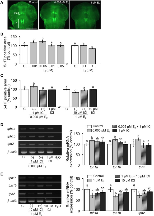
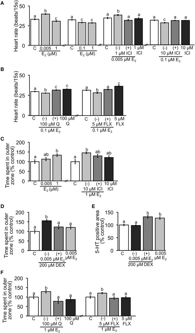
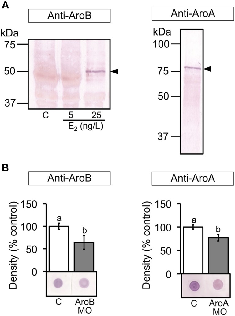
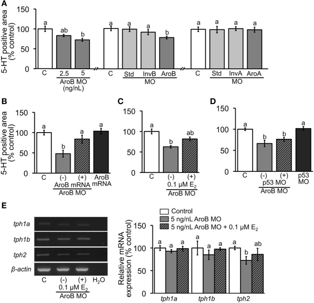
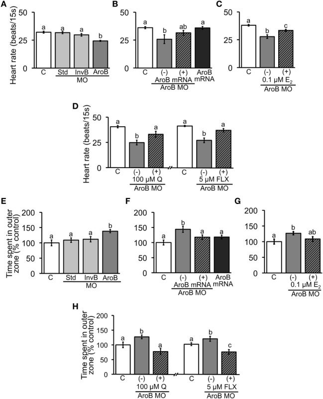
Teleost fish are known to express two isoforms of P450 aromatase, a key enzyme for estrogen synthesis. One of the isoforms, brain aromatase (AroB), cyp19a1b, is highly expressed during early development of zebrafish, thereby suggesting its role in brain development. On the other hand, early development of serotonergic neuron, one of the major monoamine neurons, is considered to play an important role in neurogenesis. Therefore, in this study, we investigated the role of AroB in development of serotonergic neuron by testing the effects of (1) estradiol (E2) exposure and (2) morpholino (MO)-mediated AroB knockdown. When embryos were exposed to E2, the effects were biphasic. The low dose of E2 (0.005 µM) significantly increased serotonin (5-HT) positive area at 48 hour post-fertilization (hpf) detected by immunohistochemistry and relative mRNA levels of tryptophan hydroxylase isoforms (tph1a, tph1b, and tph2) at 96 hpf measured by semi-quantitative PCR. To test the effects on serotonin transmission, heart rate and thigmotaxis, an indicator of anxiety, were analyzed. The low dose also significantly increased heart rate at 48 hpf and decreased thigmotaxis. The high dose of E2 (1 µM) exhibited opposite effects in all parameters. The effects of both low and high doses were reversed by addition of estrogen receptor (ER) blocker, ICI 182,780, thereby suggesting that the effects were mediated through ER. When AroB MO was injected to fertilized eggs, 5-HT-positive area was significantly decreased, while the significant decrease in relative tph mRNA levels was found only with tph2 but not with two other isoforms. AroB MO also decreased heart rate and increased thigmotaxis. All the effects were rescued by co-injection with AroB mRNA and by exposure to E2. Taken together, this study demonstrates the role of brain aromatase in development of serotonergic neuron in zebrafish embryos and larvae, implying that brain-formed estrogen is an important factor to sustain early development of serotonergic neuron.

Keywords: biphasic manner; brain aromatase; early development; estradiol; serotonergic neuron; zebrafish.


Figures

Figure 1
Figure 1
Effect of E2 on 5-HT-positive area and relative expression of tph isoforms. (A) Representative images of ventral view of head region showing 5-HT-positive cells in pretectal and thalamic complex (ptc) and raphe (r) at 2 dpf. Scale bar: 200 µm. (B,C) Measurements of the area of 5-HT-positive neurons in the experiment of E2 exposure and co-incubation of E2 and ICI, respectively. (D,E) Semi-quantitative PCR for expression of tph isoforms in 4 dpf larvae in the experiments of exposure to low and high doses of E2 and co-incubation with ICI, respectively. Data are presented as a mean ± SEM. Different letters in each graph indicate significant differences (p < 0.05).
Figure 2
Figure 2
Effect of E2 on heart rate and thigmotaxis. Heart rate was measured in 2-dpf embryos; (A) exposure to low and high doses of E2 and co-incubation with ICI; (B) co-incubation of E2 and Q or FLX to examine involvement of serotonin signaling. Thigmotaxis assay was performed to evaluate anxiety level in 6-dpf larvae; (C) exposure to low and high doses of E2 and co-incubation with ICI; (D) anxiolytic effect of low dose of E2 when larvae were exposed to 200 µM DEX. (E) Measurements of 5-HT-positive neuron area in 2-dpf embryos exposed to DEX and low dose of E2. (F) Effects of co-incubation of E2 and Q or FLX on thigmotaxis to examine involvement of serotonin signaling. Data are presented as a mean ± SEM. Different letters in each graph indicate significant differences (p < 0.05).
Figure 3
Figure 3
Validation of MO-mediated knockdown on translation. (A) Western blots of brain extracts from control and E2 exposed fish and ovarian extract from untreated fish were stained with the antiserum to AroB and AroA, respectively, showing a positive band at the expected size for each aromatase as indicated by arrowheads. (B) Dot blots of 6 and 2 dpf larval extracts using the antiserum to AroB and AroA, respectively, were analyzed. Representative blots are shown in each graph. Data are presented as a mean ± SEM. Different letters in each graph indicate significant differences (p < 0.05).
Figure 4
Figure 4
AroB MO-mediated effect on serotonergic neuron. 5-HT-positive neuron area was measured in 2-dpf embryos; (A) injection of AroB MO and control MOs (Std MO, InvB MO and AroA MO); (B,C) co-injection of AroB mRNA and exposure to E2, respectively, to rescue the effect of AroB MO; (D) co-injection of p53 MO to test off-target effect of AroB MO. (E) Semi-quantitative PCR measurement of expression of tph isoforms in 6-dpf larvae injected with AroB MO with and without exposure to E2. Data are presented as a mean ± SEM. Different letters in each graph indicate significant differences (p < 0.05).
Figure 5
Figure 5
AroB MO-mediated effect on heart rate and thigmotaxis. Heart rate was measured in 2-dpf embryos; (A) injection of AroB MO and control MOs; (B,C) co-injection of AroB mRNA and exposure to E2, respectively, to rescue the effect of AroB MO; (D) injection of AroB MO with and without exposure to Q and FLX to examine involvement of serotonin signaling. Thigmotaxis assay was conducted using 6-dpf larvae; (E) injection of AroB MO and control MOs; (F,G) co-injection of AroB mRNA and exposure to E2, respectively, to rescue the effect of AroB MO; (H) injection of AroB MO with and without exposure to Q and FLX to examine involvement of serotonin signaling. Data are presented as a mean ± SEM. Different letters in each graph indicate significant differences (p < 0.05).

References

    1. Simpson ER, Davis SR. Minireview: aromatase and the regulation of estrogen biosynthesis-some new perspectives. Endocrinology (2001) 142(11):4589–94.10.1210/endo.142.11.8547 - DOI - PubMed
    1. Cui J, Shen Y, Li R. Estrogen synthesis and signaling pathways during ageing: from periphery to brain. Trends Mol Med (2013) 19(3):197–209.10.1016/j.molmed.2012.12.007 - DOI - PMC - PubMed
    1. Kishida M, Callard GV. Distinct cytochrome P450 aromatase isoforms in zebrafish (Danio rerio) brain and ovary are differentially programmed and estrogen regulated during early development. Endocrinology (2001) 142(2):740–50.10.1210/endo.142.2.7928 - DOI - PubMed
    1. Callard GV, Greytak SR, Novillo A, Cotter CA, Mayer RK. Brain aromatase in fish: perspective and comparative approaches. In: Balthazart J, Ball G, editors. Brain Aromatase, Estrogen, and Behavior. New York: Oxford University Press; (2013). p. 13–42.
    1. Pasmanik M, Callard GV. Changes in brain aromatase and 5 alpha-reductase activities correlate significantly with seasonal reproductive cycles in goldfish (Carassius auratus). Endocrinology (1988) 122(4):1349–56.10.1210/endo-122-4-1349 - DOI - PubMed

LinkOut - more resources