Placental Inflammation and Fetal Injury in a Rare Zika Case Associated With Guillain-Barré Syndrome and Abortion
- PMID: 29867903
- PMCID: PMC5964188
- DOI: 10.3389/fmicb.2018.01018
Placental Inflammation and Fetal Injury in a Rare Zika Case Associated With Guillain-Barré Syndrome and Abortion
Abstract
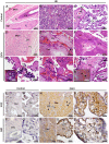
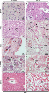
Zika virus (ZIKV) is an emerging virus involved in recent outbreaks in Brazil. The association between the virus and Guillain-Barré syndrome (GBS) or congenital disorders has raised a worldwide concern. In this work, we investigated a rare Zika case, which was associated with GBS and spontaneous retained abortion. Using specific anti-ZIKV staining, the virus was identified in placenta (mainly in Hofbauer cells) and in several fetal tissues, such as brain, lungs, kidneys, skin and liver. Histological analyses of the placenta and fetal organs revealed different types of tissue abnormalities, which included inflammation, hemorrhage, edema and necrosis in placenta, as well as tissue disorganization in the fetus. Increased cellularity (Hofbauer cells and TCD8+ lymphocytes), expression of local pro-inflammatory cytokines such as IFN-γ and TNF-α, and other markers, such as RANTES/CCL5 and VEGFR2, supported placental inflammation and dysfunction. The commitment of the maternal-fetal link in association with fetal damage gave rise to a discussion regarding the influence of the maternal immunity toward the fetal development. Findings presented in this work may help understanding the ZIKV immunopathogenesis under the rare contexts of spontaneous abortions in association with GBS.
Keywords: Guillain-Barré syndrome; Zika virus; fetal infection; histopathology; immune response.
Figures


Similar articles
-
A Stillborn Multiple Organs' Investigation from a Maternal DENV-4 Infection: Histopathological and Inflammatory Mediators Characterization.Viruses. 2019 Apr 2;11(4):319. doi: 10.3390/v11040319. Viruses. 2019. PMID: 30986974 Free PMC article.
-
Zika virus infection as a cause of congenital brain abnormalities and Guillain-Barré syndrome: From systematic review to living systematic review.F1000Res. 2018 Feb 15;7:196. doi: 10.12688/f1000research.13704.1. eCollection 2018. F1000Res. 2018. PMID: 30631437 Free PMC article.
-
Zika Virus Infects Early- and Midgestation Human Maternal Decidual Tissues, Inducing Distinct Innate Tissue Responses in the Maternal-Fetal Interface.J Virol. 2017 Jan 31;91(4):e01905-16. doi: 10.1128/JVI.01905-16. Print 2017 Feb 15. J Virol. 2017. PMID: 27974560 Free PMC article.
-
Zika virus infection and risk of Guillain-Barré syndrome: A meta-analysis.J Neurol Sci. 2019 Aug 15;403:99-105. doi: 10.1016/j.jns.2019.06.019. Epub 2019 Jun 21. J Neurol Sci. 2019. PMID: 31255970
-
Guillain-Barré syndrome in patients with a recent history of Zika in Cúcuta, Colombia: A descriptive case series of 19 patients from December 2015 to March 2016.J Crit Care. 2017 Feb;37:19-23. doi: 10.1016/j.jcrc.2016.08.016. Epub 2016 Aug 18. J Crit Care. 2017. PMID: 27610587
Cited by
-
Placental Macrophage (Hofbauer Cell) Responses to Infection During Pregnancy: A Systematic Scoping Review.Front Immunol. 2022 Feb 11;12:756035. doi: 10.3389/fimmu.2021.756035. eCollection 2021. Front Immunol. 2022. PMID: 35250964 Free PMC article.
-
The Innate Defense in the Zika-Infected Placenta.Pathogens. 2022 Nov 24;11(12):1410. doi: 10.3390/pathogens11121410. Pathogens. 2022. PMID: 36558744 Free PMC article. Review.
-
Rescue of Replication-Competent ZIKV Hidden in Placenta-Derived Mesenchymal Cells Long After the Resolution of the Infection.Open Forum Infect Dis. 2019 Oct 9;6(10):ofz342. doi: 10.1093/ofid/ofz342. eCollection 2019 Oct. Open Forum Infect Dis. 2019. PMID: 31660333 Free PMC article.
-
Liver immunopathogenesis in fatal cases of dengue in children: detection of viral antigen, cytokine profile and inflammatory mediators.Front Immunol. 2023 Jun 30;14:1215730. doi: 10.3389/fimmu.2023.1215730. eCollection 2023. Front Immunol. 2023. PMID: 37457689 Free PMC article.
-
Absence of Zika virus among pregnant women in Vietnam in 2008.Trop Dis Travel Med Vaccines. 2023 Mar 1;9(1):4. doi: 10.1186/s40794-023-00189-7. Trop Dis Travel Med Vaccines. 2023. PMID: 36855197 Free PMC article.
References
-
- Brito Ferreira M. L., Antunes de Brito C. A., Moreira Á.J. P., de Morais Machado M. Í., Henriques-Souza A., Cordeiro M. T., et al. . (2017). Guillain–Barré syndrome, acute disseminated encephalomyelitis and encephalitis associated with zika virus infection in brazil: detection of viral, RNA and isolation of virus during late infection. Am. J. Trop. Med. Hyg. 97, 1405–1419. 10.4269/ajtmh.17-0106 - DOI - PMC - PubMed
Publication types
LinkOut - more resources
Full Text Sources
Other Literature Sources

