. 2018 May 8:9:973.
doi: 10.3389/fimmu.2018.00973. eCollection 2018.

NoxO1 Controls Proliferation of Colon Epithelial Cells

Affiliations

NoxO1 Controls Proliferation of Colon Epithelial Cells

Franziska Moll et al. Front Immunol. .

Abstract

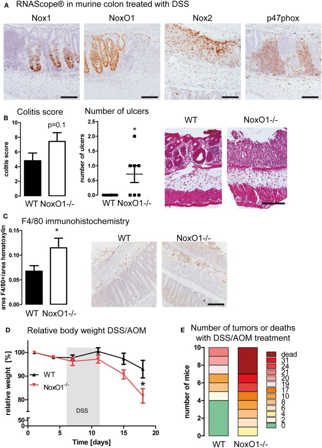
Aim: Reactive oxygen species (ROS) produced by enzymes of the NADPH oxidase family serve as second messengers for cellular signaling. Processes such as differentiation and proliferation are regulated by NADPH oxidases. In the intestine, due to the exceedingly fast and constant renewal of the epithelium both processes have to be highly controlled and balanced. Nox1 is the major NADPH oxidase expressed in the gut, and its function is regulated by cytosolic subunits such as NoxO1. We hypothesize that the NoxO1-controlled activity of Nox1 contributes to a proper epithelial homeostasis and renewal in the gut.

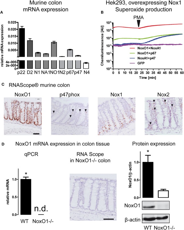
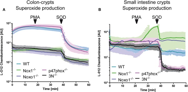
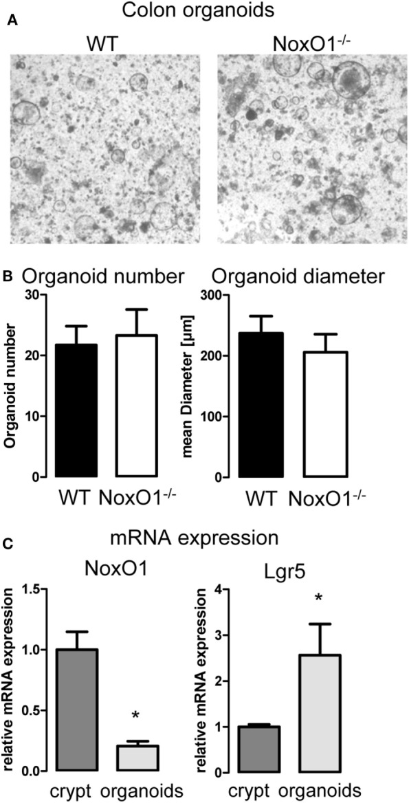
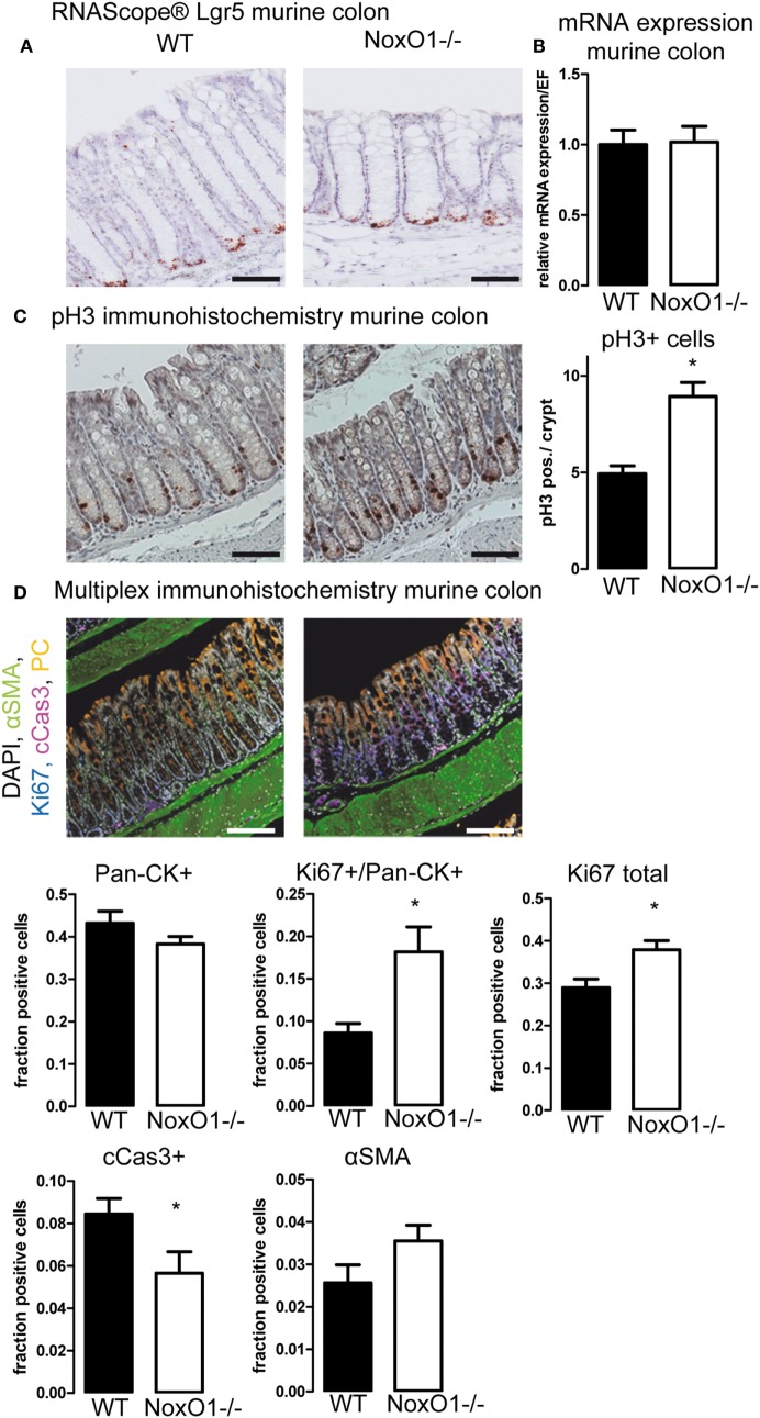
Results: NoxO1 is highly expressed in the colon. Knockout of NoxO1 reduces the production of superoxide in colon crypts and is not subsidized by an elevated expression of its homolog p47phox. Knockout of NoxO1 increases the proliferative capacity and prevents apoptosis of colon epithelial cells. In mouse models of dextran sulfate sodium (DSS)-induced colitis and azoxymethane/DSS induced colon cancer, NoxO1 has a protective role and may influence the population of natural killer cells.

Conclusion: NoxO1 affects colon epithelium homeostasis and prevents inflammation.

Keywords: Nox1; NoxO1; colon; inflammation; proliferation; reactive oxygen species.


Figures

Figure 1
Figure 1
NADPH oxidase expression in the colon. (A) Analysis of mRNA expression in murine colon tissue. Relative expression to housekeeping gene EF. Genes indicated are p22phox (p22), Duox2 (D2), Nox1 (N1), NoxA1 (NA1), NoxO1 (N01), Nox2 (N2) p67phox (p67), p47phox (p47), and Nox4 (N4). n = 7–8. (B) HEK293 cells overexpressing Nox1 were transfected with cytosolic subunits of the NADPH oxidase complex as indicated. Reactive oxygen species were measured with L012 (200 μmol/L). Activation of p47phox was triggered by phorbol myristate acetate (100 nmol/L). (C) In situ hybridization (RNAScope® DAB staining) showing the expression of NADPH oxidase subunits NoxO1, p47phox, Nox1, and Nox2 in murine colon tissue. Nuclei were counterstained with hematoxylin. Scale bars indicate 100 μm. (D) Quantitative RT-PCR, RNAScpoe®, and immunoblotting analysis of Nox1 protein expression and in colon tissue of wild-type (WT) and NoxO1 knockout (NoxO1−/−), n = 6, *p < 0.05 WT vs. NoxO1−/−.
Figure 2
Figure 2
NoxO1-dependent reactive oxygen species (ROS) formation in colon crypts. A + B Crypts from colon (A) or small intestine (B) were isolated. ROS were measured by chemiluminescence with L-012 (200 μmol/L). Activation of p47phox was triggered by phorbol myristate acetate (PMA, 100 nmol/L). Superoxide anions were decomposed with superoxide dismutase (SOD, 300 U/mL); n = 4.
Figure 3
Figure 3
Colon organoid growth. (A) 3D culture of organoids generated from colon crypts isolated from wild-type (WT) and NoxO1 knockout (NoxO1−/−) animals. Representative pictures of organoids after 7 days in culture. (B) Analysis of organoid number and mean diameter of organoids cultured for 7 days. Organoids were counted per field of vision. Per animal two wells were analyzed. n = 5. (C) mRNA expression relative to housekeeping gene EF of NoxO1 and stem cell marker Lgr5 in freshly isolated crypts and organoids cultured for 7 days. n = 3, *p < 0.05.
Figure 4
Figure 4
Immunohistochemistry of colons. (A) In situ hybridization (RNAScope®) of Lgr5 in murine colon. Nuclei were counterstained with hematoxylin, scale bars indicate 100 μm (B) Lgr5 mRNA expression relative to housekeeping gene EF in colon tissue from wild-type (WT) and NoxO1 knockout (NoxO1−/−) mice. n = 7. (C) Immunohistochemistry staining of mitosis marker phospho-Histone 3 (pH3). Representative pictures and analysis of pH3-positive cells per crypt. n = 3, *p < 0.05. Scale bars indicate 200 μm. (D) Multiplex immunohistochemistry of murine colon. Tissue was stained for DAPI (white), alpha-smooth muscle actin (aSMA, green), Ki67 (blue), cleaved Caspase 3 (cCas3, magenta), and pan-cytokeratin (Pan-CK, orange); scale bars indicate 100 μm. Positive cell fractions for indicated phenotypes in the mucosa were analyzed using the in Form 2.0 software. n = 5, *p < 0.05.
Figure 5
Figure 5
NoxO1 has a protective role in dextran sulfate sodium (DSS)-induced colitis and DSS/azoxymethane (AOM) colon carcinoma. (A) In situ hybridization (RNAScope®) showing the expression of Nox1, NoxO1, Nox2, and p47phox in murine colon tissue upon treatment with DSS. Nuclei were counterstained with hematoxylin, scale bars indicate 100 μm. (B) Quantification of histological damage and number of ulcers on day 8 of the colitis model. n = 7–8, *p < 0,05, Mann–Whitney exact test. Representative pictures of hematoxylin and eosin (H&E) staining for wild-type (WT) and NoxO1−/− are shown. Scale bar indicates 200 μm. (C) Immunohistochemistry staining of macrophage marker F4/80. Representative pictures and analysis of F4/80-positive staining per nuclei. n = 7–8, *p < 0.05. Scale bars indicate 500 μm. (D) Body weight relative to day 0 of WT and NoxO1−/− mice treated with AOM at day 0 and 3 cycles of DSS in drinking water. n = 9, *p < 0.05. (E) Tumor burden at day 70 of DSS/AOM-treated animals. n = 8–10.

References

    1. Deschner EE. Cell turnover and colon tumor development. Prev Med (1987) 16(4):580–5.10.1016/0091-7435(87)90075-2 - DOI - PubMed
    1. Cliffe LJ, Humphreys NE, Lane TE, Potten CS, Booth C, Grencis RK. Accelerated intestinal epithelial cell turnover: a new mechanism of parasite expulsion. Science (2005) 308(5727):1463–5.10.1126/science.1108661 - DOI - PubMed
    1. Di Marco E, Gray SP, Kennedy K, Szyndralewiez C, Lyle AN, Lassègue B, et al. NOX4-derived reactive oxygen species limit fibrosis and inhibit proliferation of vascular smooth muscle cells in diabetic atherosclerosis. Free Radic Biol Med (2016) 97:556–67.10.1016/j.freeradbiomed.2016.07.013 - DOI - PMC - PubMed
    1. Goettsch C, Babelova A, Trummer O, Erben RG, Rauner M, Rammelt S, et al. NADPH oxidase 4 limits bone mass by promoting osteoclastogenesis. J Clin Invest (2013) 123(11):4731–8.10.1172/JCI67603 - DOI - PMC - PubMed
    1. Schröder K, Wandzioch K, Helmcke I, Brandes RP. Nox4 acts as a switch between differentiation and proliferation in preadipocytes. Arterioscler Thromb Vasc Biol (2009) 29(2):239–45.10.1161/ATVBAHA.108.174219 - DOI - PubMed

Publication types

MeSH terms

LinkOut - more resources