. 2018 May 16:9:1046.
doi: 10.3389/fimmu.2018.01046. eCollection 2018.

Distribution and Interaction of Murine Pulmonary Phagocytes in the Naive and Allergic Lung

Affiliations

Distribution and Interaction of Murine Pulmonary Phagocytes in the Naive and Allergic Lung

Franziska M Hoffmann et al. Front Immunol. .

Abstract

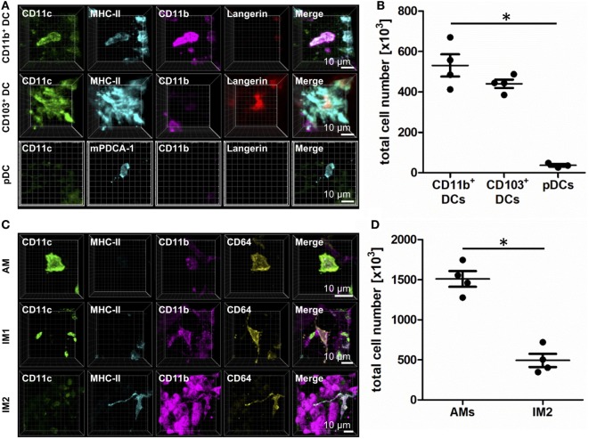
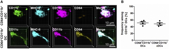
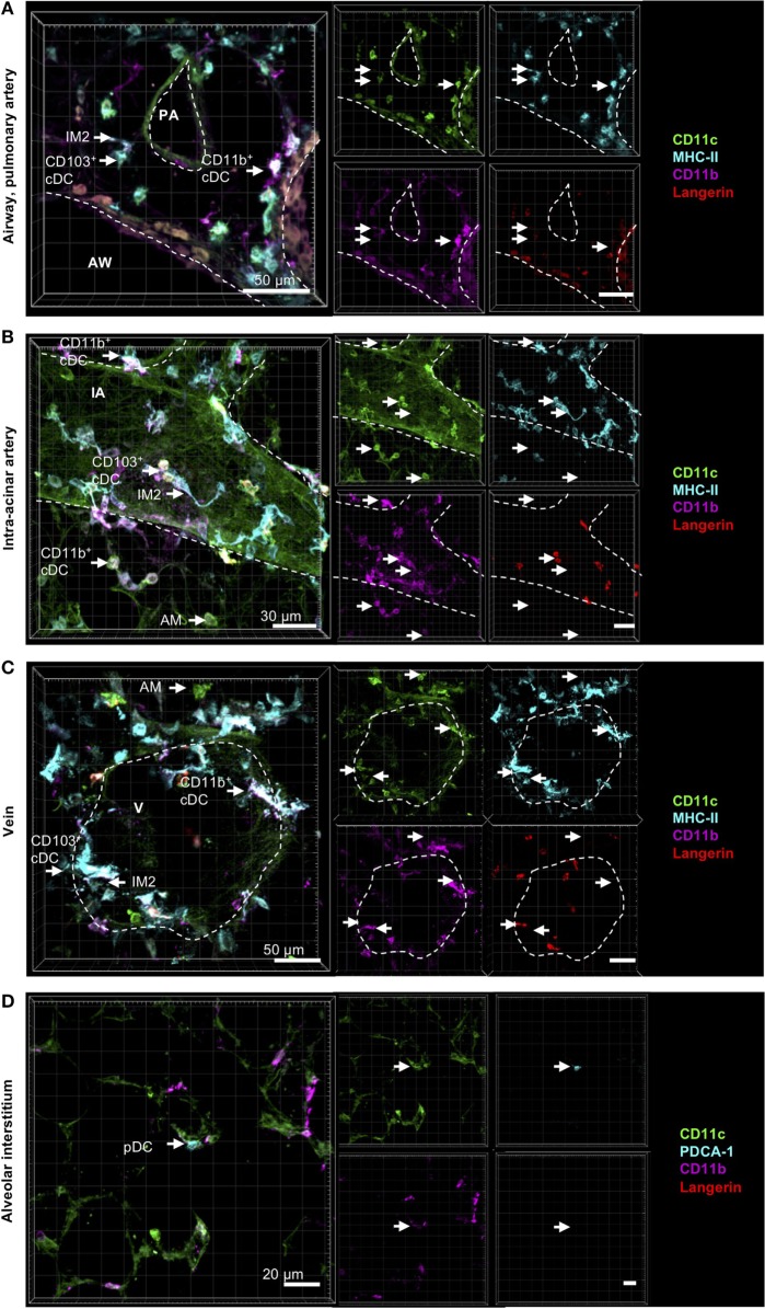
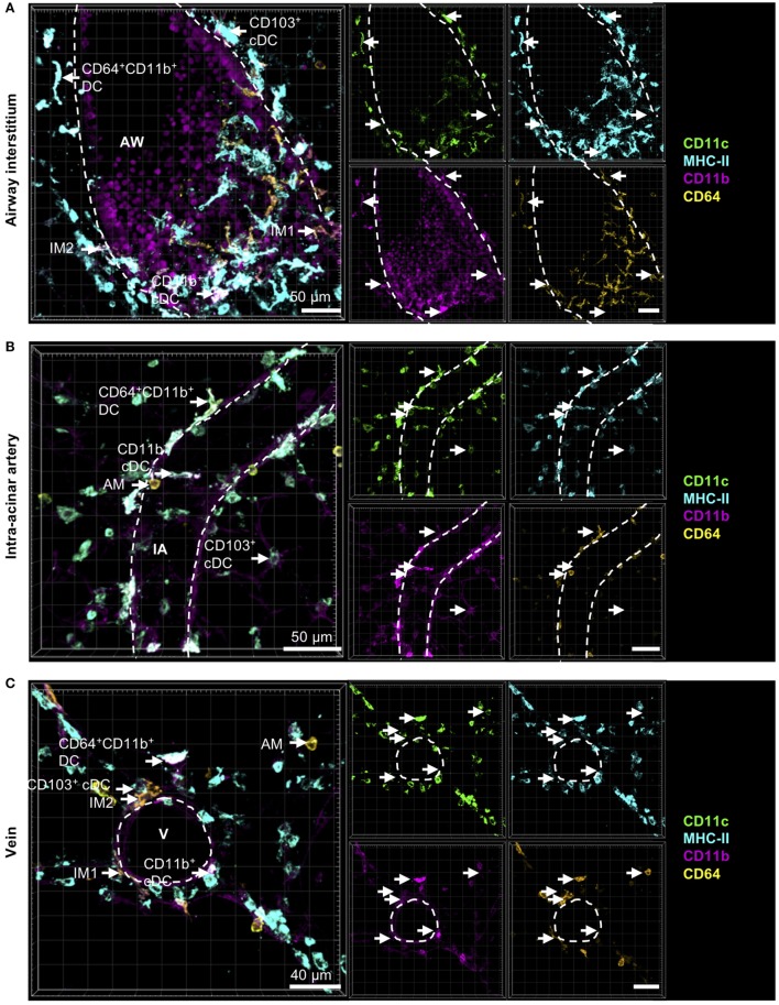
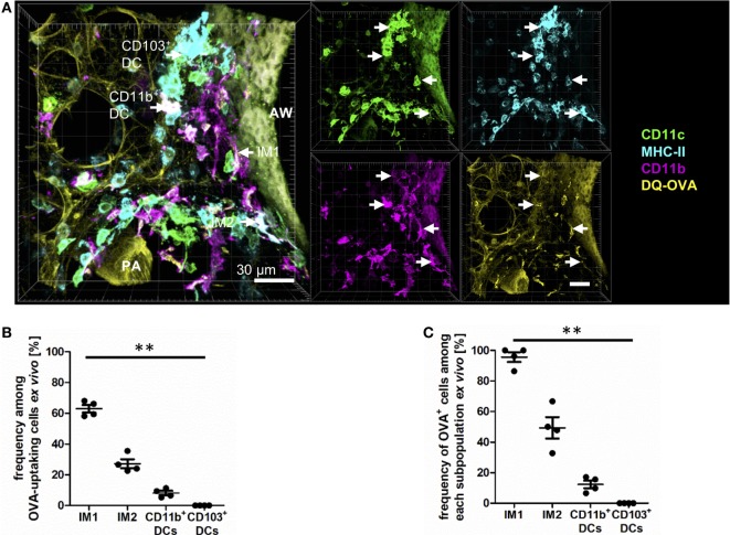
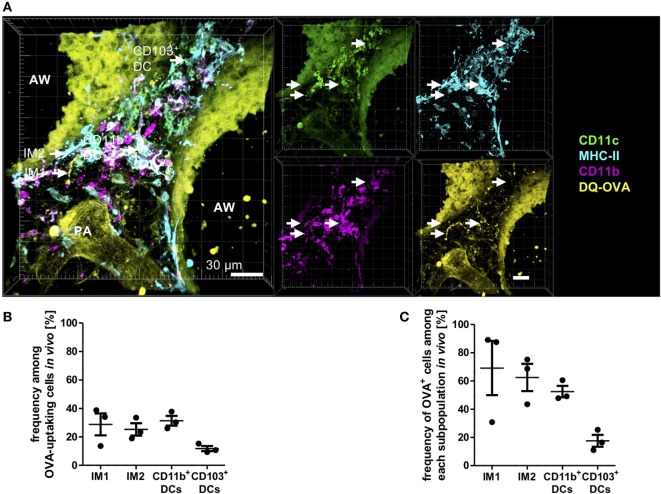
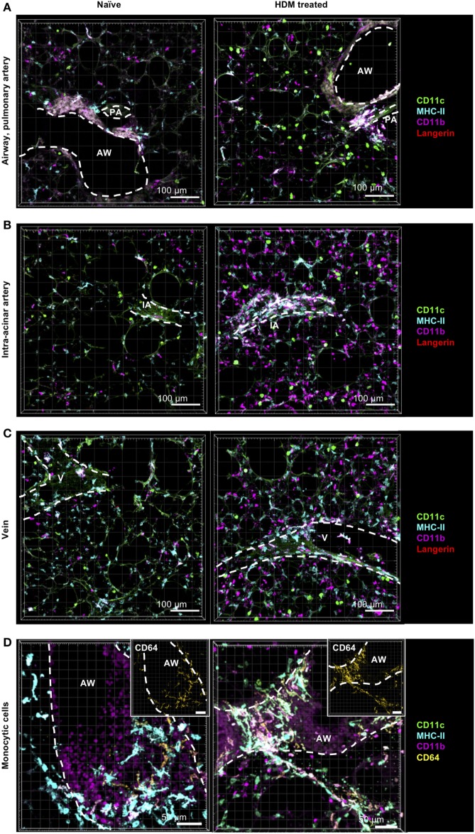
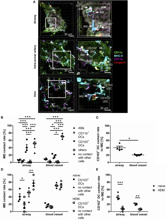
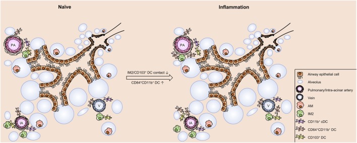
The division of labor between pulmonary phagocytic subsets [macrophage/monocyte and dendritic cell (DC) subpopulations] has been described at the functional level. However, whether these lung phagocytes also display unique spatial distribution remains unclear. Here, to analyze cellular distribution in lung compartments and contacts between phagocyte subpopulations, we established an immunohistochemistry (IHC)-based method to clearly identify murine lung phagocyte subsets in situ based on differential expression of CD11c, CD11b, MHC-II, Langerin and mPDCA-1. Furthermore, we investigated subset-specific functional differences in antigen uptake and spatial changes upon allergic sensitization. Our staining allowed the distinction between alveolar macrophages (AMs), interstitial macrophage (IM) subpopulations, CD11b+ DC subpopulations, CD103+ DCs, and plasmacytoid DCs (pDCs). We identified interstitial regions between airways and around airways as regions of IM/CD11b+ DC/CD103+ DC clusters, where a subset of IMs (IM2) and CD103+ DCs formed intense contacts that decreased upon allergic sensitization. These data indicate functional interactions between both cell types either in steady state or after antigen encounter affecting the development of allergies or tolerance. Furthermore, we observed major antigen uptake in AMs and IMs rather than DC subpopulations that was not restricted to airways and adjacent areas. This will enable to focus future studies to immunologically relevant cellular interactions and to unravel which cells are tipping the balance between pro-inflammatory immune responses or tolerance.

Keywords: allergic airway disease; antigen uptake; dendritic cells; immunohistochemistry; lung; macrophages; spatial distribution.


Figures

Figure 1
Figure 1
Identification of lung phagocyte subsets by immunohistochemistry (IHC). Precision cut lung slices (PCLS) (300 µm) from naive C57BL/6 mice were generated and stained with anti-CD11c, anti-MHC-II or anti-mPDCA-1, anti-CD11b, and anti-Langerin (A,B) or anti-CD64 ABs (C,D). Stained slices were evaluated with confocal microscopy. IHC of CD11b+ dendritic cells (DCs), CD103+ DCs, and plasmacytoid DCs (pDCs) (A) or alveolar macrophages (AMs), interstitial macrophages (IM)1, and IM2 (C). Single-color and merged color display with CD11c (green), MHC-II or mPDCA-1 (turquois), CD11b (purple), and Langerin (red) or CD64 (yellow). Data are representative of at least three independent experiments. Quantification of CD11b+ DCs, CD103+ DCs, and pDCs (B) and AMs and IM2 (D) in total lungs. Lines indicate mean ± SEM. Differences between groups were tested by Kruskal–Wallis test (b) or Mann–Whitney U-test (d) for significance; *p < 0.05.
Figure 2
Figure 2
CD11b+ dendritic cells (DCs) differ in CD64 expression based on their ontogenetic origin. Precision cut lung slices (PCLS) (300 µm) from naive C57BL/6 mice were generated and stained with anti-CD11c, anti-MHC-II, anti-CD11b, and anti-CD64 mABs. Stained slices were evaluated with confocal microscopy. (A) Immunohistochemistry (IHC) of CD11b+ conventional DCs (cDCs) and CD64+CD11b+ DCs. Single-color and merged color display with CD11c (green), MHC-II (turquois), CD11b (purple), and CD64 (yellow). Data are representative of four independent experiments. (B) Frequency of CD11b+ cDCs and monocyte-derived DCs (moDCs) among CD11b+ DCs. Lines indicate mean ± SEM. Differences between groups were tested by Mann–Whitney U-test (B) for significance.
Figure 3
Figure 3
Localization of pulmonary dendritic cell (DC) and macrophage subsets by immunohistochemistry (IHC). Precision cut lung slices (PCLS) (300 µm) from naive C57BL/6 mice were generated and stained with anti-CD11c (green), anti-MHC-II or mPDCA-1 (turquois), anti-CD11b (purple), and anti-Langerin (red) ABs. Stained slices were evaluated with confocal microscopy. Conventional DCs (cDCs) and interstitial macrophages (IMs) were localized in the interstitium around AWs and pulmonary arteries (A), around IAs (B), and Vs (C), while alveolar macrophages (AMs) were only located in the alveolar lumen (A–C). Plasmacytoid DCs (pDCs) were primarily located in the alveolar interstitium (D). Dashed lines indicate AWs and/or vessels. Abbreviations: AW, airway; PA, pulmonary artery; V, vein; IA, intra-acinar artery. Data are representative of at least three independent experiments.
Figure 4
Figure 4
Cells with monocytic origin are located around AWs and blood vessels. Precision cut lung slices (PCLS) (300 µm) from naive C57BL/6 mice were generated and stained with anti-CD11c (green), anti-MHC-II (turquois), anti-CD11b (purple), and anti-CD64 (yellow) mABs. Stained slices were evaluated with confocal microscopy. Cells with monocytic origin were observed in the interstitium around AWs (A), IAs (B), Vs (C), and in the alveolar lumen (A–C). Dashed lines indicate structures of AWs and vessels. Abbreviations: AW, airway; V, vein; IA, intra-acinar artery. Data are representative of at least three independent experiments.
Figure 5
Figure 5
Interstitial macrophages (IM)1 and IM2 are major antigen-uptaking cells in the lung in vitro. Precision cut lung slices (PCLS) (300 µm) from naive C57BL/6 mice were generated and incubated in vitro with 100 µg house dust mite extract (HDM) mixed with 40 µg DQ-ovalbumin (OVA). After 30 min of incubation, PCLS were fixed and stained with anti-CD11c, anti-MHC-II, and anti-CD11b mABs. Stained slices were evaluated with confocal microscopy. Fluorescence of DQ-OVA was measured in the FITC-channel. (A) Single-color and merged color display with CD11c (orange), MHC-II (red), CD11b (blue), and DQ-OVA (green). Data are representative of four independent experiments. (B) Frequency of IM1, IM2, CD11b+ conventional DCs (cDCs), and CD103+ dendritic cell (DCs) among OVA-uptaking cells. (C) Frequency of DQ-OVA+ cells within each phagocyte subset. Lines indicate mean ± SEM. Differences between groups were tested by Kruskal–Wallis test (B,C) for significance; **p < 0.01.
Figure 6
Figure 6
Interstitial macrophages (IM)1, IM2, and CD11b+ dendritic cells (DCs) equally contribute to antigen uptake in the lung in vivo. C57BL/6 mice were anesthetized and immunized intratracheally with 100 µg house dust mite extract (HDM) mixed with 40 µg DQ-ovalbumin (OVA). Four hours after immunization, mice were sacrificed, and precision cut lung slices (PCLS) (300 µm) were generated and stained with anti-CD11c, anti-MHC-II, and anti-CD11b mABs. Stained slices were evaluated with confocal microscopy. Fluorescence of DQ-OVA was measured in the FITC-channel. (A) Single-color and merged color display with CD11c (orange), MHC-II (red), CD11b (blue), and DQ-OVA (green). Data are representative of four independent experiments. (B) Frequency of IM1, IM2, CD11b+ conventional DCs (cDCs), and CD103+ DCs among OVA-uptaking cells. (C) Frequency of DQ-OVA+ cells within each phagocyte subset. Lines indicate mean ± SEM. Differences between groups were tested by Kruskal–Wallis test (B,C) for significance.
Figure 7
Figure 7
Allergic sensitization induces cellular accumulation of CD11b+ monocyte-derived dendritic cells (moDCs) around AWs and blood vessels. C57BL/6 mice were anesthetized and immunized intratracheally with house dust mite extract (HDM) (100 µg). A total of 24 h after immunization mice were sacrificed, precision cut lung slices (PCLS) (300 µm) were generated and stained with anti-CD11c (green), anti-MHC-II (turquois), anti-CD11b (purple), and anti-Langerin (red) or anti-CD64 (yellow) ABs (right panel). Naive C57/BL6 mice served as controls (left panel). Stained slices were evaluated with confocal microscopy. Localization of conventional DCs (cDCs), interstitial macrophages (IMs), and alveolar macrophages (AMs) was determined in the interstitium around AWs and pulmonary arteries (A), around IAs (B), Vs (C) and in the alveolar lumen (A–C). Accumulation of CD64+CD11b+ DCs was determined around AWs (D). Dashed lines indicate AWs and/or vessels. Abbreviations: AW, airway; V, vein; IA, intra-acinar artery. Data are representative of at least three independent experiments.
Figure 8
Figure 8
Interstitial macrophages (IM)2 and CD103+ dendritic cells (DCs) form specific contacts around AWs. Naive C57BL/6 mice were anesthetized and immunized intratracheally with house dust mite extract (HDM) (100 µg). A total of 24 h after immunization, mice were sacrificed, precision cut lung slices (PCLS) (300 µm) were generated and stained with anti-CD11c (green), anti-MHC-II (turquois), anti-CD11b (purple), and anti-Langerin (red) ABs. Naive C57/BL6 mice served as controls (A). Stained slices were evaluated with confocal microscopy. Contact rates at the AW epithelium and blood vessels of naive mice were determined between IM2 and other phagocytes (B) as well as between CD103+ DCs and IM2 (C). Furthermore, the contact rates after HDM immunization were assessed at the AW epithelium and blood vessels between IM2 and CD103+ DCs (D) and CD103+ DCs and IM2 (E). Abbreviations: AW, airway; IA, intra-acinar artery; V, vein. Data are representative of at least four independent experiments. Lines indicate mean ± SEM. Differences between groups were tested by two-way ANOVA (B,D,E) or Mann–Whitney U-test (C) for significance; *p < 0.05, **p < 0.01, ***p < 0.001.
Figure 9
Figure 9
Schematic view on cell distribution and interaction in naive mice and upon allergic sensitization. In the naive lung of C57BL/6 mice clusters of phagocytes are found around IAs, pulmonary arteries, and Vs. These clusters comprise dendritic cells (DCs) (CD103+ DCs, CD11b+ DCs, and CD64+CD11b+ DCs) and interstitial macrophages 2 (IM2). Within the clusters contacts between CD103+, DCs and IM2 were observed. Allergic sensitization induces an increase in CD64+CD11b+ DCs without changing the distribution pattern of the investigated cell types. However, allergic immunization reduces the frequency of interactions between CD103+ DCs and IM2. In contrast to the aforementioned cells, alveolar macrophages (AMs) are localized both under naive and asthmatic conditions in the alveolar lumen without any close contact to other phagocytes. Abbreviations: AW, airway; IA, intra-acinar artery; V, vein.

Similar articles

Cited by

References

    1. MacLean JA, Xia W, Pinto CE, Zhao L, Liu HW, Kradin RL. Sequestration of inhaled particulate antigens by lung phagocytes. A mechanism for the effective inhibition of pulmonary cell-mediated immunity. Am J Pathol (1996) 148:657–66. - PMC - PubMed
    1. Soroosh P, Doherty TA, Duan W, Mehta AK, Choi H, Adams YF, et al. Lung-resident tissue macrophages generate Foxp3+ regulatory T cells and promote airway tolerance. J Exp Med (2013) 210:775–88.10.1084/jem.20121849 - DOI - PMC - PubMed
    1. Huber-Lang M, Younkin EM, Sarma JV, Riedemann N, McGuire SR, Lu KT, et al. Generation of C5a by phagocytic cells. Am J Pathol (2002) 161:1849–59.10.1016/S0002-9440(10)64461-6 - DOI - PMC - PubMed
    1. Westphalen K, Gusarova GA, Islam MN, Subramanian M, Cohen TS, Prince AS, et al. Sessile alveolar macrophages communicate with alveolar epithelium to modulate immunity. Nature (2014) 506:503–6.10.1038/nature12902 - DOI - PMC - PubMed
    1. Gibbings SL, Thomas SM, Atif SM, McCubbrey AL, Desch AN, Danhorn T, et al. Three unique interstitial macrophages in the murine lung at steady state. Am J Respir Cell Mol Biol (2017) 57:66–76.10.1165/rcmb.2016-0361OC - DOI - PMC - PubMed

Publication types