. 2018 May 9:9:1053.
doi: 10.3389/fimmu.2018.01053. eCollection 2018.

Protective Role of Myeloid Cells Expressing a G-CSF Receptor Polymorphism in an Induced Model of Lupus

Affiliations

Protective Role of Myeloid Cells Expressing a G-CSF Receptor Polymorphism in an Induced Model of Lupus

Ramya Sivakumar et al. Front Immunol. .

Abstract

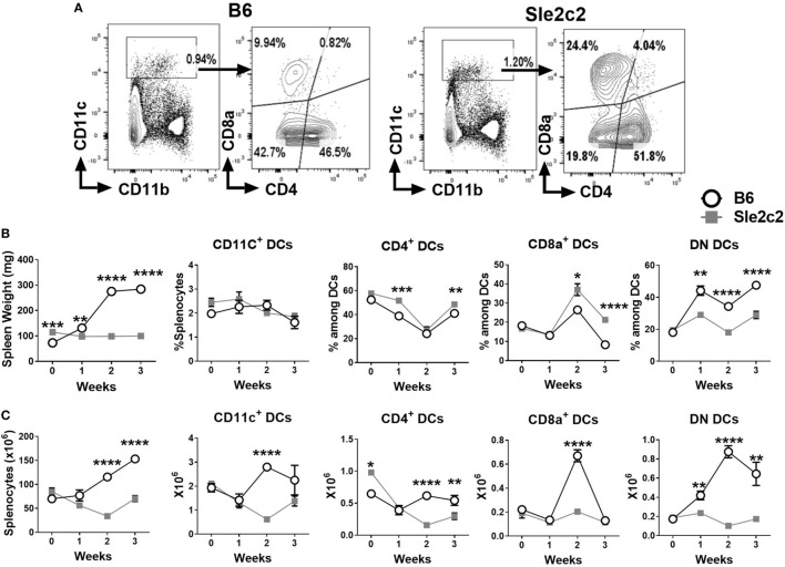
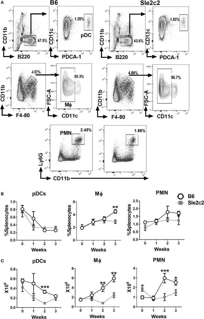
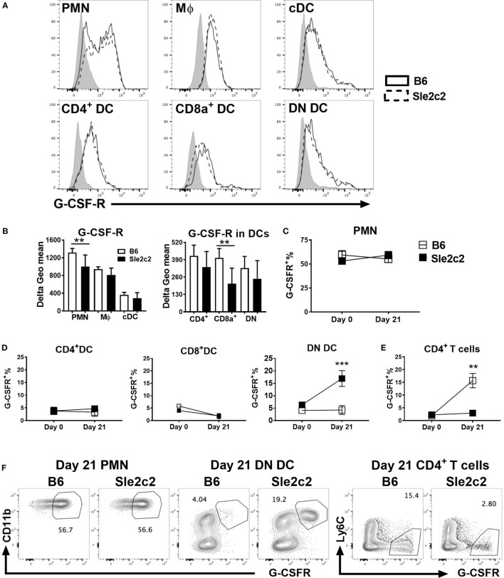
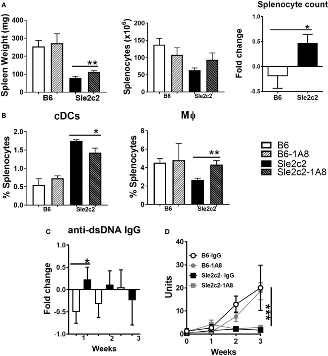
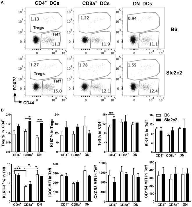
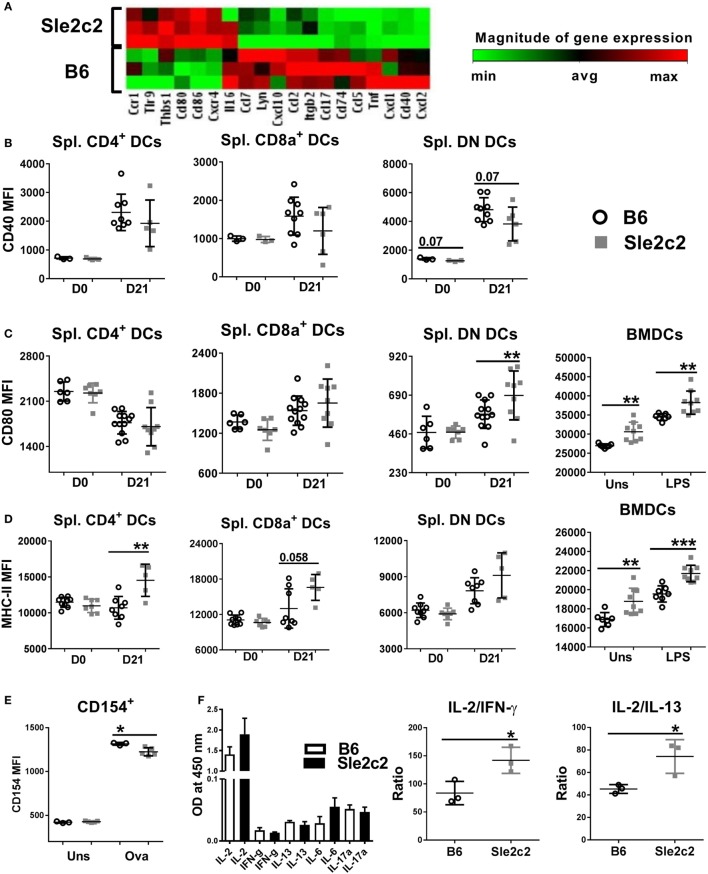
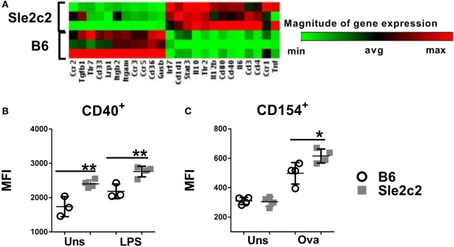
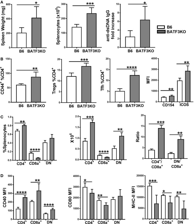
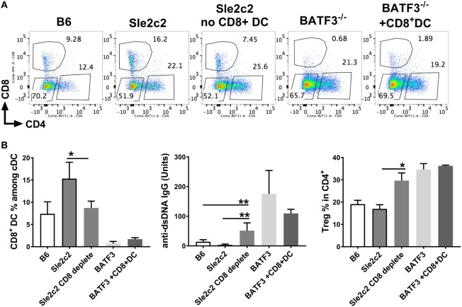
The genetic analysis of the lupus-prone NZM2410 mouse has identified a suppressor locus, Sle2c2, which confers resistance to spontaneous lupus in combination with NZM2410 susceptibility loci, or in the chronic graft-versus-host disease (cGVHD) induced model of lupus in the B6.Sle2c2 congenic strain. The candidate gene for Sle2c2, the Csf3r gene encoding the granulocyte colony-stimulating factor receptor (G-CSF-R/CD114), was validated when cGVHD was restored in B6.Sle2c2 mice after treatment with G-CSF. The goal of the project reported herein was to investigate the myeloid cells that confer resistance to cGVHD and to ascertain if the mechanism behind their suppression involves the G-CSF pathway. We showed that despite expressing the highest levels of G-CSF-R, neutrophils play only a modest role in the autoimmune activation induced by cGVHD. We also found reduced expression levels of G-CSF-R on the surface of dendritic cells (DCs) and a differential distribution of DC subsets in response to cGVHD in B6.Sle2c2 versus B6 mice. The CD8α+ DC subset, known for its tolerogenic phenotype, was expanded upon induction of cGVHD in B6.Sle2c2 mice. In addition, the deficiency of CD8α+ DC subset enhanced the severity of cGVHD in B6.Batf3-/- and B6.Sle2c2 mice, confirming their role in suppression of cGVHD. B6.Sle2c2DCs presented lowered activation and antigen presentation abilities and expressed lower levels of genes associated with DC activation and maturation. Exposure to exogenous G-CSF reversed the majority of these phenotypes, suggesting that tolerogenic DCs maintained through a defective G-CSF-R pathway mediated the resistance to cGVHD in B6.Sle2c2 mice.

Keywords: G-CSF-R; lupus; neutrophils; suppressive allele; tolerogenic dendritic cells.


Figures

Figure 1
Figure 1
Splenic dendritic cell (DC) distribution during the course of chronic graft-versus-host disease (cGVHD) in B6 and B6.Sle2c2 mice. (A) Gating strategy used to identify CD4+, CD8α+, and DN DCs from CD11c+ gated cells. (B) Spleen weight and frequency of the gated populations at the indicated time points after cGVHD induction. (C) Splenocytes numbers and absolute numbers of total gated populations (N = 5–12 per time point, *p < 0.05; **p < 0.01, ***p < 0.001, ****p < 0.0001).
Figure 2
Figure 2
Splenic plasmacytoid dendritic cell (pDC), macrophages (Mϕ), and polymorphonuclear neutrophil (PMN) distribution during the course of chronic graft-versus-host disease (cGVHD) in B6 and B6.Sle2c2 mice. (A) Gating strategy used to identify pDCs (top), Mϕ (middle), and PMN (bottom panel) from live cells. Frequency (B) and numbers (C) of the gated populations at the indicated time points after cGVHD induction (N = 5–12 per time point, *p < 0.05; **p < 0.01, ***p < 0.001).
Figure 3
Figure 3
B6.Sle2c2 mice express lower levels of G-CSF-R on polymorphonuclear neutrophils (PMNs) and dendritic cells (DCs). (A) Representative histogram overlays showing G-CSF-R expression on splenic PMNs, macrophages (Mϕ), conventional DCs (cDCs), and CD4+, CD8α+, and DN DC subsets from B6 (plain) and B6.Sle2c2 (dashed) mice at steady state with the control shown in gray. (B) G-CSF-R expression (delta geometric mean) on myeloid cell subsets. Percentage of G-CSFRhi cells among PMNs (C), CD4+, CD8α+, and DN DCs (D), as well as CD4+ T cells (E) from B6 (open square) and B6.Sle2c2 (black-squares) mice at days 0 and 21 after chronic graft-versus-host disease (cGVHD) induction. (F) Representative FACS dot plots showing G-CSF-Rhi population within splenic PMNs, DN DCs and CD4+ T cells at day 21 post-cGVHD induction (N > 3 per subset and strain, **p < 0.01, ***p < 0.001).
Figure 4
Figure 4
Polymorphonuclear neutrophil (PMN) depletion partially rescued chronic graft-versus-host disease (cGVHD) in B6.Sle2c2 mice. (A) Spleen weight and splenocyte numbers 3 weeks after cGVHD induction in B6 and B6.Sle2c2 mice with and without PMN depletion with the 1A8 antibody. The graph of the right show relative between mice treated with 1A8 and controls for each strain. (B) Frequency and numbers of conventional DCs (cDCs) and Mϕ at week 3. Fold changes were calculated as [(depleted − controls)/controls] values. *p < 0.05 indicates significant differences versus the B6 value. Serum anti-dsDNA IgG fold changes at week 1 (C) and time course analysis (D) in the four groups (N = 3 per treatment per strain) ***p < 0.001 two-way ANOVA between strains either treated or untreated with 1A8.
Figure 5
Figure 5
B6.Sle2c2 dendritic cells (DCs) are more tolerogenic in antigen-specific CD4+ T cell cocultures. chronic graft-versus-host disease-induced B6 and B6.Sle2c2 mice were immunized with NP-OVA on d 20. One-day later, splenic CD4+, CD8α+, and DN DCs were sorted from each strain. Each DC subset was cocultured individually with CD4+ OT-II cells for 72 h. (A) Representative FACS plots showing CD4+CD44+FOXP3 effector T cells (Teff) and CD4+FOXP3+ regulatory T cells (Tregs) 72 h post coculture. (B) Top row: frequencies of Tregs and Teffs and percentages of Ki-67+ proliferating cells in each compartment. Bottom row: frequencies of KLRG-1+ cells and expression of ICOS, CXCR3, CD154 in the Teff population (N = 2–5 per subset and strain, *p < 0.05, **p < 0.01, ***p < 0.001).
Figure 6
Figure 6
B6.Sle2c2 bone marrow-derived DCs (BMDCs) display a less inflammatory gene expression signature than B6 BMDCs. (A) Differential gene expression of B6 and B6.Sle2c2 BMDCs stimulated with 1 µg/ml LPS for 24 h (N = 3). CD40 (B), CD80 (C), and MHC-II (D) protein expression on unstimulated and LPS-stimulated BMDCs and the three splenic dendritic cell (DC) subsets from B6 and B6.Sle2c2 mice before and after chronic graft-versus-host disease induction. (E) CD154 expression on OT-II cells cocultured with BMDCs with or without OVA peptide for 72 h. (F) Cytokine levels in the supernatants of the cocultures, assessed by multi-analyte ELISA. The two graphs on the right show the relative ratios of IL-2 to IFNγ and IL-13 (N = 3–8, *p < 0.05; **p < 0.01).
Figure 7
Figure 7
Exogenous G-CSF enhanced B6.Sle2c2 dendritic cells functions. (A) Differential gene expression in bone marrow-derived DCs (BMDCs) differentiated from B6 and B6.Sle2c2 mice treated with G-CSF (N = 3). (B) CD40 protein expression in unstimulated and LPS-stimulated BMDCs from G-CSF-treated B6 and B6.Sle2c2 mice. (C) CD154 protein expression on OT-II T cells cocultured with BMDCs from G-CSF treted B6 and B6.Sle2c2 mice, with or without OVA peptide for 72 h (N = 4 per group for (B,C), *p < 0.05, **p < 0.01).
Figure 8
Figure 8
Reduction of CD8α+ dendritic cells (DCs) by Batf3-deficiency enhances bm12.cGVHD autoimmune phenotypes. Phenotypes were analyzed 21 days after chronic graft-versus-host disease (cGVHD) induction with bm12 splenocytes in B6 and B6.Batf3−/− mice. (A) Spleen weight, splenocyte numbers and serum anti-dsDNA IgG fold increase from day 0. (B) Frequencies of effector T cell (Teff), regulatory T cells (Treg), and Tfh cells and MFI of CD154 and ICOS on Teff cells. (C) Frequencies, absolute numbers, and ratios of the three DC subsets. (D) CD40, CD80, and MHC-II expression on splenic DC subsets (N = 6, *p < 0.05; **p < 0.01, ***p < 0.001, ****p < 0.0001).
Figure 9
Figure 9
CD8α+ dendritic cells (DCs) are sufficient to induce resistance against chronic graft-versus-host disease (cGVHD). cGVHD was induced in indicated mice for 21 days. A sub-group of B6.Sle2c2 was depleted with anti-CD8a depleting antibody (300 µg/mouse/i.p.) every 3 days and until day 17 while a sub-group of B6.Batf3−/− mice was i.v. transferred with 50–70 × 103 CD8α+ DC on days 2, 7, and 14 post-cGVHD induction. (A) Representative FACS plots of spleen DC subsets within each group. (B) Frequencies of CD8α+ DC among spleen DCs, anti-dsDNA IgG titers, and regulatory T cells (Treg) in CD4+ Tcells at day 21 post-cGVHD induction (N = 2–9 mice/group, *p < 0.05; **p < 0.01).

References

    1. Rahman A, Isenberg DA. Systemic lupus erythematosus. N Engl J Med (2008) 358:929–39. 10.1056/NEJMra071297 - DOI - PubMed
    1. Tsokos GC. Systemic lupus erythematosus. N Engl J Med (2011) 365:2110–21. 10.1056/NEJMra1100359 - DOI - PubMed
    1. Choi J, Kim ST, Craft J. The pathogenesis of systemic lupus erythematosus-an update. Curr Opin Immunol (2012) 24:651–7. 10.1016/j.coi.2012.10.004 - DOI - PMC - PubMed
    1. Liu Z, Davidson A. Taming lupus—a new understanding of pathogenesis is leading to clinical advances. Nat Med (2012) 18:871–82. 10.1038/nm.2752 - DOI - PMC - PubMed
    1. Coutant F, Miossec P. Altered dendritic cell functions in autoimmune diseases: distinct and overlapping profiles. Nat Rev Rheumatol (2016) 12:703–15. 10.1038/nrrheum.2016.147 - DOI - PubMed

Publication types

Substances

LinkOut - more resources