. 2018 May 8:9:626.
doi: 10.3389/fpls.2018.00626. eCollection 2018.

Alternaria Brassicae Induces Systemic Jasmonate Responses in Arabidopsis Which Travel to Neighboring Plants via a Piriformsopora Indica Hyphal Network and Activate Abscisic Acid Responses

Affiliations

Alternaria Brassicae Induces Systemic Jasmonate Responses in Arabidopsis Which Travel to Neighboring Plants via a Piriformsopora Indica Hyphal Network and Activate Abscisic Acid Responses

Khabat Vahabi et al. Front Plant Sci. .

Abstract

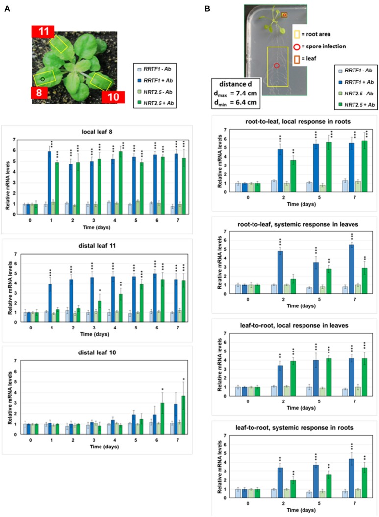
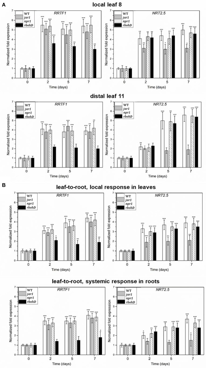
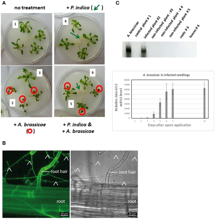
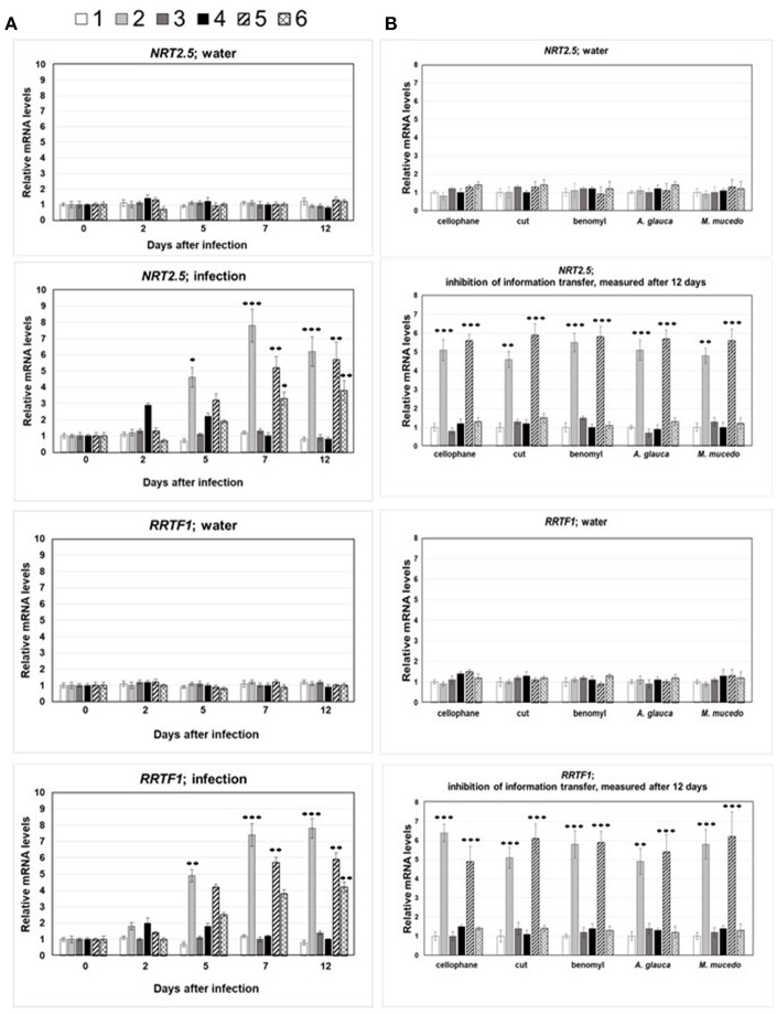
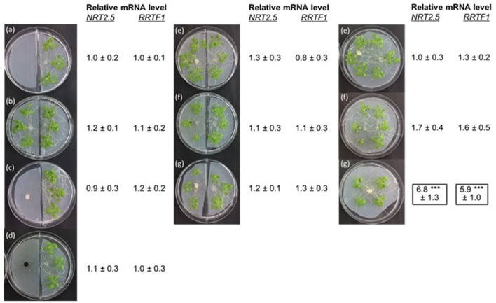
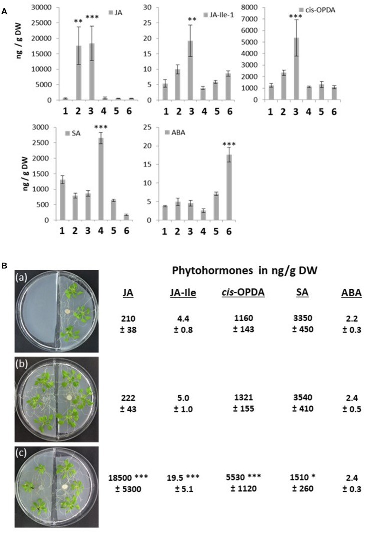
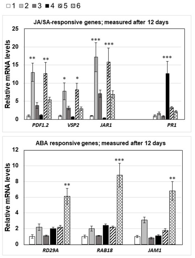
Stress information received by a particular local plant tissue is transferred to other tissues and neighboring plants, but how the information travels is not well understood. Application of Alternaria Brassicae spores to Arabidopsis leaves or roots stimulates local accumulation of jasmonic acid (JA), the expression of JA-responsive genes, as well as of NITRATE TRANSPORTER (NRT)2.5 and REDOX RESPONSIVE TRANSCRIPTION FACTOR1 (RRTF1). Infection information is systemically spread over the entire seedling and propagates radially from infected to non-infected leaves, axially from leaves to roots, and vice versa. The local and systemic NRT2.5 responses are reduced in the jar1 mutant, and the RRTF1 response in the rbohD mutant. Information about A. brassicae infection travels slowly to uninfected neighboring plants via a Piriformospora Indica hyphal network, where NRT2.5 and RRTF1 are up-regulated. The systemic A. brassicae-induced JA response in infected plants is converted to an abscisic acid (ABA) response in the neighboring plant where ABA and ABA-responsive genes are induced. We propose that the local threat information induced by A. brassicae infection is spread over the entire plant and transferred to neighboring plants via a P. indica hyphal network. The JA-specific response is converted to a general ABA-mediated stress response in the neighboring plant.

Keywords: Alternaria brassicae; NITRATE TRANSPORTER2.5; Piriformospora indica; REDOX RESPONSIVE TRANSCRIPTION FACTOR1; abscisic acid; interplant communication; jasmonic acid; systemic signaling.


Figures

Figure 1
Figure 1
Local and systemic induction of RRTF1 (blue bars) and NRT2.5 (green bars) mRNA levels by A. brassicae spore infection. (A) The picture shows the experimental set-up. The leaf numbers of 4-week-old seedlings, the A. brassicae infection area (o) and the sections used for RNA extraction (yellow squares) are shown. RRTF1 and NRT2.5 mRNA levels in the local infected leaf no. 8 and the distal leaves no. 11 and 10. At day 0, infection was performed with an A. brassicae spore suspension and the mRNA accumulation was followed in infected (darker bars) and mock (water)-treated (lighter bars) seedlings over a period of 7 days. (B) The picture shows 2-week-old Arabidopsis seedlings grown in square Petri dishes for the measurements of RRTF1 and NRT2.5 mRNA levels in local and systemic tissues. The spore infection occurred either at the leaf or at the root (red o). The local and systemic leaf areas harvested for RNA isolation are indicated (red and yellow squares). The graphs show RRTF1 (blue bars) and NRT2.5 (green bars) mRNA levels in local and systemic tissues 0, 2, 5, and 7 days after application of an A. brassicae spore suspension (darker bars), lighter bars show water controls. All RNA data are based on 6 independent experiments with 10 seedlings for each treatment. The mRNA levels for the 4 datasets at day 0 were set as 1.0 and all other values were expressed relative to them (±SEs). Asterisks indicate significant differences of the values for Alternaria-treated tissue compared to the corresponding water control at the same time point, as determined by Student's t-test (*P ≤ 0.1; **P ≤ 0.01; ***P ≤ 0.001). *** >, all higher values have P ≤ 0.001 compared to mock-treated controls.
Figure 2
Figure 2
(A) Induction of RRTF1 and NRT2.5 mRNA levels in the A. brassicae-infected leaf no. 8 and the systemic leaf no. 11 of WT, jar1, npr1, and rbohD plants. The treatment was the same as described in the legend to Figure 1A. (B) Induction of RRTF1 and NRT2.5 mRNA levels in the A. brassicae-infected leaves and the non-infected roots of WT, jar1, npr1, and rbohD seedlings on agar plates, as shown in the Figure 1B. The treatment was the same as described in the legend to Figures 1A,B. The mRNA levels at day 0 were set as 1.0 and all other values were expressed relative to them (±SEs). Asterisks indicate significant differences of the values compared to the mock-treated controls (which are not shown, but comparable to the results shown for WT material in Figure 1), as determined by Student's t-test (*P ≤ 0.1; **P ≤ 0.01; ***P ≤ 0.001).
Figure 3
Figure 3
(A) Experimental set-up for interplant communication. Four 2-week-old Arabidopsis seedlings were positioned in a fresh Petri dish. The roots were not connected (left two Petri dishes) or connected (right two Petri dishes) to each other via P. indica hyphae; P. indica was inoculated 1 week before transfer of the seedlings to the plates (cf. section Methods and Materials). The leaves of six seedlings were inoculated with an A. brassicae spore suspension (circled in red). The leaves of the seedlings 1–6 were harvested 0, 2, 5, 7, and 12 days after infection for RNA extraction and qPCR analyses. (1, no treatment; 2, A. brassicae infected material; 3, seedlings grown next to A. brassicae-infected seedlings, 4–6, as 1–3, except that seedlings were connected by a P. indica hyphal network) (B) Confocal image of an Arabidopsis root grown on the P. indica hyphal lawn for 12 days (end of experiment). The signal detected with the GFP channel is shown on the left and a bright field image on the right; root hairs are indicated by the label and fungal hyphae by arrow heads. (C) Quantification of A. brassicae AbreATr1 mRNA by qPCR. The gel shows amplified cDNA fragments from mRNAs of the seedlings #1–6 after 12 days of co-cultivation which are shown in panel A. A. brassicae: PCR product from RNA of an A. brassicae culture was used as positive control. The graph shows relative AbreATr1 mRNA levels of the infected seedlings #2 between 0 and 12 days after spore application. Based on 6 independent experiments with 10 seedlings each. Error bars are SEs.
Figure 4
Figure 4
(A) NRT2.5 and RRTF1 mRNA levels in infected and systemic leaves following A. brassicae spore application at day 0. The seedlings were either mock-treated (water) or inoculated with A. brassicae spores (infection). The numbers 1–6 refer to the seedlings shown in Figure 3A: 1, no treatment; 2, A. brassicae infected material; 3, seedlings grown next to A. brassicae-infected seedlings, 4–6, as 1–3, except that seedlings were connected by a P. indica hyphal network. The mRNA levels at the time point of infection (t = 0) was set as 1.0 and all other values are expressed relative to them. (B) NRT2.5 and RRTF1 mRNA levels in leaves of neighboring non-infected plants requires P. indica hyphal connection. Same experiment as in (A), but the connection between the roots via P. indica hyphae were interrupted by the insertion of a cellophane membrane (cellophane), or the hyphal connections were cut with a razor blade every 2nd day (cut), or P. indica were treated with benomyl at day 0, 2, 5, and 7 (benomyl). A. glauca, M. mucedo; P. indica was replaced by these fungi. All measurements were performed 12 dai and are based on 6 independent experiments with 10 plants each. Asterisks indicate significant differences of the values for A. brassicae-treated tissue compared to the corresponding water control at the same time point, as determined by Student's t-test (*P ≤ 0.1; **P ≤ 0.01; *** P ≤ 0.001). The data for the water control did not change significantly within the 12 days and were below 1.5 ± 0.4.
Figure 5
Figure 5
NRT2.5 and RRTF1 mRNA levels in Arabidopsis seedlings grown in the right chamber of a split Petri dish. The other chamber contained either PNM medium alone (a), or PNM medium with Arabidopsis seedlings (b), P. indica (c), or A. brassicae (d) hyphae or Arabidopsis seedlings co-cultivated with the fungi, as described in the legends to the Figures 2, 3 (e, P. indica alone, f, Arabidopsis infected with A. brassicae spores, g, Arabidopsis seedlings exposed to both fungi). The organisms grown in the three Petri dishes on the right site were not separated from each other. RNA extraction was performed 12 days. The mRNA levels of Arabidopsis seedlings with only PNM medium in the neighboring chamber was set as 1.0 and all other values are expressed relative to them. Based on 6 independent experiments with 10 seedlings for each treatment.
Figure 6
Figure 6
(A) Phytohormone concentrations in ng/g dry weight (DW) in the 6 seedlings (number 1–6) shown in Figure 3A. 1, no treatment; 2, A. brassicae infected material; 3, seedlings grown next to A. brassicae-infected seedlings, 4–6, as 1–3, except that seedlings were connected by a P. indica hyphal network. The experimental set-up is shown in Figure 3A. Based on 5 independent experiments, errors represent SEs. Asterisks indicate significant differences to the values for the untreated seedling number 1, by Student's t-test (**P ≤ 0,01; ***P ≤ 0.001). (B) Split Petri dish experiment with Arabidopsis seedlings. Left chamber: (a) empty, (b) Arabidopsis seedlings, (c) Arabidopsis seedlings infected with A. brassicae spores for 12 days. The hormone levels were determined for the seedlings grown on the right site in the Petri dish, which were grown in the presence of P. indica for 12 days. Based on 5 independent experiments, errors represent SEs. Asterisks indicate significant differences to the values of experiment (a), by Student's t-test (*P ≤ 0.1, ***P ≤ 0.001).
Figure 7
Figure 7
mRNA levels of JA-, SA-, and ABA-responsive genes in the 6 seedlings shown in Figure 3A, 12 days after spore infection: 1, no treatment; 2, A. brassicae infected material; 3, seedlings grown next to A. brassicae-infected seedlings, 4–6, as 1–3, except that seedlings were connected by a P. indica hyphal network. The experimental conditions were the same as described in the legend to Figure 4. Asterisks indicate significant differences of the values for Alternaria-treated tissue compared to the corresponding control (1, no treatment) at the same time point, as determined by Student's t-test (*P ≤ 0.1; **P ≤ 0.01; ***P ≤ 0.001).

Similar articles

Cited by

References

    1. Agrawal A. A. (2000). Communication between plants: this time it's real. Trends Ecol. Evol. 15:446. 10.1016/S0169-5347(00)01987-X - DOI - PubMed
    1. Akum F. N., Steinbrenner J., Biedenkopf D., Imani J., Kogel K.-H. (2015). The Piriformospora indica effector PIIN_08944 promotes the mutualistic Sebacinalean symbiosis. Front. Plant Sci. 6:906. 10.3389/fpls.2015.00906 - DOI - PMC - PubMed
    1. Arimura G., Ozawa R., Shimoda T., Nishioka T., Boland W., Takabayashi J. (2000). Herbivory-induced volatiles elicit defence genes in lima bean leaves. Nature 406, 512–515. 10.1038/35020072 - DOI - PubMed
    1. Baldwin I. T., Halitschke R., Paschold A., von Dahl C. C., Preston C. A. (2006). Volatile signaling in plant-plant interactions: “talking trees” in the genomics era. Science 311, 812–815. 10.1126/science.1118446 - DOI - PubMed
    1. Baldwin I. T., Kessler A., Halitschke R. (2002). Volatile signaling in plant-plant-herbivore interactions: what is real? Curr. Opin. Plant Biol. 5, 351–354. 10.1016/S1369-5266(02)00263-7 - DOI - PubMed

LinkOut - more resources