. 2018 May 15:9:159.
doi: 10.3389/fgene.2018.00159. eCollection 2018.

Heterosis Is a Systemic Property Emerging From Non-linear Genotype-Phenotype Relationships: Evidence From in Vitro Genetics and Computer Simulations

Affiliations

Heterosis Is a Systemic Property Emerging From Non-linear Genotype-Phenotype Relationships: Evidence From in Vitro Genetics and Computer Simulations

Julie B Fiévet et al. Front Genet. .

Abstract

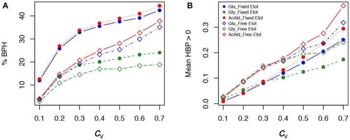
Heterosis, the superiority of hybrids over their parents for quantitative traits, represents a crucial issue in plant and animal breeding as well as evolutionary biology. Heterosis has given rise to countless genetic, genomic and molecular studies, but has rarely been investigated from the point of view of systems biology. We hypothesized that heterosis is an emergent property of living systems resulting from frequent concave relationships between genotypic variables and phenotypes, or between different phenotypic levels. We chose the enzyme-flux relationship as a model of the concave genotype-phenotype (GP) relationship, and showed that heterosis can be easily created in the laboratory. First, we reconstituted in vitro the upper part of glycolysis. We simulated genetic variability of enzyme activity by varying enzyme concentrations in test tubes. Mixing the content of "parental" tubes resulted in "hybrids," whose fluxes were compared to the parental fluxes. Frequent heterotic fluxes were observed, under conditions that were determined analytically and confirmed by computer simulation. Second, to test this model in a more realistic situation, we modeled the glycolysis/fermentation network in yeast by considering one input flux, glucose, and two output fluxes, glycerol and acetaldehyde. We simulated genetic variability by randomly drawing parental enzyme concentrations under various conditions, and computed the parental and hybrid fluxes using a system of differential equations. Again we found that a majority of hybrids exhibited positive heterosis for metabolic fluxes. Cases of negative heterosis were due to local convexity between certain enzyme concentrations and fluxes. In both approaches, heterosis was maximized when the parents were phenotypically close and when the distributions of parental enzyme concentrations were contrasted and constrained. These conclusions are not restricted to metabolic systems: they only depend on the concavity of the GP relationship, which is commonly observed at various levels of the phenotypic hierarchy, and could account for the pervasiveness of heterosis.

Keywords: enzyme variability; genotype-phenotype map; heterosis; mathematical modeling; metabolic network; non-linear processes.


Conflict of interest statement

The authors declare that the research was conducted in the absence of any commercial or financial relationships that could be construed as a potential conflict of interest.

Figures

Figure 1
Figure 1
The two most common types of GP relationships with possible inheritance. (A) The S-shaped relationship. There is convexity below the abscissa of the inflection point (red point) and concavity above. The three phenotypes P1, F1, and P2 are associated with the three genotypes A1A1, A1A2, and A2A2, respectively. MP: mid-parental value. In the convex region of the curve (mauve background), the low allele is dominant over the high allele (negative dominance), while in the concave region of the curve (yellow background) the high allele is dominant over the low allele (positive dominance). (B) The hyperbolic concave relationship. Whatever the genotypic values, there is positive dominance. In all cases there is additive inheritance of the genotypic parameters.
Figure 2
Figure 2
The upstream part of glycolysis reconstructed in vitro. HK, hexokinase (E.C. 2.7.1.1); PGI, phosphoglucose isomerase (E.C. 5.3.1.9); PFK, phosphofructokinase (E.C. 2.7.1.11); FBA, fructose-1,6-bisphosphate aldolase (E.C. 4.1.2.13); TPI, triosephosphate isomerase (E.C. 5.3.1.1); G3PDH, glycerol 3-phosphate dehydrogenase (E.C. 1.1.1.8); CK, creatine phosphokinase (E.C. 2.7.3.2). Variable enzymes are in red. Reaction rate was measured from the decrease in NADH concentration.
Figure 3
Figure 3
The simplified yeast glycolysis/fermentation network. Variable enzymes are in orange. Blue arrows point to the input flux, glucose, and to the output fluxes, glycerol and acetaldehyde.
Figure 4
Figure 4
Flux response upon variation of two enzymes in a hyperbolic GP relationship. The two enzymes have the same arbitrary values of their kinetic parameters. Red and blue curves show two types of crosses between parents P1 and P2, or P3 and P4, respectively (from Fiévet et al., 2010). The positions of hybrids F1 (light blue and orange points) were determined assuming additivity of enzyme concentrations. (A) In the “red” cross, where parent P2 has a flux close to the maximum (high concentration of both enzymes), there is positive mid-parent heterosis (+MPH) for the flux. In the “blue” cross, where parents have low flux values due to low concentrations of enzyme 1 (parent P3) or enzyme 2 (parent P4), the hybrid displays best-parent heterosis (BPH). (B) If there is a constraint on the total enzyme amount, the “red” cross also displays BPH because the concavity of the surface is increased.
Figure 5
Figure 5
In vitro heterosis. Hybrids were created by mixing 1:1 the content of parental tubes. #cross refers to the cross numbers of Table S1. (A) Parental flux values (blue and orange points) relative to the hybrid flux value used as a reference (vertical red line) for the 61 crosses (in μM.s−1). Gray points indicate the mid-parental values. Crosses were ranked by decreasing HBP values, the index of best parent heterosis. (B) HBP values. (C) Inheritance: BPH, Best-Parent Heterosis; +MPH, positive Mid-Parent Heterosis; ADD, additivity; –MPH, negative Mid-Parent Heterosis. (D,E) Parental (blue and orange bars) and hybrid (gray bars) flux values, alongside the corresponding parental enzyme concentrations for six crosses, identified by broken lines, displaying different types of inheritance. From top to bottom: the two crosses with the highest significant BPH values, the two crosses that are the closest to additivity and the two crosses with the smallest significant –MPH values. Red points correspond to the optimal enzyme concentrations.
Figure 6
Figure 6
In vitro heterosis predictors. (A) Relationship between HBP and Denz, the Euclidean distance between parents computed from enzyme concentrations (r = 0.15, p < 0.25). (B) Relationship between HBP and Dflux, the flux difference between parents (r = −0.33, p < 0.01). (C) Relationship between HBP and Hreg, the heterosis value computed from the equation of the multiple linear regression performed with Denz and Dflux as predictor variables (r = 0.46, p < 0.001). (D) Relationship between HBP and HBPmod, the expected heterosis value computed from the model (see text) (r = 0.77, p < 2.5.10−13). Red points correspond to positive HBP, i.e., to BPH.
Figure 7
Figure 7
Effect of the coefficient of variation (cv) of enzyme concentrations on heterosis. (A) Percentage of BPH; (B) Mean of the positive HBP values (HBP+¯) over a range of cv's of parental enzyme concentrations, from 0.1 to 1.2. –/–: equidistributed mean enzyme concentrations and free Etot, –/+: equidistributed and fixed Etot, +/–: mean enzyme concentrations centered on their optimum and free Etot, +/+: optimum centered means and fixed Etot.
Figure 8
Figure 8
Relationship between parental fluxes J1 and J2 and hybrid fluxes under four simulation conditions. Each point corresponds to a hybrid, with warm colors when there is BPH (HBP > 0). +/+, +/–, –/+, and –/– have the same meaning as in Figure 7. Note that when Etot is fixed, the flux value is limited. (cv = 0.6).
Figure 9
Figure 9
Heterosis predictors. Relationship between HBP and Dflux (A), and between HBP and Denz (B) when cv = 0.6. Red points: positive HBP values. (C) Square grid of percentages of BPH as a function of Denz (x-axis) and Dflux (y-axis). Colors range from blue (minimum % BPH) to yellow (maximum % BPH). Empty squares are white. (D) Same as (C) with HBP+¯ values. The additional white squares correspond to cases where there was no BPH (0 in C). –/–, –/+, +/–, and +/+ have the same meaning as in Figure 7.
Figure 10
Figure 10
Heterosis for the three fluxes in the glycolytic/fermentation network. Percentages of different types of inheritance over the 0.1–0.7 cv range of parental enzyme concentrations, for free and fixed Etot.
Figure 11
Figure 11
Effect of the coefficient of variation (cv) of enzyme concentrations on heterosis in the glycolysis/fermentation network. Percentage of BPH (A) and HBP+¯ (B) over the cv range of parental enzyme concentrations.
Figure 12
Figure 12
Heterosis predictors in the glycolysis/fermentation network: scatter plots. Relationships between HBP and Dflux (A,C) and between HBP and Denz (B,D) with free Etot (A,B) and fixed Etot (C,D) for the glucose flux (A1–D1), glycerol flux (A2–D2) and acetaldehyde flux (A3—D3) (cv = 0.4).
Figure 13
Figure 13
Heterosis predictors in the glycolysis/fermentation network: square grids. (A,B) Square grid with % BPH (A) and HBP+¯ (B) as a function of Denz (x-axis) and Dflux (y-axis) for glucose (1st row), glycerol (2nd row) and acetaldehyde (3rd row) when Etot is free. (C,D) Same as (A,B) with fixed Etot.
Figure 14
Figure 14
The geometry of heterosis when the enzymatic distance between parents is constant. (A) Parental enzyme concentrations are on a circle of diameter Denz = 76 centered on the hybrid point, the coordinates of which are x = y = 40. Four pairs of parents are highlighted by yellow, blue, red and green diameters. The closed curve on the surface shows the flux values corresponding to the circle of concentrations. The equation of the flux surface is J=1/(1E1+1E2+0.2), in arbitrary units. (B) Same as (A), except that Denz = 50 and hybrid coordinates are x = 30 and y = 70. (C) Variation of parental fluxes J1 and J2 (dotted and long-dashed gray curves), of Dflux (blue curve) and of HBP (orange curve) over the rotation of a segment joining the parents from α = 0 (parallel to the x-axis, yellow case) to α = π (reversed positions of the parents). The two-dashed horizontal gray line corresponds to the constant hybrid flux value. The yellow, blue, red, and green vertical lines correspond to the pairs of parents of the 3D vignette in (A), with barplots showing corresponding heterosis. (D) Same representation as in (C), for the case shown in (B). (E,F): Same as in (C,D), with HMP instead of HBP.

References

    1. Agrawal A. F., Whitlock M. C. (2011). Inferences about the distribution of dominance drawn from yeast gene knockout data. Genetics 187, 553–U274. 10.1534/genetics.110.124560 - DOI - PMC - PubMed
    1. Albertin W., Marullo P., Bely M., Aigle M., Bourgais A., Langella O., et al. . (2013). Linking post-translational modifications and variation of phenotypic traits. Mol. Cell. Proteom. 12, 720–735. 10.1074/mcp.M112.024349 - DOI - PMC - PubMed
    1. Allison A. (1954). Protection afforded by sickle-cell trait against subtertian malarial infection. Br. Med. J. 1, 290–294. 10.1136/bmj.1.4857.290 - DOI - PMC - PubMed
    1. Barkai N., Leibler S. (1997). Robustness in simple biochemical networks. Nature 387, 913–917. 10.1038/43199 - DOI - PubMed
    1. Barth S., Busimi A. K., Utz H. F., Melchinger A. E. (2003). Heterosis for biomass yield and related traits in five hybrids of Arabidopsis thaliana L. Heynh. Heredity 91, 36–42. 10.1038/sj.hdy.6800276 - DOI - PubMed