

Improved prediction of outcome in Parkinson's disease using radiomics analysis of longitudinal DAT SPECT images
- PMID: 29868437
- PMCID: PMC5984570
- DOI: 10.1016/j.nicl.2017.08.021
Improved prediction of outcome in Parkinson's disease using radiomics analysis of longitudinal DAT SPECT images
Abstract
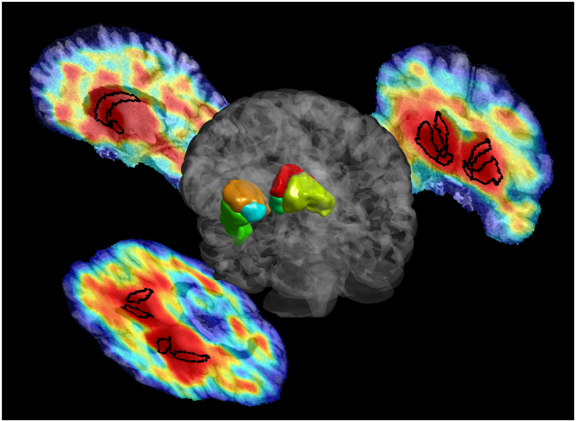
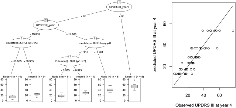
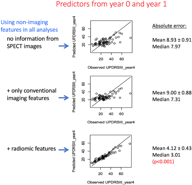
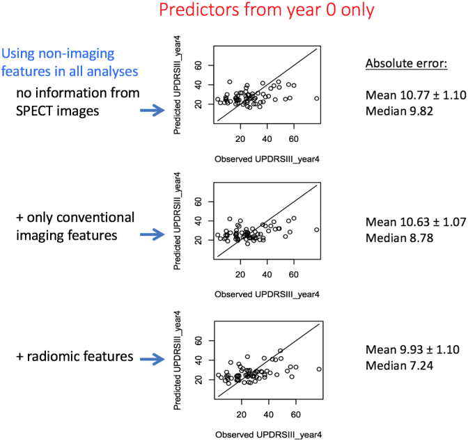
No disease modifying therapies for Parkinson's disease (PD) have been found effective to date. To properly power clinical trials for discovery of such therapies, the ability to predict outcome in PD is critical, and there is a significant need for discovery of prognostic biomarkers of PD. Dopamine transporter (DAT) SPECT imaging is widely used for diagnostic purposes in PD. In the present work, we aimed to evaluate whether longitudinal DAT SPECT imaging can significantly improve prediction of outcome in PD patients. In particular, we investigated whether radiomics analysis of DAT SPECT images, in addition to use of conventional non-imaging and imaging measures, could be used to predict motor severity at year 4 in PD subjects. We selected 64 PD subjects (38 male, 26 female; age at baseline (year 0): 61.9 ± 7.3, range [46,78]) from the Parkinson's Progressive Marker Initiative (PPMI) database. Inclusion criteria included (i) having had at least 2 SPECT scans at years 0 and 1 acquired on a similar scanner, (ii) having undergone a high-resolution 3 T MRI scan, and (iii) having motor assessment (MDS-UPDRS-III) available in year 4 used as outcome measure. Image analysis included automatic region-of-interest (ROI) extraction on MRI images, registration of SPECT images onto the corresponding MRI images, and extraction of radiomic features. Non-imaging predictors included demographics, disease duration as well as motor and non-motor clinical measures in years 0 and 1. The image predictors included 92 radiomic features extracted from the caudate, putamen, and ventral striatum of DAT SPECT images at years 0 and 1 to quantify heterogeneity and texture in uptake. Random forest (RF) analysis with 5000 trees was used to combine both non-imaging and imaging variables to predict motor outcome (UPDRS-III: 27.3 ± 14.7, range [3,77]). The RF prediction was evaluated using leave-one-out cross-validation. Our results demonstrated that addition of radiomic features to conventional measures significantly improved (p < 0.001) prediction of outcome, reducing the absolute error of predicting MDS-UPDRS-III from 9.00 ± 0.88 to 4.12 ± 0.43. This shows that radiomics analysis of DAT SPECT images has a significant potential towards development of effective prognostic biomarkers in PD.
Keywords: DAT SPECT; Longitudinal; Outcome prediction; Parkinson's disease; Radiomics; Textural features.
Figures
References
-
- Aerts H.J.W.L., Velazquez E.R., Leijenaar R.T.H., Parmar C., Grossmann P., Carvalho S., Bussink J., Monshouwer R., Haibe-Kains B., Rietveld D., Hoebers F., Rietbergen M.M., Leemans C.R., Dekker A., Quackenbush J., Gillies R.J., Lambin P. Decoding tumour phenotype by noninvasive imaging using a quantitative radiomics approach. Nat. Commun. 2014;5:4006. - PMC - PubMed
-
- Ashrafinia S., DiGianvittorio M., Rowe S., Gorin M., Lu L., Lodge M., Pomper M., Allaf M., Rahmim A. Reproducibility and reliability of radiomic features in 18F-DCFPyL PET/CT imaging of prostate cancer. J. Nucl. Med. 2017;58:503.
-
- Asselin M.C., O'Connor J.P.B., Boellaard R., Thacker N.A., Jackson A. Quantifying heterogeneity in human tumours using MRI and PET. Eur. J. Cancer. 2012;48:447–455. - PubMed
-
- Badiavas K., Molyvda E., Iakovou I., Tsolaki M., Psarrakos K., Karatzas N. SPECT imaging evaluation in movement disorders: far beyond visual assessment. Eur. J. Nucl. Med. Mol. Imaging. 2011;38:764–773. - PubMed
Publication types
MeSH terms
Substances
LinkOut - more resources
Full Text Sources
Other Literature Sources
Medical
Research Materials
Miscellaneous
