

Fixel-based analysis reveals alterations is brain microstructure and macrostructure of preterm-born infants at term equivalent age
- PMID: 29868441
- PMCID: PMC5984576
- DOI: 10.1016/j.nicl.2018.01.003
Fixel-based analysis reveals alterations is brain microstructure and macrostructure of preterm-born infants at term equivalent age
Abstract
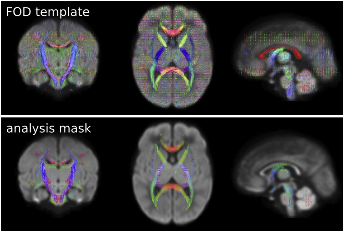
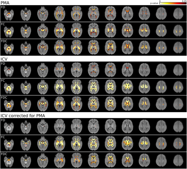
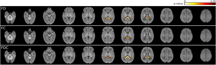
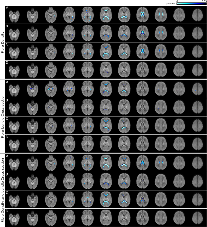
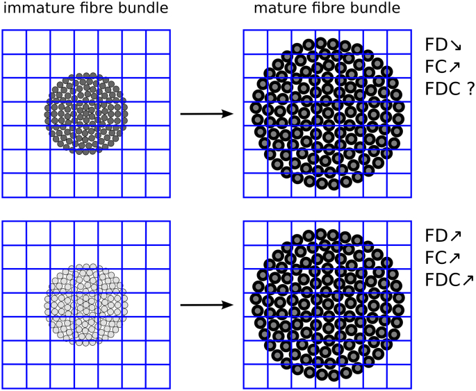
Preterm birth causes significant disruption in ongoing brain development, frequently resulting in adverse neurodevelopmental outcomes. Brain imaging using diffusion MRI may provide valuable insight into microstructural properties of the developing brain. The aim of this study was to establish whether the recently introduced fixel-based analysis method, with its associated measures of fibre density (FD), fibre bundle cross-section (FC), and fibre density and bundle cross-section (FDC), is suitable for the investigation of the preterm infant brain at term equivalent age. High-angular resolution diffusion weighted images (HARDI) of 55 preterm-born infants and 20 term-born infants, scanned around term-equivalent age, were included in this study (3 T, 64 directions, b = 2000 s/mm2). Postmenstrual age at the time of MRI, and intracranial volume (FC and FDC only), were identified as confounding variables. Gestational age at birth was correlated with all fixel measures in the splenium of the corpus callosum. Compared to term-born infants, preterm infants showed reduced FD, FC, and FDC in a number of regions, including the corpus callosum, anterior commissure, cortico-spinal tract, optic radiations, and cingulum. Preterm infants with minimal macroscopic brain abnormality showed more extensive reductions than preterm infants without any macroscopic brain abnormality; however, little differences were observed between preterm infants with no and with minimal brain abnormality. FC showed significant reductions in preterm versus term infants outside regions identified with FD and FDC, highlighting the complementary role of these measures. Fixel-based analysis identified both microstructural and macrostructural abnormalities in preterm born infants, providing a more complete picture of early brain development than previous diffusion tensor imaging (DTI) based approaches.
Keywords: Diffusion; Fixel-based analysis; Neonate; Prematurity.
Figures
Similar articles
-
Brain microstructure and morphology of very preterm-born infants at term equivalent age: Associations with motor and cognitive outcomes at 1 and 2 years.Neuroimage. 2020 Nov 1;221:117163. doi: 10.1016/j.neuroimage.2020.117163. Epub 2020 Jul 11. Neuroimage. 2020. PMID: 32663645
-
Fixel-based analysis of the preterm brain: Disentangling bundle-specific white matter microstructural and macrostructural changes in relation to clinical risk factors.Neuroimage Clin. 2019;23:101820. doi: 10.1016/j.nicl.2019.101820. Epub 2019 Apr 10. Neuroimage Clin. 2019. PMID: 30991305 Free PMC article.
-
Long-term development of white matter fibre density and morphology up to 13 years after preterm birth: A fixel-based analysis.Neuroimage. 2020 Oct 15;220:117068. doi: 10.1016/j.neuroimage.2020.117068. Epub 2020 Jun 22. Neuroimage. 2020. PMID: 32585342
-
Diffusion Tensor Imaging in Very Preterm, Moderate-Late Preterm and Term-Born Neonates: A Systematic Review.J Pediatr. 2021 May;232:48-58.e3. doi: 10.1016/j.jpeds.2021.01.008. Epub 2021 Jan 13. J Pediatr. 2021. PMID: 33453200
-
Fixel-based Analysis of Diffusion MRI: Methods, Applications, Challenges and Opportunities.Neuroimage. 2021 Nov 1;241:118417. doi: 10.1016/j.neuroimage.2021.118417. Epub 2021 Jul 21. Neuroimage. 2021. PMID: 34298083 Review.
Cited by
-
Brain tissue microstructure in a prospective, longitudinal, population-based cohort of preterm and term-born young adults.J Child Psychol Psychiatry. 2025 May;66(5):635-649. doi: 10.1111/jcpp.14069. Epub 2024 Nov 19. J Child Psychol Psychiatry. 2025. PMID: 39561978 Free PMC article.
-
Brain structural connectome in neonates with prenatal opioid exposure.Front Neurosci. 2022 Sep 16;16:952322. doi: 10.3389/fnins.2022.952322. eCollection 2022. Front Neurosci. 2022. PMID: 36188457 Free PMC article.
-
Reduced apparent fiber density in the white matter of premature-born adults.Sci Rep. 2020 Oct 14;10(1):17214. doi: 10.1038/s41598-020-73717-6. Sci Rep. 2020. PMID: 33057208 Free PMC article.
-
A framework for multi-component analysis of diffusion MRI data over the neonatal period.Neuroimage. 2019 Feb 1;186:321-337. doi: 10.1016/j.neuroimage.2018.10.060. Epub 2018 Nov 2. Neuroimage. 2019. PMID: 30391562 Free PMC article.
-
Applications of advanced diffusion MRI in early brain development: a comprehensive review.Brain Struct Funct. 2023 Mar;228(2):367-392. doi: 10.1007/s00429-022-02605-8. Epub 2022 Dec 31. Brain Struct Funct. 2023. PMID: 36585970 Free PMC article. Review.
References
-
- Akazawa K., Chang L., Yamakawa R., Hayama S., Buchthal S., Alicata D., Andres T., Castillo D., Oishi K., Skranes J., Ernst T., Oishi K. Probabilistic maps of the white matter tracts with known associated functions on the neonatal brain atlas: application to evaluate longitudinal developmental trajectories in term-born and preterm-born infants. NeuroImage. 2015;128:167–179. - PMC - PubMed
-
- Allen M.C., Cristofalo E.A., Kim C. Outcomes of preterm infants: morbidity replaces mortality. Clin. Perinatol. 2011;38(3):441–454. - PubMed
-
- Cohen Y., Assaf Y. Diffusion MRI: Theory, Methods, and Applications. 2010. Extracting geometric properties of white matter with q-space diffusion MRI; pp. 125–151.
-
- Dubois J., Dehaene-Lambertz G., Perrin M., Mangin J.F., Cointepas Y., Duchesnay E., Le Bihan D., Hertz-Pannier L. Asynchrony of the early maturation of white matter bundles in healthy infants: quantitative landmarks revealed noninvasively by diffusion tensor imaging. Hum. Brain Mapp. 2008;29(1):14–27. - PMC - PubMed
Publication types
MeSH terms
LinkOut - more resources
Full Text Sources
Other Literature Sources
