. 2018 May 8:8:130.
doi: 10.3389/fcimb.2018.00130. eCollection 2018.

FcpB Is a Surface Filament Protein of the Endoflagellum Required for the Motility of the Spirochete Leptospira

Affiliations

FcpB Is a Surface Filament Protein of the Endoflagellum Required for the Motility of the Spirochete Leptospira

Elsio A Wunder Jr et al. Front Cell Infect Microbiol. .

Abstract

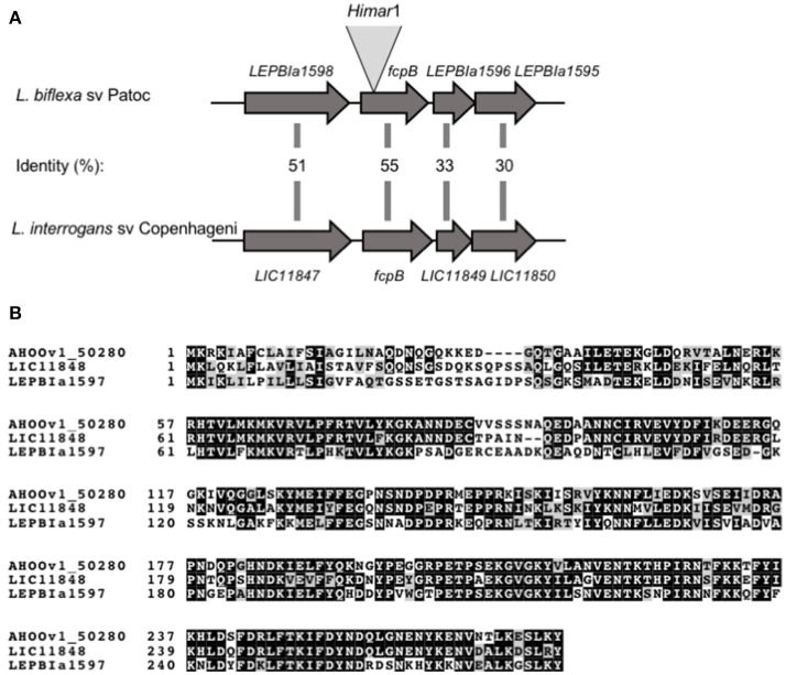
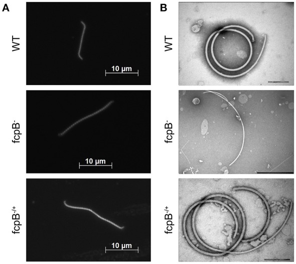
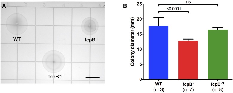
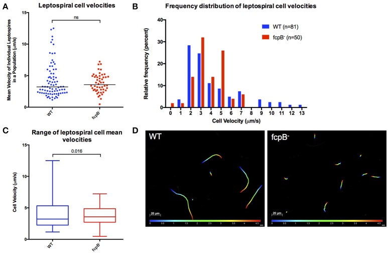
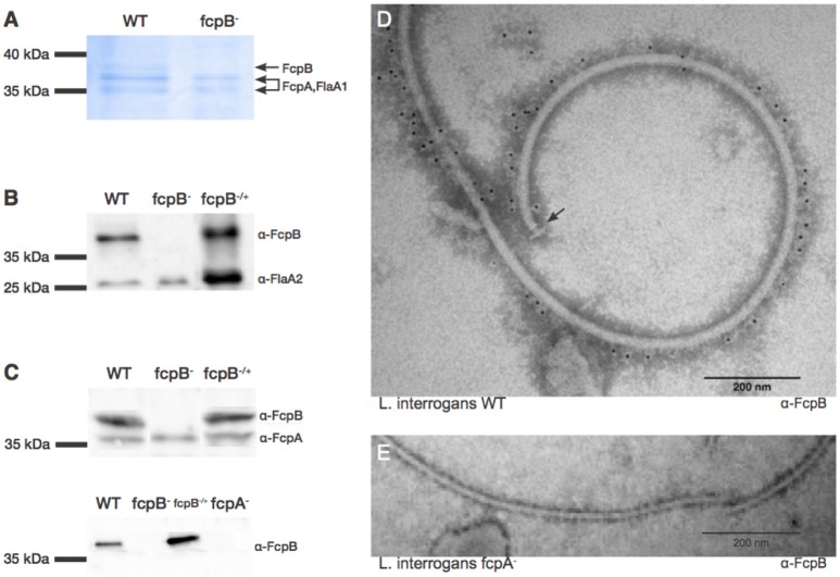
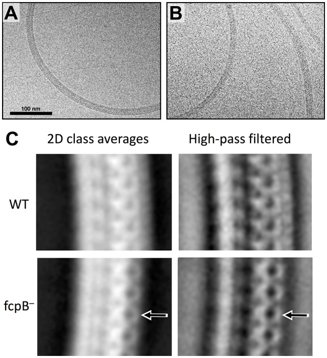
The spirochete endoflagellum is a unique motility apparatus among bacteria. Despite its critical importance for pathogenesis, the full composition of the flagellum remains to be determined. We have recently reported that FcpA is a novel flagellar protein and a major component of the sheath of the filament of the spirochete Leptospira. By screening a library of random transposon mutants in the spirochete Leptospira biflexa, we found a motility-deficient mutant harboring a disruption in a hypothetical gene of unknown function. Here, we show that this gene encodes a surface component of the endoflagellar filament and is required for typical hook- and spiral-shaped ends of the cell body, coiled structure of the endoflagella, and high velocity phenotype. We therefore named the gene fcpB for flagellar-coiling protein B. fcpB is conserved in all members of the Leptospira genus, but not present in other organisms including other spirochetes. Complementation of the fcpB- mutant restored the wild-type morphology and motility phenotypes. Immunoblotting with anti-FcpA and anti-FcpB antisera and cryo-electron microscopy of the filament indicated that FcpB assembled onto the surface of the sheath of the filament and mostly located on the outer (convex) side of the coiled filament. We provide evidence that FcpB, together with FcpA, are Leptospira-specific novel components of the sheath of the filament, key determinants of the coiled and asymmetric structure of the endoflagella and are essential for high velocity. Defining the components of the endoflagella and their functions in these atypical bacteria should greatly enhance our understanding of the mechanisms by which these bacteria produce motility.

Keywords: Leptospira; flagella; flagellar system; motility; spirochetes.


Figures

Figure 1
Figure 1
Schematic representation of fcpB in Leptospira spp. (A) Genetic locus of fcpB in L. biflexa (LEPBIa1597) and L. interrogans (LIC11848) with the corresponding amino acid identities between the two species. The insertion site of Himar1 in the chromosome of the fcpB mutant is indicated. (B) Alignment of FcpB protein sequences from the saprophyte species L. biflexa (LEPBIa1597), the intermediate L. licerasiae (AH00v1_50280), and the pathogenic L. interrogans (LIC11848). Identical and similar residues are shaded in black and gray, respectively.
Figure 2
Figure 2
Determination of phenotypes in cell morphology and purified flagella caused by deletion of fcpB. (A) The L. biflexa fcpB mutant is deficient in its ability to form hook- and spiral-shaped ends, which are characteristic of wild-type cells. Cells were examined by dark-field microscopy with a 100X oil-immersion objective. (B) Periplasmic filaments purified from the L. biflexa fcpB mutant lack the super-coiled morphology, which is characteristic of purified wild-type flagellar filaments when examined in vitro. Complementation of fcpB mutant restores wild-type morphology and coiled flagellar filament structure. Bar scales for negative staining images are 200 nm for WT and fcpB−/+ complemented strain, and 500 nm for fcpB mutant.
Figure 3
Figure 3
Motility phenotype of the fcpB mutant in semisolid media. Soft agar plates were inoculated with 105 leptospires of the wild-type, fcpB mutant, and fcpB−/+ complemented strains. (A) The diameter of the zone of spread for each colony was measured using image analysis software. Bar scale, 10 mm. (B) The average colony diameter measured for the fcpB mutant was significantly smaller (p < 0.0001) than for the wild-type. The difference in average colony diameter between the fcpB−/+ complemented and wild-type strains was not significant.
Figure 4
Figure 4
Analysis of velocity of the fcpB mutant cells in liquid viscous media. Dark-field video microscopy and tracking analysis were used to measure the average velocities of individual cells in viscous liquid media containing 1% methyl-cellulose. (A) Scatter plot of the velocities of individual cells shows that the difference in median velocity (horizontal bar) between the wild-type and mutant populations is not statistically significant. (B) Histogram showing the relative frequency of the velocities of individual cells. Note that depletion of FcpB affects the proportion of highly motile leptospires in a population. (C) The range of leptospiral cell velocities in a population is reduced in the fcpB mutant, with a lower maximum velocity, when compared to the wild-type strain. (D) Representative trajectories of wild-type and fcpB mutant over 5 s, illustrating that the fcpB mutant population is deficient in highly-motile leptospires.
Figure 5
Figure 5
Characterization and localization of FcpB in Leptospira cells. (A) Coomassie Blue-stained SDS-PAGE gel of purified periplasmic flagella. FcpA, FcpB, and FlaA1 are indicated, according to MS-MS identification. (B) Western blot of purified flagella using anti-FcpB antibodies showing that FcpB is expressed in wild-type and fcpB−/+ complemented strains, but not in the fcpB mutant. Anti-FlaA2 antibodies were used as a positive control. (C) Western blots of whole-cell lysates using anti-FcpA and anti-FcpB antibodies, showing that FcpB is not detected in the L. biflexa fcpA mutant. (D) Immunogold labeling with anti-FcpB antibody on negatively-stained periplasmic flagella purified from wild-type L. interrogans, showing that FcpB is surface-exposed on the supercoiled flagellar filament and is distributed all along its length. The flagellar basal body is labeled with an arrow. (E) Immunogold labeling with anti-FcpB antibody on negatively-stained periplasmic flagella purified from a L. interrogans fcpA mutant, detecting no FcpB.
Figure 6
Figure 6
Cryo-EM imaging reveals morphologic differences near the outer diameter of curved flagellar filaments. (A) Example micrograph of purified wild-type Leptospira flagella, exhibiting a characteristic coiled shape. (B) Example micrograph of flagella purified from the fcpB mutant. (C) 2D class averages representing the dominant views of flagella found in wild-type (top) and fcpB mutant (bottom) of L. biflexa cells. Periodic features corresponding to a subunit repeat distance of ~52 Å are apparent. Arrows denote density features present near the outer diameter of the wild-type flagella that are not seen in the fcpB mutant flagellum. To better visualize fine structural details, the right-hand panels depict high-pass filtered (100 Å) versions of the same class averages.

References

    1. Charon N. W., Goldstein S. F. (2002). Genetics of motility and chemotaxis of a fascinating group of bacteria: the spirochetes. Annu. Rev. Genet. 36, 47–73. 10.1146/annurev.genet.36.041602.134359 - DOI - PubMed
    1. Costa F., Hagan J. E., Calcagno J., Kane M., Torgerson P., Martinez-Silveira M. S., et al. . (2015). Global Morbidity and Mortality of Leptospirosis: a systematic Review. PLoS Negl. Trop. Dis. 9:e0003898. 10.1371/journal.pntd.0003898 - DOI - PMC - PubMed
    1. Fontana C., Lambert A., Benaroudj N., Gasparini D., Gorgette O., Cachet N., et al. . (2016). Analysis of a spontaneous non-motile and avirulent mutant shows that FliM is Required for full endoflagella assembly in Leptospira interrogans. PLoS ONE 11:e0152916. 10.1371/journal.pone.0152916 - DOI - PMC - PubMed
    1. Fouts D. E., Matthias M. A., Adhikarla H., Adler B., Berg D. E., Bulach D., et al. . (2016). What makes a bacterial species pathogenic? Comparative genomic analysis of the genus Leptospira PLoS Negl. Trop. Dis. 10:e0004403. 10.1371/journal.pntd.0004403 - DOI - PMC - PubMed
    1. Goldstein S. F., Charon N. W. (1988). Motility of the spirochete Leptospira. Cell Motil. Cytoskeleton 9, 101–110. 10.1002/cm.970090202 - DOI - PubMed

Publication types

LinkOut - more resources