. 2018 Mar 30;50(3):e464.
doi: 10.1038/emm.2017.308.

Toxoplasma gondii GRA8 induces ATP5A1-SIRT3-mediated mitochondrial metabolic resuscitation: a potential therapy for sepsis

Affiliations

Toxoplasma gondii GRA8 induces ATP5A1-SIRT3-mediated mitochondrial metabolic resuscitation: a potential therapy for sepsis

Ye-Ram Kim et al. Exp Mol Med. .

Abstract

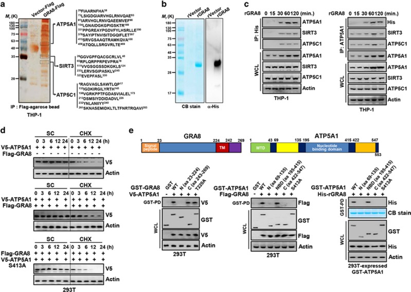
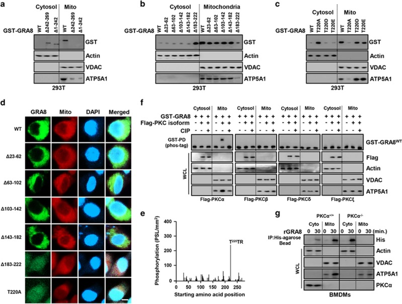
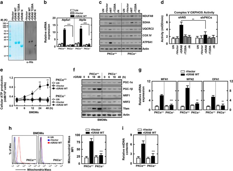
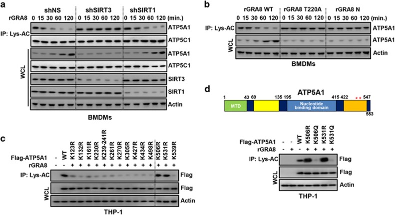
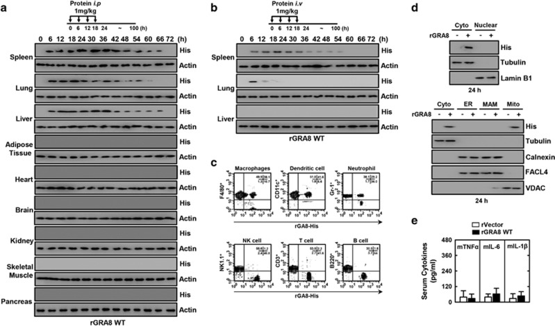
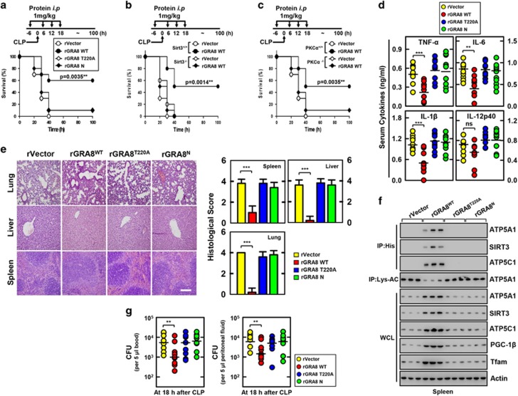
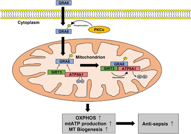
The intracellular parasite Toxoplasma gondii has unique dense granule antigens (GRAs) that are crucial for host infection. Emerging evidence suggests that GRA8 of T. gondii is a promising serodiagnostic marker in toxoplasmosis. However, little is known about the intracellular regulatory mechanisms involved in GRA8-induced host responses. We found that GRA8 interacts with host proteins involved in mitochondria activation and might be useful as a therapeutic strategy for sepsis. Here, we show that protein kinase-Cα (PKCα)-mediated phosphorylation of T. gondii GRA8 (Thr220) is required for mitochondrial trafficking and regulates the interaction of C terminal of GRA8 with nucleotide binding domain of ATP5A1. Furthermore, GRA8 interacts with SIRT3 in mitochondria, facilitating ATP5A1 deacetylation (K506 and K531), adenosine triphosphate production and subsequent anti-septic activity in vivo. Taken together, these results demonstrate a new anti-sepsis therapeutic strategy using T. gondii GRA8-induced mitochondrial metabolic resuscitation. This strategy represents an urgently needed paradigm shift for therapeutic intervention.


Conflict of interest statement

The authors declare no conflict of interest.

Figures

Figure 1
Figure 1
GRA8 interacts with ATP5A1 and SIRT3. (a) Identification of ATP5A1, SIRT3 and ATP5C1 by mass spectrometry analysis in THP-1 cell lysates expressed with GRA8 or vector. (b) Bacterially purified 6xHis-GRA8 were analyzed by Coomassie blue staining (left) or immunoblotting (IB) with αHis (right). (c) THP-1 cells were stimulated with rGRA8 (5 μg ml−1) for the indicated times, followed by immunoprecipitation (IP) with αHis-agarose bead or αATP5A1 and IB with αATP5A1, αSIRT3, αATP5C1, αHis and αActin. (d) GRA8-mediated increases of ATP5A1 stability. At 24 h after transfection with V5-ATP5A1and/or Flag-GRA8, 293T cells were treated with solvent control (SC) or cyclohexamide (CHX, 1 μg ml−1) for indicated times and cell lysates were used for IB with αV5 and αActin. (e) Binding mapping. Schematic diagrams of the structures of GRA8 and ATP5A1 (upper). At 48 h after transfection with mammalian glutathione S-transferase (GST) or GST-GRA8 and truncated mutant constructs together with V5-ATP5A1, or GST-ATP5A1 constructs together with Flag or His-GRA8, 293T cells were used for GST pull down, followed by IB with αV5, αFlag or αHis. Whole cell lysates (WCLs) were used for IB with αGST, αV5, αFlag, αHis or αActin. The data are representative of four independent experiments with similar results (ae).
Figure 2
Figure 2
Protein kinase-Cα (PKCα)-dependent phosphorylation of GRA8 was essential for mitochondrial localization. (ac) Subcellular fractionation of 293T cells stably expressing either GST-GRA8 WT, Δ23–62, Δ63–102, Δ103–142, Δ143–182, Δ183–222, Δ242–269, Δ1–242 or GST T220A/D/E. Mitochondrial and cytosolic fractions were fractionated and analyzed for expression of glutathione S-transferase (GST) by immunoblotting (IB). Purity of the fractions was assessed by blotting for VDAC (voltage-dependent anion channel; mitochondrial protein) and actin (cytosolic protein). (d) Representative immunofluorescence images of 293-GRA8-GFP cells expressing wild-type (WT) and deletion mutants were colocalized with MitotrackerDeep Red FM (100 nM). Scale bar, 20 μm. (e) Mapping of PKCα phosphorylation sites on GRA8 by tiled peptide array analysis using purified recombinant PKCα. Phosphorylation intensity of 15-amino acid peptides that span full-length GRA8 and are each shifted by 3 amino acids was detected using MultiGauge version 3.0. The threonine in the peptides that showed a phosphorylation signal stronger than 100 PSL mm−2 is indicated above the corresponding peaks. (f) Phos-tag and SDS–polyacrylamide gel electrophoresis (SDS-PAGE) analyses of GST-GRA8 together with Flag-tagged isoform of PKC in 293T cells left untreated (CIP−) or treated with calf-intestinal alkaline phosphatase (CIP+), and subcellular fractionation as the experimental conditions follow the same pattern in (a). (g) Bone marrow-derived macrophages (BMDMs) from PKCα+/+ and PKCα−/− were stimulated with rGRA8 for 30 min, followed by subcellular fractionation as the experimental conditions follow the same pattern in (a). The data are representative of four independent experiments with similar results (ag).
Figure 3
Figure 3
The rGRA8 treatment increases the induction of mitochondrial activity and biogenesis via protein kinase-Cα (PKCα). (a) Bacterially purified 6xHis-GRA8-WT and its mutants were analyzed by Coomassie blue staining (left) or immunoblotting (IB) with αHis (right). (b–d) Bone marrow-derived macrophages (BMDMs) from PKCα+/+ and PKCα−/− (b, c) or THP-1 cells were transduced with lentivirus-shRNA-NS or lentivirus-shRNA-PKCα (multiplicity of infection (MOI)=100) with polybrene (8 μg ml−1) (right) for 2 days (d), the cells were stimulated with rGRA8 (1 μg ml−1) and its mutants for 6 h (b) and 24 h (c, d) and subjected to quantitative real-time PCR (b), IB (c) or enzymatic activity (d) of oxidative phosphorylation (OXPHOS) genes. (eg) BMDMs from PKCα+/+ and PKCα−/− were stimulated with rGRA8 for the indicated times and subjected to cellular adenosine triphosphate (ATP) production (e), IB analysis with αPGC-1, αNRF1, αNRF2, αTfam and αActin (f) or quantitative real-time PCR of fusion genes (g). (h) Mitotracker fluorescence signals assessed by a flow cytometric analysis. (Left) Representative histograms from seven independent replicates. (Right) Bar graph indicates the mitochondrial mass mean fluorescence intensities (MFIs). Results are expressed as means±s.d. of seven experiments. (i) Mitochondrial DNA (mtDNA) content in BMDMs measured by quantitative real-time PCR. The mtDNA content was normalized to nuclear DNA. Significant differences (**P<0.01; ***P<0.001) compared with PKCα+/+ or shRNA-NS (Non-specific) (b, d, e and gi).The data are representative of five independent experiments with similar results (ag and i).
Figure 4
Figure 4
The rGRA8 treatment increases the induction of deacetylation of ATP5A1 via SIRT3. Bone marrow-derived macrophages (BMDMs) were transduced with lentivirus-shRNA-NS (Non-specific) or lentivirus-shRNA-SIRT1 or SIRT3 (multiplicity of infection (MOI)=100) with polybrene (8 μg ml−1) (right) for 2 days (a), BMDMs (b) or THP-1 cells stably expressing either ATP5A1 or its mutants (c, d), the cells were stimulated with rGRA8 (1 μg ml−1) and its mutants for the indicated times and subjected to immunoprecipitation (IP) with αLys-AC and immunoblotting (IB) with αATP5A1, αATP5C1, αSIRT1, αSIRT3, αFlag and αActin. Schematic diagrams of the structures of ATP5A1 (d, upper).The data are representative of five independent experiments with similar results.
Figure 5
Figure 5
Therapeutic rGRA8 proteins are uptaken by cells of the reticuloendothelial system in mice. (a, b) Schematic of the pharmacokinetic analysis in mice treated with rGRA8 (upper). Pharmacokinetic analysis of proteins in the various organs, followed by immunoblotting (IB) with αHis and αActin. The data are representative of three independent experiments with similar results. Fluorescence-activated cell sorting (FACS) analysis in spleen (c), tissue subcellular fractionation in spleen (d), and (e) serum cytokine levels were evaluated 24 h after being injected with rGRA8 (10 mice per group). Significant differences compared with rVector-treated mice.
Figure 6
Figure 6
The rGRA8 protects mice from cecal ligation and puncture (CLP)-induced polymicrobial sepsis. (ac) Schematic of the CLP model treated with rGRA8 or its mutants (upper). The survival of mice was monitored for 7 days; mortality was measured for n=25 mice per group (lower). Statistical differences compared with the rVector-treated mice are indicated (log-rank test). The data are representative of two independent experiments with similar results. (d) Serum cytokine levels and (e) representative hematoxylin and eosin (H&E) staining of the lung, liver and spleen (left) from 10 mice per group. Histopathology scores were obtained from H&E stained as described in Methods (right) were determined at 30 h in CLP mice were treated with rGRA8 or its mutants. Scale bar, 200 μm. (f) Splenocytes were used for immunoprecipitation (IP) with αHis or αLys-AC, followed by immunoblotting (IB) with αATP5A1, αATP5C1 or αSIRT3. Whole cell lysates (WCLs) were used for IB with αATP5A1, αATP5C1, αSIRT3, αPGC-1, αTfam or αActin. The data are representative of three independent experiments with similar results. (g) The bacterial burden was evaluated 18 h after treatment of CLP mice with rGRA8 or its mutants (n=10 mice per group). Results are expressed as means±s.d. (10 mice per group (d, g). Significant differences (**P<0.01; ***P<0.001) compared with rVector-treated mice (d, e right, and g).
Figure 7
Figure 7
Schematic model for the roles of GRA8 and GRA8-mediated regulatory pathways against sepsis. Please see the Discussion for detail.

References

    1. Koethe M, Pott S, Ludewig M, Bangoura B, Zoller B, Daugschies A et al. Prevalence of specific IgG-antibodies against Toxoplasma gondii in domestic turkeys determined by kinetic ELISA based on recombinant GRA7 and GRA8. Vet Parasitol 2011; 180: 179–190. - PubMed
    1. Koh HJ, Kim YR, Kim JS, Yun JS, Jang K, Yang CS. Toxoplasma gondii GRA7-targeted ASC and PLD1 promote antibacterial host defense via PKCalpha. PLoS Pathog 2017; 13: e1006126. - PMC - PubMed
    1. Costa JG, Dure AB. Immunochemical evaluation of two Toxoplasma gondii GRA8 sequences to detect acute toxoplasmosis infection. Microb Pathog 2016; 100: 229–236. - PubMed
    1. Ahn HJ, Kim S, Kim HE, Nam HW. Interactions between secreted GRA proteins and host cell proteins across the paratitophorous vacuolar membrane in the parasitism of Toxoplasma gondii. Korean J Parasitol 2006; 44: 303–312. - PMC - PubMed
    1. Kim YR, Hwang J, Koh HJ, Jang K, Lee JD, Choi J et al. The targeted delivery of the c-Src peptide complexed with schizophyllan to macrophages inhibits polymicrobial sepsis and ulcerative colitis in mice. Biomaterials 2016; 89: 1–13. - PubMed

Publication types

MeSH terms