. 2018 Aug 3;293(31):11971-11983.
doi: 10.1074/jbc.M117.802975. Epub 2018 Jun 5.

The CCCH-type zinc finger transcription factor Zc3h8 represses NF-κB-mediated inflammation in digestive organs in zebrafish

Affiliations

The CCCH-type zinc finger transcription factor Zc3h8 represses NF-κB-mediated inflammation in digestive organs in zebrafish

Qingliang Zou et al. J Biol Chem. .

Abstract

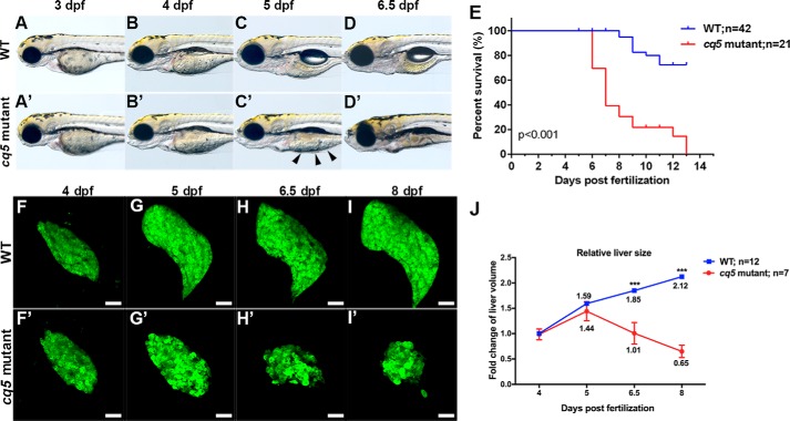
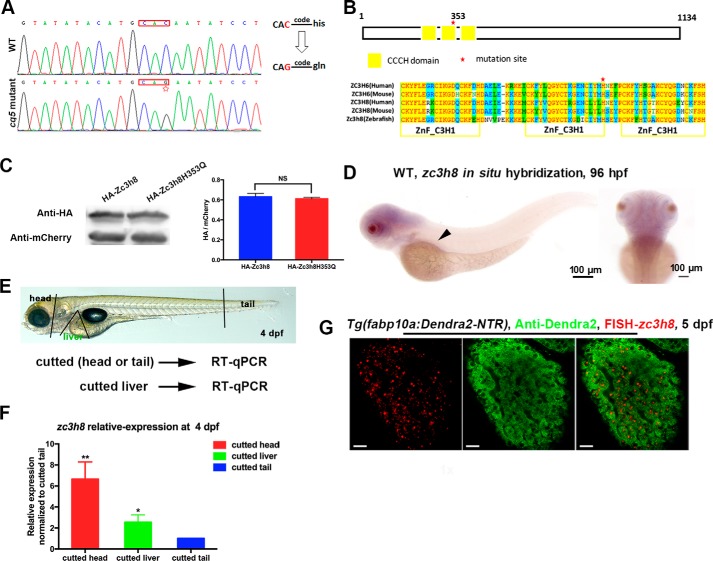
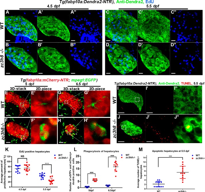
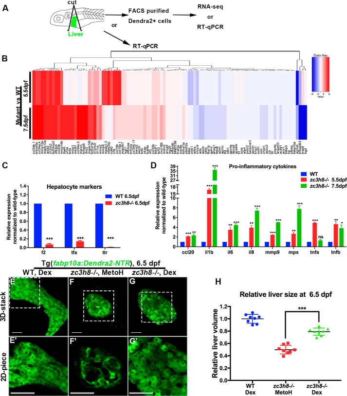
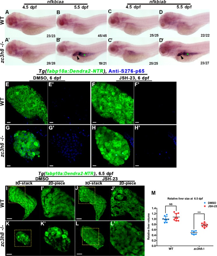
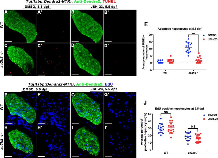
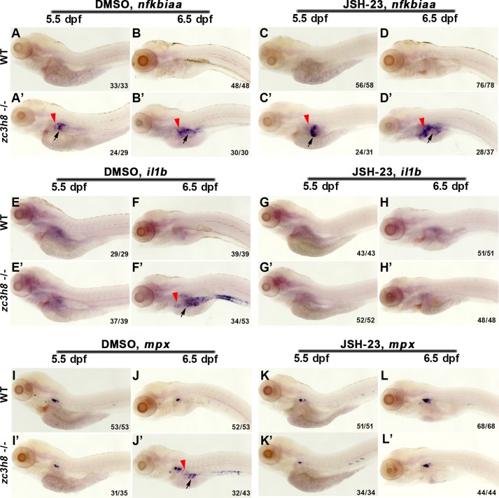
Degenerative diseases of organs lead to their impaired function. The cellular and molecular mechanisms underlying organ degeneration are therefore of great research and clinical interest but are currently incompletely characterized. Here, using a forward-genetic screen for genes regulating liver development and function in zebrafish, we identified a cq5 mutant that exhibited a liver-degeneration phenotype at 5 days postfertilization, the developmental stage at which a functional liver develops. Positional cloning revealed that the liver degeneration was caused by a single point mutation in the gene zc3h8 (zinc finger CCCH-type containing 8), changing a highly conserved histidine to glutamine at position 353 of the Zc3h8 protein. The zc3h8 mutation-induced liver degeneration in the mutant was accompanied by reduced proliferation, increased apoptosis, and macrophage phagocytosis of hepatocytes. Transcriptional profile analyses revealed up-regulation and activation of both proinflammatory cytokines and the NF-κB signaling pathway in the zc3h8 mutant. Suppression of NF-κB signaling activity efficiently rescued the proinflammatory cytokine response, as well as the inflammation-mediated liver degeneration phenotype of the mutant. Of note, the zc3h8 mutation-induced degeneration of several other organs, including the gut and exocrine pancreas, indicating that Zc3h8 is a general repressor of inflammation in zebrafish. Collectively, our findings demonstrate that Zc3h8 maintains organ homeostasis by inhibiting the NF-κB-mediated inflammatory response in zebrafish and that Zc3h8 dysfunction causes degeneration of multiple organs, including the liver, gut, and pancreas.

Keywords: NF-κ B (NF-κB); cell signaling; degeneration; digestive organ; inflammation; zebrafish; zinc finger.


Conflict of interest statement

The authors declare that they have no conflicts of interest with the contents of this article

Figures

Figure 1.
Figure 1.
Cq5 is a liver degeneration mutant. A–D and A′–D′, WT (n = 14) and cq5 mutant (n = 18) embryos were imaged from 3 to 6.5 dpf; unabsorbed yolk is indicated by black arrowheads. E, cumulative survival of cq5 mutant larvae (n = 21) and WT siblings (n = 42). p < 0.001 by the log-rank Mantel-Cox test. F–I and F′–I′, the cq5 mutants showed significant liver degeneration (57 of 59), as normalized to the WT (62 of 62). Scale bar, 50 μm. J, the relative fold change of liver size was measured in WT (n = 12) and mutant (n = 7) larvae from 4 to 8 dpf. Liver volume was measured with Imaris software and normalized to the liver size of WT larvae at 4 dpf. The error bars represent S.E. ***, p < 0.001 by Student's t test.
Figure 2.
Figure 2.
zc3h8 is required for the maintenance of liver homeostasis. A, sequencing of zc3h8 from cq5 mutant and WT embryos identified a C-to-G point mutation (red star marking the mutation site), resulting in a coding change from histidine to glutamine at position 353. B, amino acid sequence alignment of Zc3h8 homologs/orthologs according to the three conserved zinc finger domains (red star marking the cq5 amino acid mutation site, ZnF_C3H1 domain indicated by yellow frame) in human, mouse, and zebrafish. C, Western blotting of FASC sorted mCherry-positive cells showing that mutation of cq5 (Zc3h8H353Q) does not alter protein stability in zebrafish embryos. mCherry levels were used as loading controls. Ratio of the anti-HA with anti-mCherry densitometry was used as the relative repression level. The error bars represent S.D. N.S. represents no significance and p value by Student's t test. D, zc3h8 expression pattern in WT embryos determined via whole-mount in situ hybridization at 96 hpf. Arrowheads indicate the region of digestive organ expression. E, scheme for determining the expression levels of zc3h8 in different regions in WT larvae via qPCR. F, quantitative real-time PCR analysis of zc3h8 expression in different areas (head, liver, and tail) in WT embryos at 4 dpf. The error bars indicate S.E. *, p < 0.05; **, p < 0.01 by Student's t test. G, double fluorescence labeling of zc3h8 RNA probe and anti-Dendra2 antibody staining at 5 dpf. Note the zc3h8 transcripts expressed in the hepatocytes. Scale bars, 20 μm.
Figure 3.
Figure 3.
Reduced proliferation and increased apoptosis of hepatocytes are associated with zc3h8 mutation. A–D and A′–D″, confocal images of EdU and anti-Dendra2 antibody labeling in WT and mutant larvae from 4.5 (29 of 29, 25 of 27) to 5.5 dpf (28 of 29, 25 of 28). Scale bars, 20 μm. E–H and E′–H′, Tg(fabp10a:mCherry-NTRcq2;mpeg1:EGFP) double-transgenic line fish were visualized via confocal microscopy from 5 (E-E′ and F-F′; 53 of 53, 48 of 52) to 6.5 dpf (G-G′ and H-H′; 54 of 54, 55 of 59) for WT and zc3h8 mutant larvae. White arrowheads highlight macrophages engulfing degenerated hepatocytes. Scale bars, 20 μm. I, I′, J, and J″, confocal images of TUNEL-labeled apoptotic cells in zc3h8 mutant and WT livers at 5.5 dpf (38 of 40, 42 of 45). Scale bars, 50 μm. K, quantification of EdU-labeled hepatocytes in the zc3h8 mutant and the WT at 4.5 dpf (n = 5, n = 9) and 5.5 dpf (n = 8, n = 8). The error bars indicate S.D. N.S. represents no significance. ***, p < 0.001 by Student's t test. L, quantification of macrophages engulfing degenerated hepatocytes in zc3h8 mutant and WT larvae at 5 dpf (n = 10, n = 10) and 6.5 dpf (n = 9, n = 10). The error bars indicate S.D. ***p < 0.001 by Student's t test. M, quantification of TUNEL-positive hepatocytes in zc3h8 mutant and WT (n = 12) larvae at 5.5 dpf. The error bars indicate S.D. ***, p < 0.001 by Student's t test.
Figure 4.
Figure 4.
zc3h8 mutant hepatocyte degeneration is inflammatory response–dependent. A, schematic representation of the process of transcriptome sequencing or qPCR analysis of sorted hepatocytes and direct qPCR analysis of liver tissues from WT and zc3h8 mutants. B, heat map of the log10-transformed expression of statistically significant, differentially expressed genes between sorted hepatocytes of WT and zc3h8 mutants. Red indicates up-regulation, and blue indicates down-regulation of mRNA compared with WT. C, quantitative real-time PCR of hepatocyte-specific markers for sorted hepatocytes from zc3h8 mutant and WT larvae at 6.5 dpf. The error bars represent S.E. *, p < 0.05; **, p < 0.01; ***, p < 0.001 by Student's t test. D, quantitative real-time PCR analysis of proinflammatory cytokine expression in liver tissues at 5.5 and 7.5 dpf. The error bars represent S.E. ns represents no significance. *, p < 0.05; **, p < 0.01; ***, p < 0.001 by Student's t test. E–G and E′–G′, fluorescence confocal microscopy of Tg(fabp10a:Dendra2-NTR)cq1 in zc3h8 mutant (31 of 44) and WT (49 of 56) livers treated with Dex or methanol (56 of 56) at 6.5 dpf. Note that the zc3h8 mutant degeneration phenotype could be partially rescued by treatment with the anti-inflammatory drug Dex. Scale bars, 50 μm. H, quantification effects of Dex on zc3h8 mutant degeneration liver at 6.5 dpf. The error bars represent S.D. ***, p < 0.001 by Student's t test.
Figure 5.
Figure 5.
The NF-κB signaling pathway is activated and contributes to liver degeneration in zc3h8 mutants. A–D and A–D′, whole-mount in situ hybridization showed increased expression of NF-κB components (nfkbiaa and nfkbiab) in the livers (arrowhead) and intestines (green asterisks) of zc3h8 mutant larvae at 4.5 and 5.5 dpf. E–H and E′–H′, antibody staining of phosphorylated p65 (S273-p65) in Tg(fabp10a:Dendra2-NTR)cq1 zc3h8 mutant (38 of 43) and WT (45 of 47) larvae treated with DMSO and an NF-κB inhibitor (JSH-23) (zc3h8 mutant (28 of 33) and WT (32 of 32)) at 6 dpf. Scale bars, 20 μm. I–L and I′–L′, fluorescence confocal microscopy of Tg(fabp10a:Dendra2-NTR)cq1 in zc3h8 mutant (17 of 20) and WT (24 of 24) livers treated with an NF-κB inhibitor (JSH-23) and DMSO (zc3h8 mutant (17 of 19) and WT (15 of 17)) at 6.5 dpf. Note that the mutant degeneration phenotype could also be partially rescued by treatment with an NF-κB signaling pathway inhibitor. Scale bars, 50 μm. M, quantification effects of JSH-23 on zc3h8 mutant degeneration liver at 6.5 dpf. The error bars represent S.D. NS represents no significance. ***, p < 0.001 by Student's t test.
Figure 6.
Figure 6.
JSH-23 rescues zc3h8 mutant hepatocyte apoptosis, but not proliferation. A–D and A′–D′, double fluorescence labeling with TUNEL and anti-Dendra2 antibody staining in control larvae (n = 23) or larvae treated with JSH-23 (n = 25) at 5.5 dpf. Scale bars, 50 μm. E, quantification of apoptotic hepatocytes in livers from zc3h8 mutant and WT larvae (n = 13) treated with JSH-23. The error bars indicate S.D. ***, p < 0.001 by Student's t test. F–I and F′–I′, double labeling with EdU and anti-Dendra2 antibody staining in WT and zc3h8 mutant larvae (28 of 32 and 24 of 28) or larvae treated with JSH-23 (23 of 28 and 19 of 22) at 5.5 dpf. Scale bars, 50 μm. J, quantification of hepatocyte proliferation in livers from zc3h8 mutant and WT larvae (n = 13) treated with JSH-23. The error bars indicate S.D. NS represents no significance, p value test by Student's t test.
Figure 7.
Figure 7.
Uncontrolled inflammation is induced by activated NF-κB signaling. A–D and A′–D′, whole-mount in situ hybridization of an NF-κB component gene (nfkbiaa) in WT and zc3h8 mutant larvae at 5.5 and 6.5 dpf after pretreatment with DMSO or JSH-23. Red arrowheads point to the liver, and the arrows point to the intestine. E–L and E′–L′, whole-mount in situ hybridization of proinflammatory cytokines (il1b and mpx) in WT and zc3h8 mutant larvae at 5.5 and 6.5 dpf after pretreatment with DMSO or JSH-23. Red arrowheads point to the liver, and the black arrows point to the intestine.

References

    1. Kawamoto S., Matsumoto Y., Mizuno K., Okubo K., and Matsubara K. (1996) Expression profiles of active genes in human and mouse livers. Gene 174, 151–158 10.1016/0378-1119(96)00512-4 - DOI - PubMed
    1. Pack M., Solnica-Krezel L., Malicki J., Neuhauss S. C., Schier A. F., Stemple D. L., Driever W., and Fishman M. C. (1996) Mutations affecting development of zebrafish digestive organs. Development 123, 321–328 - PubMed
    1. Field H. A., Ober E. A., Roeser T., and Stainier D. Y. (2003) Formation of the digestive system in zebrafish: I. liver morphogenesis. Dev. Biol. 253, 279–290 10.1016/S0012-1606(02)00017-9 - DOI - PubMed
    1. Janeway C. A. (1989) Approaching the asymptote: evolution and revolution in immunology. Cold Spring Harbor Symp. Quant. Biol. 54, 1–13 10.1101/SQB.1989.054.01.003 - DOI - PubMed
    1. Matzinger P. (1994) Tolerance, danger, and the extended family. Annu. Rev. Immunol. 12, 991–1045 10.1146/annurev.iy.12.040194.005015 - DOI - PubMed

Publication types

MeSH terms

LinkOut - more resources