

High-resolution μCT of a mouse embryo using a compact laser-driven X-ray betatron source
- PMID: 29871946
- PMCID: PMC6016801
- DOI: 10.1073/pnas.1802314115
High-resolution μCT of a mouse embryo using a compact laser-driven X-ray betatron source
Abstract
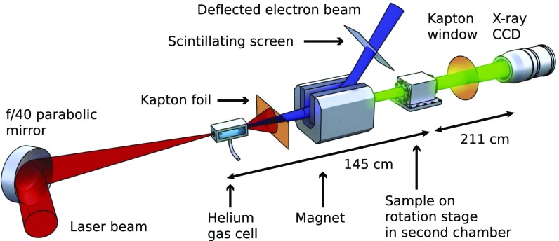
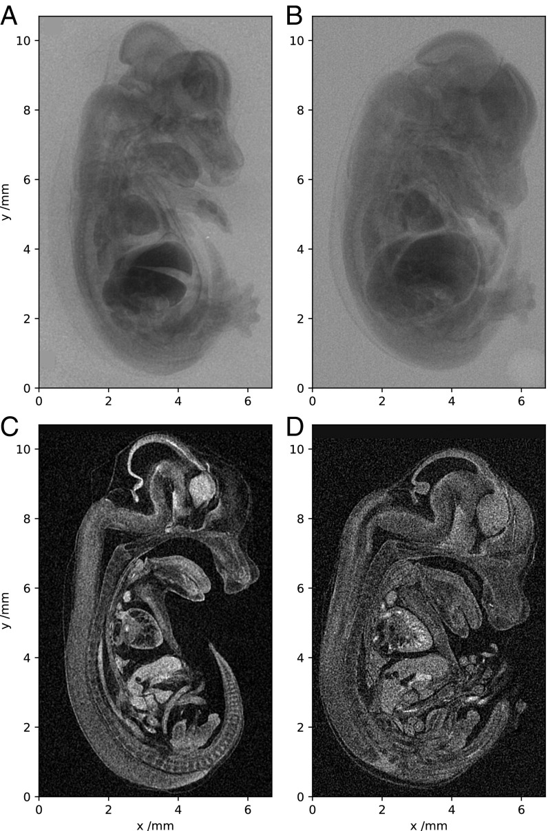
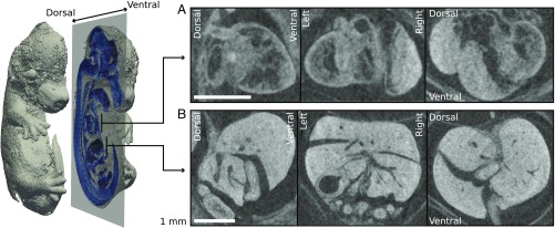
In the field of X-ray microcomputed tomography (μCT) there is a growing need to reduce acquisition times at high spatial resolution (approximate micrometers) to facilitate in vivo and high-throughput operations. The state of the art represented by synchrotron light sources is not practical for certain applications, and therefore the development of high-brightness laboratory-scale sources is crucial. We present here imaging of a fixed embryonic mouse sample using a compact laser-plasma-based X-ray light source and compare the results to images obtained using a commercial X-ray μCT scanner. The radiation is generated by the betatron motion of electrons inside a dilute and transient plasma, which circumvents the flux limitations imposed by the solid or liquid anodes used in conventional electron-impact X-ray tubes. This X-ray source is pulsed (duration <30 fs), bright (>1010 photons per pulse), small (diameter <1 μm), and has a critical energy >15 keV. Stable X-ray performance enabled tomographic imaging of equivalent quality to that of the μCT scanner, an important confirmation of the suitability of the laser-driven source for applications. The X-ray flux achievable with this approach scales with the laser repetition rate without compromising the source size, which will allow the recording of high-resolution μCT scans in minutes.
Keywords: X-ray imaging; laser–plasma acceleration; microcomputed tomography.
Copyright © 2018 the Author(s). Published by PNAS.
Conflict of interest statement
The authors declare no conflict of interest.
Figures


References
-
- Mangles SPD, et al. Monoenergetic beams of relativistic electrons from intense laser-plasma interactions. Nature. 2004;431:535–538. - PubMed
-
- Geddes CGR, et al. High-quality electron beams from a laser wakefield accelerator using plasma-channel guiding. Nature. 2004;431:538–541. - PubMed
-
- Faure J, et al. A laser-plasma accelerator producing monoenergetic electron beams. Nature. 2004;431:541–544. - PubMed
-
- Tajima T, Dawson JM. Laser electron accelerator. Phys Rev Lett. 1979;43:267–270.
-
- Esarey E, Schroeder CB, Leemans WP. Physics of laser-driven plasma-based electron accelerators. Rev Mod Phys. 2009;81:1229–1285.
Publication types
MeSH terms
Grants and funding
LinkOut - more resources
Full Text Sources
Other Literature Sources
