. 2018 Jul;30(7):1543-1561.
doi: 10.1105/tpc.17.00981. Epub 2018 Jun 5.

Receptor-Like Cytoplasmic Kinases Directly Link Diverse Pattern Recognition Receptors to the Activation of Mitogen-Activated Protein Kinase Cascades in Arabidopsis

Affiliations

Receptor-Like Cytoplasmic Kinases Directly Link Diverse Pattern Recognition Receptors to the Activation of Mitogen-Activated Protein Kinase Cascades in Arabidopsis

Guozhi Bi et al. Plant Cell. 2018 Jul.

Abstract

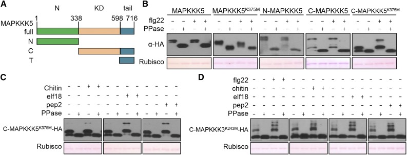
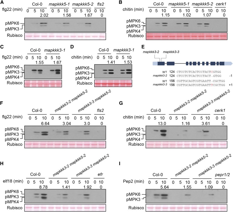
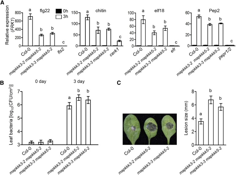
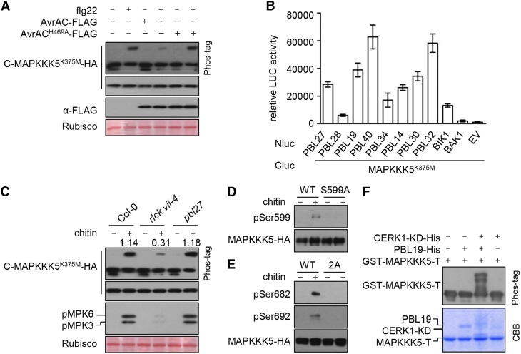
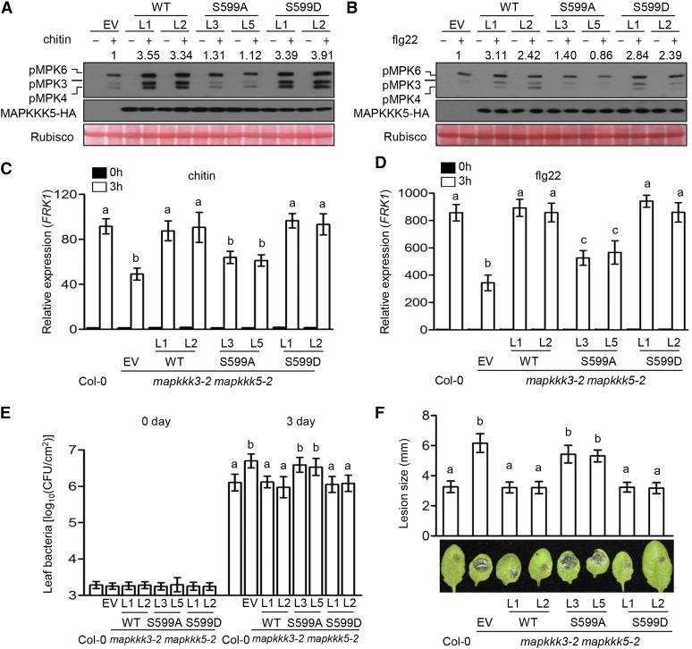
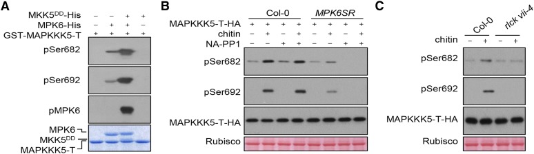
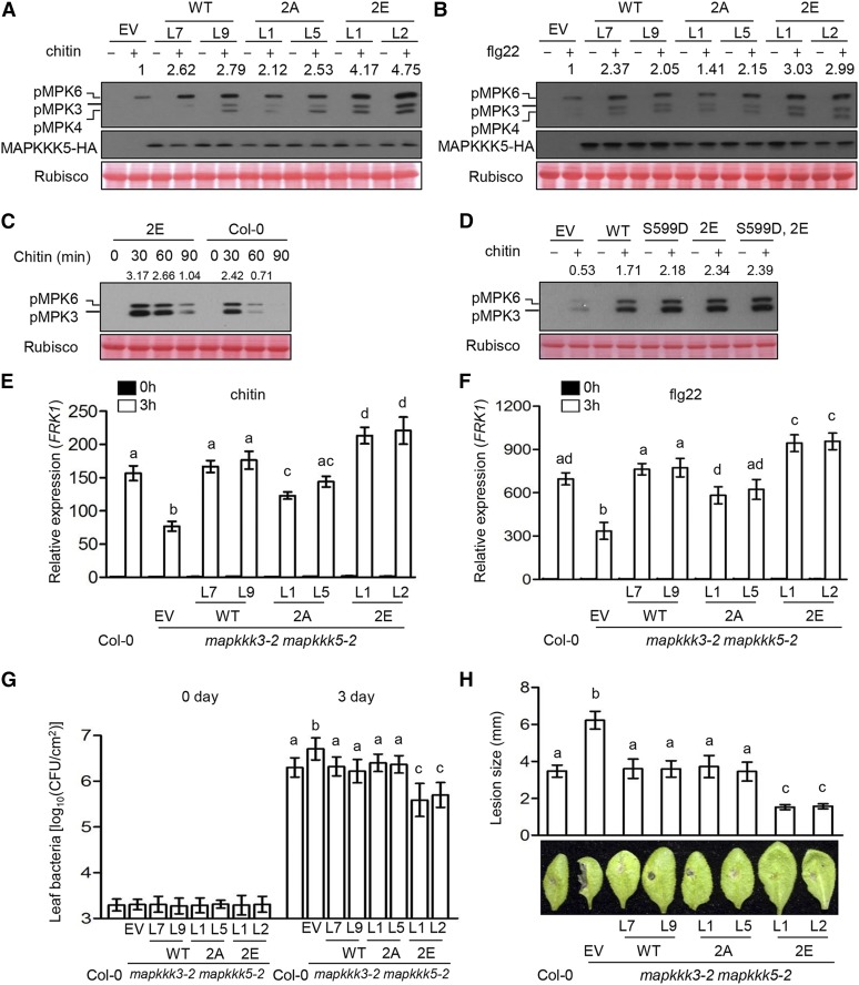
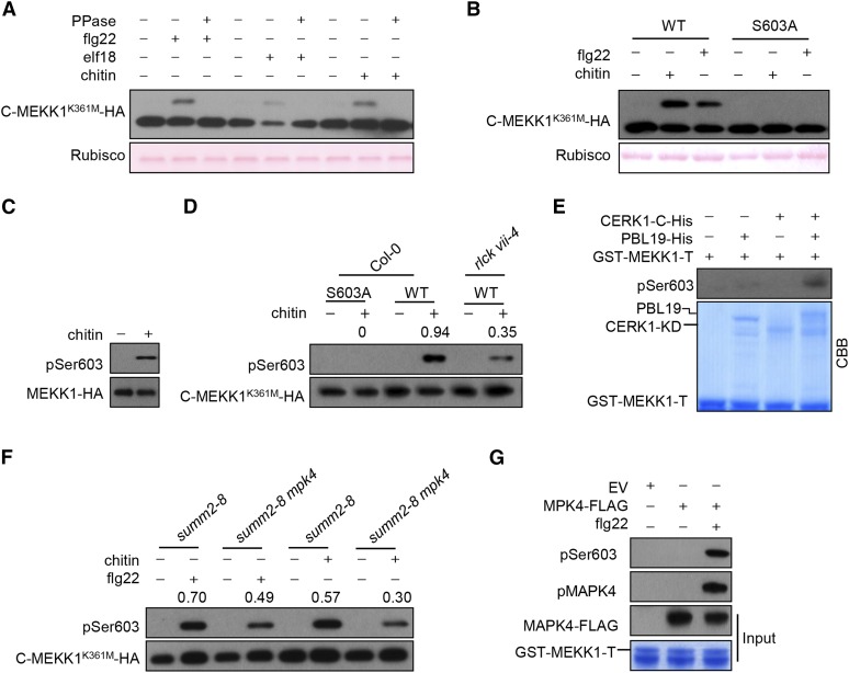
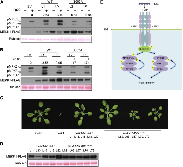
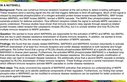
Plants deploy numerous cell surface-localized pattern-recognition receptors (PRRs) to perceive host- and microbe-derived molecular patterns that are specifically released during infection and activate defense responses. The activation of the mitogen-activated protein kinases MPK3, MPK4, and MPK6 (MPK3/4/6) is a hallmark of immune system activation by all known PRRs and is crucial for establishing disease resistance. The MAP kinase kinase kinase (MAPKKK) MEKK1 controls MPK4 activation, but the MAPKKKs responsible for MPK3/6 activation downstream of diverse PRRs and how the perception of diverse molecular patterns leads to the activation of MAPKKKs remain elusive. Here, we show that two highly related MAPKKKs, MAPKKK3 and MAPKKK5, mediate MPK3/6 activation by at least four PRRs and confer resistance to bacterial and fungal pathogens in Arabidopsis thaliana The receptor-like cytoplasmic kinases VII (RLCK VII), which act downstream of PRRs, directly phosphorylate MAPKKK5 Ser-599, which is required for pattern-triggered MPK3/6 activation, defense gene expression, and disease resistance. Surprisingly, MPK6 further phosphorylates MAPKKK5 Ser-682 and Ser-692 to enhance MPK3/6 activation and disease resistance, pointing to a positive feedback mechanism. Finally, MEKK1 Ser-603 is phosphorylated by both RLCK VII and MPK4, which is required for pattern-triggered MPK4 activation. These findings illustrate central mechanisms by which multiple PRRs activate MAPK cascades and disease resistance.


Figures

Figure 1.
Figure 1.
Patterns Trigger MAPKKK3/5 Phosphorylation. (A) Schematic representation of MAPKKK5 domain structure and deletion constructs. Green, N-terminal domain (N); orange, kinase domain (KD); aqua, C-terminal tail (T). The C terminus construct (C) contains both the KD and T. (B) Flg22-induced phosphorylation of MAPKKK5 in the C terminus. Protoplasts expressing full-length, truncated, or kinase-dead (K375M) variants of MAPKKK5 were treated with flg22. Total protein treated with (+) or without (−) λ protein phosphatase (PPase) was subjected to SDS-PAGE containing Phos-tag acrylamide and detected by immunoblotting with anti-HA antibody. Ponceau staining of Rubisco indicates equal loading. (C) and (D) Pattern-triggered phosphorylation of MAPKKK3 and MAPKKK5 C termini. Protoplasts expressing C-MAPKKK5K375M-HA (C) and C-MAPKKK3K243M-HA (D) were treated with chitin, elf18, Pep2, or flg22. Total protein was treated with (+) or without (−) λ protein phosphatase (PPase) and subjected to SDS-PAGE containing Phos-tag acrylamide and detected by immunoblotting with anti-HA antibody.
Figure 2.
Figure 2.
MAPKKK3 and MAPKKK5 Regulate Pattern-Induced MAPK Activation. (A) to (D) Pattern-triggered MAPK activation is normal in mapkkk5 ([A] and [B]) and mapkkk3 ([C] and [D]) single mutants. Ten-day-old seedlings were sprayed with flg22 or chitin, and samples were harvested at the indicated times for immunoblot analysis with anti-pERK antibody. Note that MPK4 and MPK3 are highly similar in size and were not well-separated in some experiments. Numbers indicate arbitrary densitometry units of phosphorylated MPK3/6 (pMPK3/6) bands normalized to Rubisco 10 min after pattern stimulation. All assays were performed three times, and a representative photograph is shown. (E) CRISPR-Cas9-mediated mutations in the first exon of MAPKKK3 in the mapkkk3 mapkkk5 (mapkkk3-2 mapkkk5-2 and mapkkk3-3 mapkkk5-2) double mutant lines. The numbers 124 and 156 indicate the nucleotide positions in the MAPKKK3 coding sequence. −1 and +1 indicate frame shifts in the mutant lines. (F) to (I) Pattern-triggered MPK3/6 activation is diminished in mapkkk3 mapkkk5 double mutants. Seedlings were sprayed with flg22 (F), chitin (G), elf18 (H), or Pep2 (I), and immunoblot analysis with anti-pERK antibody was performed at the indicated times. Note that MPK4 and MPK3 are highly similar in size and were not well separated in some experiments. Numbers indicate relative protein band density of pMPK3/6 normalized to the loading control (Rubisco). All assays were performed at least three times, and a representative photograph is shown. Ponceau staining of Rubisco indicates equal loading.
Figure 3.
Figure 3.
MAPKKK3/5 Are Required for Defense Gene Expression and Disease Resistance. (A) Pattern-induced FRK1 expression is impaired in mapkkk3 mapkkk5 mutants. Ten-day-old seedlings were sprayed with flg22, chitin, elf18, or Pep2, and samples were harvested at 3 h. ACTIN1 was used as the internal standard. Data are presented as mean ± sd. Different letters indicate significant difference at P < 0.05 (n = 3, one-way ANOVA, Tukey post-test, three independent experiments). (B) mapkkk3 mapkkk5 mutants display increased susceptibility to P. syringae pv tomato (Pto) DC3000. Plants were infiltrated with Pto DC3000, and bacterial population in the leaf was determined 3 d after inoculation. Data are presented as mean ± sd. Different letters indicate significant difference at P < 0.05 (n ≥ 8, one-way ANOVA, Tukey post-test, three independent experiments). (C) mapkkk3 mapkkk5 mutants display increased susceptibility to B. cinerea. Disease lesions were recorded 3 d after inoculation with B. cinerea. Lesion size represents mean ± sd. Different letters indicate significant difference at P < 0.05 (n ≥ 14, one-way ANOVA, Tukey post-test, three independent experiments).
Figure 4.
Figure 4.
RLCK VII Subfamily Members Link PRRs to MAPKKK5. (A) AvrAC inhibits flg22-induced phosphorylation of MAPKKK5 C terminus. C-MAPKKK5K375M-HA was coexpressed with AvrAC or AvrACH469A in Col-0 protoplasts, treated with flg22 for 10 min, and total protein was subjected to SDS-PAGE containing Phos-tag acrylamide and detected by immunoblotting with anti-HA antibody. (B) MAPKKK5K375M interacts with selected members of RLCK VII. The indicated Nluc and Cluc constructs were transiently expressed in N. benthamiana plants for luciferase complementation assays. Error bars indicate sd. (C) Chitin-triggered phosphorylation of MAPKKK5 C terminus and MPK3/6 is impaired in rlck vii-4 sextuple mutant, but occurs normally in pbl27. Protoplasts expressing C-MAPKKK5K375M-HA were treated with chitin. Total protein was subjected to SDS-PAGE containing Phos-tag acrylamide and detected by immunoblotting with anti-HA antibody for MAPKKK5 phosphorylation and anti-pERK antibody for MPK3/6 phosphorylation. Numbers indicate relative band density of pMPK3/6 normalized to the loading control (Rubisco). (D) and (E) Chitin-triggered phosphorylation of MAPKKK5 at Ser-599, Ser-682, and Ser-692. Stable transgenic seedlings expressing MAPKKK5-HA (WT), MAPKKK5S599A-HA (S599A), and MAPKKK5S682/692A-HA (2A) were sprayed with chitin. The proteins were affinity purified with anti-HA antibodies and detected by immunoblot analysis with anti-pSer599 (D), anti-pSer682 (E), and anti-pSer692 (E) antibodies to investigate MAPKKK5 phosphorylation. (F) In the presence of the CERK1 kinase domain (CERK1-KD), PBL19 phosphorylates MAPKKK5 C-terminal tail in vitro. GST-MAPKKK5-T was incubated with PBL19-His and/or CERK1-KD-His proteins, and total protein was subjected to SDS-PAGE containing Phos-tag acrylamide and detected by immunoblot analysis with anti-GST antibodies. The smeared appearance of CERK1-KD and PBL19 in lanes 3 and 4 of the Coomassie Brilliant Blue (CBB)-stained gel likely reflects phosphorylation of these proteins.
Figure 5.
Figure 5.
Phosphorylation of MAPKKK5 at Ser-599 Is Required for MPK3/6 Activation and Disease Resistance. (A) and (B) Phosphorylation of Ser-599 in MAPKKK5 is required for MAPK activation induced by chitin (A) and flg22 (B). Seedlings of mapkkk3-2 mapkkk5-2 mutant T2 transgenic lines complemented with EV, wild-type MAPKKK5 (WT), MAPKKK5S599A (S599A), or MAPKKK5S599D (S599D) were sprayed with chitin (A) or flg22 (B), and total protein was subjected to immunoblot analysis with anti-pERK and anti-HA antibody. Numbers indicate arbitrary densitometry units of pMPK3/6 normalized to EV. All assays were performed at least three times, and a representative photograph is shown. (C) and (D) Phosphorylation of Ser-599 in MAPKKK5 is required for pattern-induced FRK1 expression. Ten-day-old seedlings were sprayed with chitin (C) and flg22 (D), and the samples were harvested at 3 h after treatment. ACTIN1 was used as the internal standard. Data are presented as mean ± sd. Different letters indicate significant difference at P < 0.05 (n = 3, one-way ANOVA, Tukey post-test, three independent experiments). (E) Phosphorylation of Ser-599 in MAPKKK5 is required for immunity to P. syringae DC3000. Plants were infiltrated with Pto DC3000, and bacterial population in the leaf was determined 3 d after inoculation. Data are presented as mean ± sd. Different letters indicate significant difference at P < 0.05 (n ≥ 8, one-way ANOVA, Tukey post-test, three independent experiments). (F) Phosphorylation of Ser-599 in MAPKKK5 is required for immunity to B. cinerea. Plants were inoculated with B. cinerea, and disease lesions were recorded 3 d after inoculation with B. cinerea. Lesion size represents mean ± sd. Different letters indicate significant difference at P < 0.05 (n ≥ 14, one-way ANOVA, Tukey post-test, four independent experiments).
Figure 6.
Figure 6.
MPK6 Phosphorylated MAPKKK5 at Ser-682 and Ser-692. (A) MPK6 phosphorylates MAPKKK5 in vitro. GST-MAPKKK5-T was incubated with MPK6-His and/or MKK5DD-His in kinase buffer and detected by immunoblotting with anti-pSer682 and pSer692 antibodies. (B) MAPKKK5 phosphorylation induced by chitin is blocked in the conditional mpk3 mpk6 double mutant. Protoplasts of Col-0 and the conditional mpk3 mpk6 double mutant were transfected with MAPKKK5-T-HA, incubated in the presence of DMSO (solvent control) or 2 μM NA-PP1 overnight, and treated with chitin. MAPKKK5-T-HA was affinity purified with anti-HA antibodies and detected by immunoblotting with anti-pSer682 and -pSer692 antibodies. (C) Chitin-triggered phosphorylation of Ser682/692 is impaired in the rlck vii-4 sextuple mutant. Protoplasts expressing MAPKKK5-T-HA were treated with chitin. MAPKKK5-T-HA was affinity purified with anti-HA antibody and detected by immunoblotting with anti-pSer682 and pSer692 antibodies.
Figure 7.
Figure 7.
Phosphorylation of MAPKKK5 Ser-682/692 Enhances MPK3/6 Activation and Immunity. (A) and (B) Phosphorylation of Ser-682/692 in MAPKKK5 is required for MAPK activation induced by chitin (A) and flg22 (B). Seedlings of mapkkk3-2 mapkkk5-2 mutant T2 transgenic lines complemented with EV, wild-type MAPKKK5 (WT), MAPKKK5S682/S692A (2A), or MAPKKK5S682/692E (2E) were sprayed with chitin (A) or flg22 (B), and total protein was subjected to immunoblot analysis with anti-pERK and anti-HA antibodies. Numbers indicate arbitrary densitometry units of phosphorylated MPK3/6 normalized to EV. All assays were performed at least three times, and a representative photograph is shown. (C) Phospho-mimetic mutations at Ser682/692 prolong chitin-triggered MPK3/6 activation. Seedlings of mapkkk3-2 mapkkk5-2 transgenic lines complemented with MAPKKK5S682/692E (2E) and Col-0 were treated with chitin for the indicated times, and total protein was subjected to immunoblotting with anti-pERK antibodies. Numbers indicate arbitrary densitometry units of phosphorylated MPK3/6 normalized to Rubisco. All assays were performed at least three times, and a representative photograph is shown. (D) Phospho-mimetic mutations at Ser-599/682/692 do not further enhance chitin-triggered MPK3/6 activation compared with phospho-mimetic mutations at Ser-682/692. Protoplasts of the mapkkk3-2 mapkkk5-2 double mutant expressing EV, the wild type, phospho-dead (S599D), phospho-mimicking Ser682/692E (2E), and phospho-mimicking S599D, S682/692E (S599D,2E) forms of MAPKKK5 were treated with chitin. Total protein was subjected to immunoblot analysis with anti-pERK antibody. Numbers indicate relative protein band density of phosphorylated MPKs normalized to Rubisco. All assays were performed at least three times, and a representative photograph is shown. (E) and (F) Phosphorylation of Ser-682/692 in MAPKKK5 is required for pattern-induced FRK1 expression. Ten-day-old seedlings were sprayed with chitin (E) and flg22 (F), and the samples were harvested at 3 h after treatment. ACTIN1 was used as the internal standard. Data are presented as mean ± sd. Different letters indicate significant difference at P < 0.05 (n = 3, one-way ANOVA, Tukey post-test, three independent experiments). (G) Phosphorylation of Ser-682/692 in MAPKKK5 is required for immunity to P. syringae DC3000 and B. cinerea. Plants were infiltrated with Pto DC3000, and bacterial population size in the leaf and diseased lesions was determined 3 d after inoculation. Data are presented as mean ± sd. Different letters indicate significant difference at P < 0.05 (n ≥ 8, one-way ANOVA, Tukey post-test, three independent experiments). (H) Phosphorylation of Ser-682/692 in MAPKKK5 is required for immunity to B. cinerea. Plants were inoculated with B. cinerea, and disease lesions were recorded 3 d after inoculation with B. cinerea. Lesion size represents mean ± sd. Different letters indicate significant difference at P < 0.05 (n ≥ 14, one-way ANOVA, Tukey post-test, three independent experiments).
Figure 8.
Figure 8.
Both RLCK VII-4 and MPK4 Phosphorylate MEKK1 at Ser-603. (A) Pattern-triggered phosphorylation of MEKK1 at the C terminus. Protoplasts expressing C-MEKK1K361M-HA were treated with flg22, elf18, or chitin. Total protein was fractionated by SDS-PAGE containing Phos-tag acrylamide and detected by immunoblotting with anti-HA antibody. (B) Ser-603 is the major phospho-site in MEKK1 C terminus MEKK1 upon chitin and flg22 induction. C-MEKK1K361M-HA (WT) or C-MEKK1K361M/S603A-HA (S603A) was expressed in protoplasts and total protein was detected by immunoblotting with anti-HA antibody. (C) Chitin-triggered phosphorylation of MEKK1 at Ser-603. Stable transgenic seedlings expressing MEKK1-FLAG were sprayed with chitin. MEKK1-FLAG was affinity purified with anti-FLAG antibody and detected by immunoblotting with anti-pSer603 antibodies for MEKK1 phosphorylation. (D) Chitin-triggered phosphorylation of Ser-603 is impaired in the rlck vii-4 sextuple mutant. Protoplasts of the indicated genotypes transfected with the C-MEKK1 K361M-HA constructs containing a wild-type Ser603 (WT) or a Ser603Ala substitution (S603A) were treated with chitin. The C-MEKK1K361M-HA protein was immunoprecipitated with anti-HA antibodies, and Ser-603 phosphorylation was detected by immunoblotting with anti-pSer603 antibodies. Total C-MEKK1K361M-HA protein was detected by immunoblotting with anti-HA antibody. Numbers indicate arbitrary densitometry units of pSer603 normalized to total C-MEKK1 K361M-HA protein. (E) CERK1-activated PBL19 phosphorylates the MEKK1 C-terminal tail in vitro. GST-MEKK1-T was incubated with PBL19-His and/or CERK1-KD-His (CERK1-C-His) proteins, and MEKK1 phosphorylation was detected by immunoblotting with anti-pSer603 antibodies. (F) Flg22- or chitin-triggered Ser-603 phosphorylation of MEKK1 is impaired in the summ2-8 mpk4 mutant. Numbers indicate arbitrary densitometry units of pSer603 normalized to total C-MEKK1 K361M-HA protein. (G) Activated MPK4 phosphorylates MEKK1 in vitro. Protoplasts expressing MPK4-FLAG were treated with (+) or without (−) flg22. MPK4-FLAG proteins were affinity-purified with anti-FLAG antibodies and incubated with GST-MEKK1-T protein in kinase buffer, and MEKK1 phosphorylation was detected by immunoblotting with anti-pSer603 antibodies.
Figure 9.
Figure 9.
Phosphorylation of MEKK1 at Ser-603 Is Required for the Activation of MPK4 and the Suppression of Autoimmunity. (A) and (B) Phosphorylation of Ser-603 in MEKK1 is required for flg22- (A) and chitin-triggered (B) MPK4 activation. Seedlings of summ2-8 mekk1 T2 lines complemented with EV, wild-type MEKK1 (WT), or phospho-dead MEKK1S603A (S603A) transgene were sprayed with flg22 (A) and chitin (B) prior to immunoblot analysis with anti-pERK antibody. Numbers indicate arbitrary densitometry units of pMPK4 normalized to Rubisco. (C) Phosphorylation of Ser-603 in MEKK1 is required for the suppression of autoimmune phenotypes. Wild-type MEKK1-FLAG and MEKK1S603A-FLAG were transformed into heterozygous MEKK1 (+/−) plants. T2 plants homozygous for mekk1 and carrying MEKK1-FLAG and MEKK1S603A-FLAG transgenes were identified by genotyping and immunoblot analysis with anti-FLAG antibody. Plants of the indicated genotypes were scored for phenotypes, and representative photographs are shown. All plants of lines complemented with wild-type MEKK1-FLAG (mekk1/MEKK1) displayed a wild-type phenotype, whereas lines complemented with MEKK1S603A-FLAG (mekk1/MEKK1S603A) displayed a range of phenotypes from wild-type-like (approximately one-third) to partially stunted growth (approximately two-thirds) in individual plants. (D) Accumulation of MEKK1-FLAG protein in lines shown in (C). Immunoblot analysis with anti-FLAG antibody was performed on mature plants of the indicated lines. Ponceau staining indicates equal loading of protein. (E) A model for MPK3/6/4 activation triggered by chitin. LYK5 forms a receptor complex with CERK1 for chitin perception, leading to the activation of RLCK VII-4 subfamily members, which subsequently phosphorylate MAPKKK5 Ser-599 and MEKK1 Ser-603. This phosphorylation positively regulates MAPKKK3/5 and MEKK1 activity to activate MKK4/5-MPK3/6 and MKK1/2-MPK4, respectively. The activated MPK3/6 phosphorylate MAPKKK5 Ser-682/692 to further enhance MAPKKK5 activity, and the activated MPK4 phosphorylates MEKK1 to amplify the MAPK cascade.
None

Comment in

References

    1. Asai T., Tena G., Plotnikova J., Willmann M.R., Chiu W.L., Gomez-Gomez L., Boller T., Ausubel F.M., Sheen J. (2002). MAP kinase signalling cascade in Arabidopsis innate immunity. Nature 415: 977–983. - PubMed
    1. Bayer M., Nawy T., Giglione C., Galli M., Meinnel T., Lukowitz W. (2009). Paternal control of embryonic patterning in Arabidopsis thaliana. Science 323: 1485–1488. - PubMed
    1. Bethke G., Unthan T., Uhrig J.F., Pöschl Y., Gust A.A., Scheel D., Lee J. (2009). Flg22 regulates the release of an ethylene response factor substrate from MAP kinase 6 in Arabidopsis thaliana via ethylene signaling. Proc. Natl. Acad. Sci. USA 106: 8067–8072. - PMC - PubMed
    1. Boller T., Felix G. (2009). A renaissance of elicitors: perception of microbe-associated molecular patterns and danger signals by pattern-recognition receptors. Annu. Rev. Plant Biol. 60: 379–406. - PubMed
    1. Boudsocq M., Willmann M.R., McCormack M., Lee H., Shan L., He P., Bush J., Cheng S.H., Sheen J. (2010). Differential innate immune signalling via Ca2+ sensor protein kinases. Nature 464: 418–422. - PMC - PubMed

Publication types

MeSH terms

Substances