Review
. 2018 Jun 1;74(Pt 6):585-594.
doi: 10.1107/S2059798318007945. Epub 2018 Jun 1.

Insights into the evolution of bacterial flagellar motors from high-throughput in situ electron cryotomography and subtomogram averaging

Affiliations
Review

Insights into the evolution of bacterial flagellar motors from high-throughput in situ electron cryotomography and subtomogram averaging

Florian M Rossmann et al. Acta Crystallogr D Struct Biol. .

Abstract

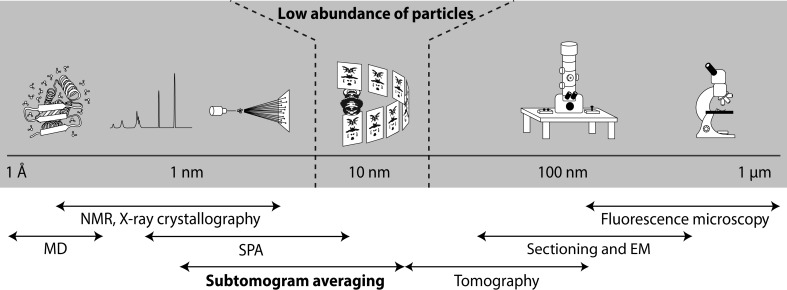
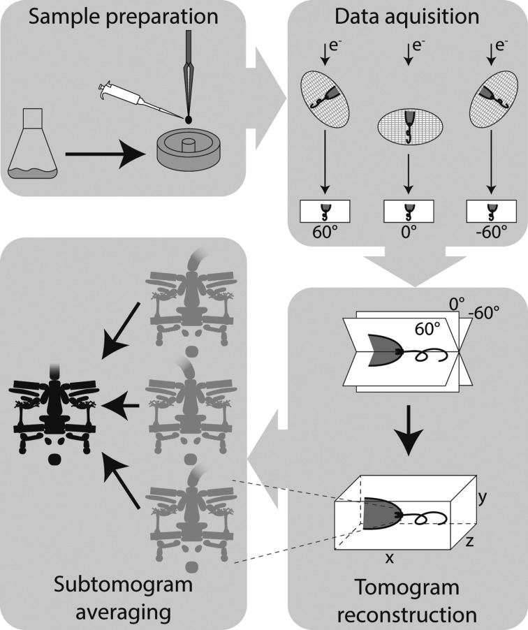
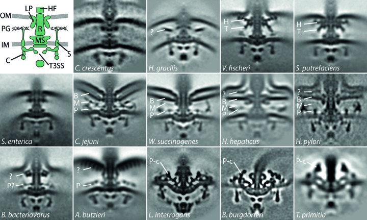
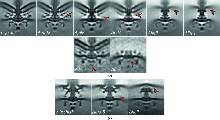
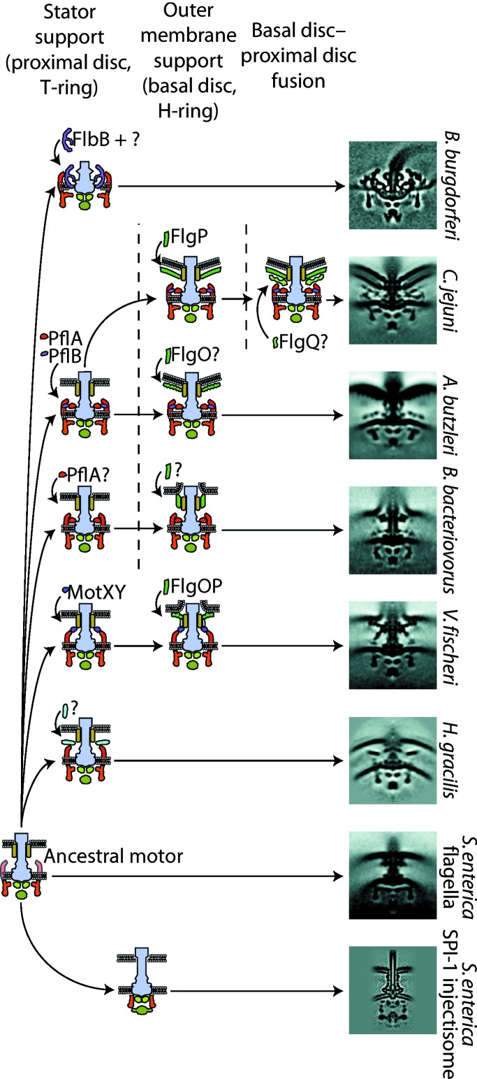
In situ structural information on molecular machines can be invaluable in understanding their assembly, mechanism and evolution. Here, the use of electron cryotomography (ECT) to obtain significant insights into how an archetypal molecular machine, the bacterial flagellar motor, functions and how it has evolved is described. Over the last decade, studies using a high-throughput, medium-resolution ECT approach combined with genetics, phylogenetic reconstruction and phenotypic analysis have revealed surprising structural diversity in flagellar motors. Variations in the size and the number of torque-generating proteins in the motor visualized for the first time using ECT has shown that these variations have enabled bacteria to adapt their swimming torque to the environment. Much of the structural diversity can be explained in terms of scaffold structures that facilitate the incorporation of additional motor proteins, and more recent studies have begun to infer evolutionary pathways to higher torque-producing motors. This review seeks to highlight how the emerging power of ECT has enabled the inference of ancestral states from various bacterial species towards understanding how, and `why', flagellar motors have evolved from an ancestral motor to a diversity of variants with adapted or modified functions.

Keywords: bacterial flagellar motors; electron cryotomography; low-abundance imaging; molecular evolution; subtomogram averaging.


Figures

Figure 1
Figure 1
Schematic depicting electron cryotomography and subtomogram averaging of rare particles in the structural biology continuum. ECT bridges the gap between high-resolution structural biology techniques requiring the presence of a high abundance of particles and the low-resolution techniques used in cell biology. (EM, electron microscopy; MD, molecular dynamics; NMR, nuclear magnetic resonance spectroscopy; SPA, single-particle analysis.)
Figure 2
Figure 2
Illustration of the general workflow of ECT and STA to study bacterial flagellar motors. Schematic showing the different steps including sample preparation, data acquisition, tomogram reconstruction and subtomogram averaging. Samples are plunge-frozen in liquid cryogen, transferred to the microscope for the acquisition of images of cells over a range of angles and computationally reconstructed to form a tomogram; finally, identical structures from different cells are superimposed and averaged to yield a subtomogram average.
Figure 3
Figure 3
The architecture of bacterial flagellar motors reveals considerable structural diversity. Top left: schematic of the flagellar motor. Top right, middle and bottom row: micrographs show central slices (100 × 100 nm) of subtomogram averages of C. crescentus, H. gracilis (Chen et al., 2011 ▸); V. fischeri (Beeby et al., 2016); S. putrefaciens; S. enterica, C. jejuni (Beeby et al., 2016 ▸); W. succinogenes (Chaban et al., 2018 ▸); H. hepaticus (Chen et al., 2011 ▸); H. pylori (Qin et al., 2016 ▸); B. bacteriovorus, A. butzleri (Chaban et al., 2018 ▸); Leptospira interrogans (Zhao et al., 2014 ▸); B. burgdorferi (Zhao et al., 2013 ▸); T. primitia (Murphy et al., 2006 ▸). Components are labelled as follows: B, basal disk; C, C-ring; H, H-ring; HF, hook/filament; IM, inner membrane; LP, L/P-ring; M, medial disk; MS, MS-ring; OM, outer membrane; P, proximal disk; P-c, P-collar; PG, peptidoglycan layer; R, rod; S, stators; T, T-ring; T3SS, type 3 secretion system.
Figure 4
Figure 4
ECT and STA of deletion mutants helps to locate individual proteins within the overall motor architecture. Micrographs show central slices (100 × 100 nm) of subtomogram averages of C. jejuni (a) and V. fischeri (b) wild type and deletion mutants. Arrows point at the putative location of the respective, deleted protein that can be determined by comparison with other mutants and established biochemical data (Beeby et al., 2016; Abrusci et al., 2013; Chen et al., 2011 ▸).
Figure 5
Figure 5
Proposed model for the evolution of the bacterial flagellar motor inferred from ECT and STA data. ECT and STA have indicated multiple pathways for the acquisition of additional accessory proteins resulting in improved stator support, increased outer membrane support and finally, in the case of C. jejuni, a basal disk–proximal disk fusion. ECT has also revealed structural and evolutionary insights into degenerate flagellar motors that have become injectisomes: virulence-factor delivery systems that are used by many pathogenic bacteria. Ancestral states have been inferred from representative subtomogram averages on the right coupled with phylogenetic studies (Chaban et al., 2018; Beeby et al., 2016 ▸).

Similar articles

Cited by

References

    1. Abby, S. S. & Rocha, E. P. C. (2012). PLoS Genet. 8, e1002983. - PMC - PubMed
    1. Abrusci, P., Vergara-Irigaray, M., Johnson, S., Beeby, M. D., Hendrixson, D. R., Roversi, P., Friede, M. E., Deane, J. E., Jensen, G. J., Tang, C. M. & Lea, S. M. (2013). Nature Struct. Mol. Biol. 20, 99–104. - PMC - PubMed
    1. Agulleiro, J. I. & Fernandez, J. J. (2011). Bioinformatics, 27, 582–583. - PubMed
    1. Altegoer, F. & Bange, G. (2015). Curr. Opin. Microbiol. 28, 98–105. - PubMed
    1. Amat, F., Moussavi, F., Comolli, L. R., Elidan, G., Downing, K. H. & Horowitz, M. (2008). J. Struct. Biol. 161, 260–275. - PubMed

Publication types

MeSH terms

Substances