. 2018 Jun 5;9(1):2184.
doi: 10.1038/s41467-018-04580-3.

In vivo base editing of post-mitotic sensory cells

Affiliations

In vivo base editing of post-mitotic sensory cells

Wei-Hsi Yeh et al. Nat Commun. .

Abstract

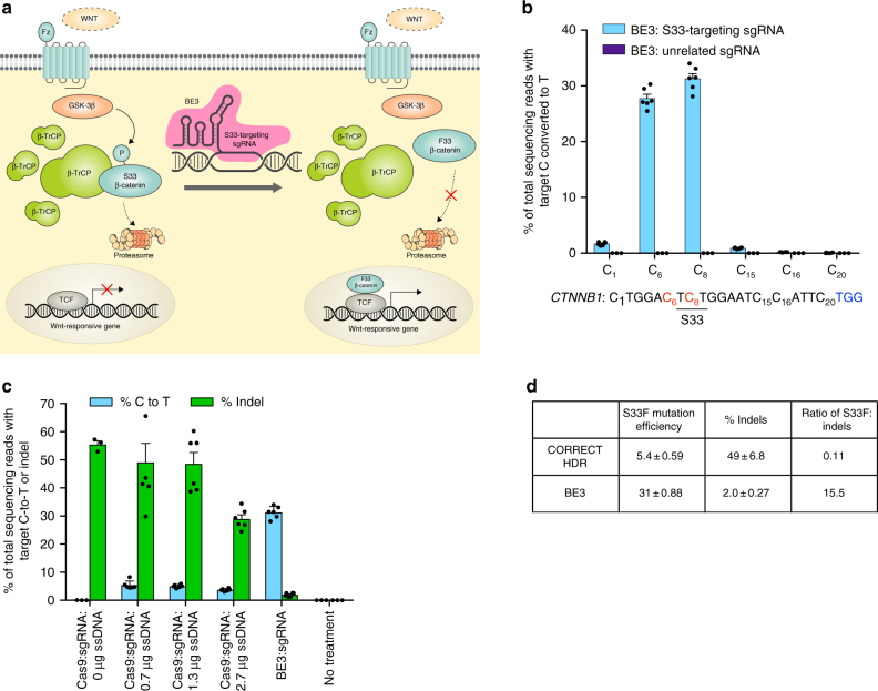
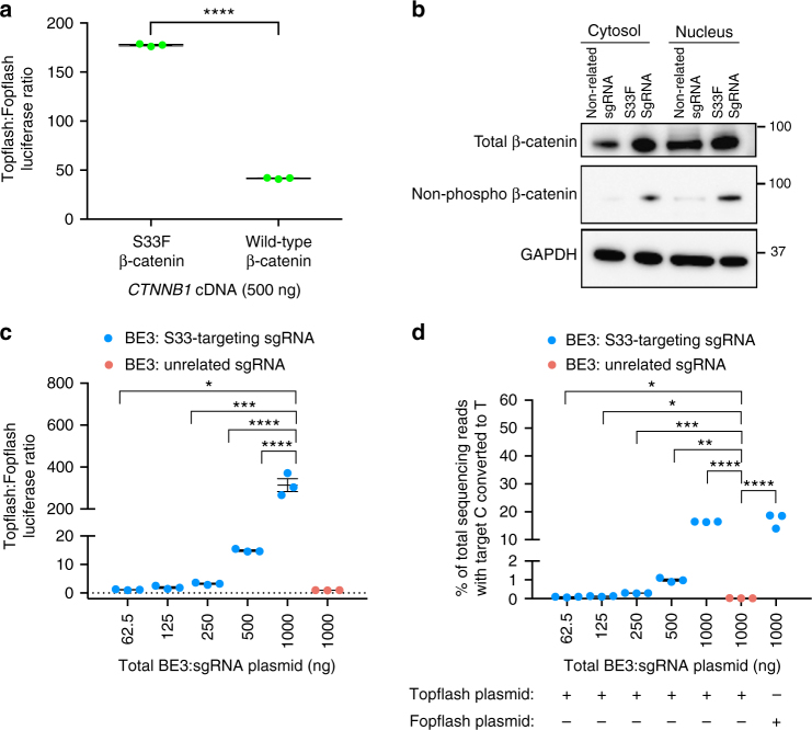
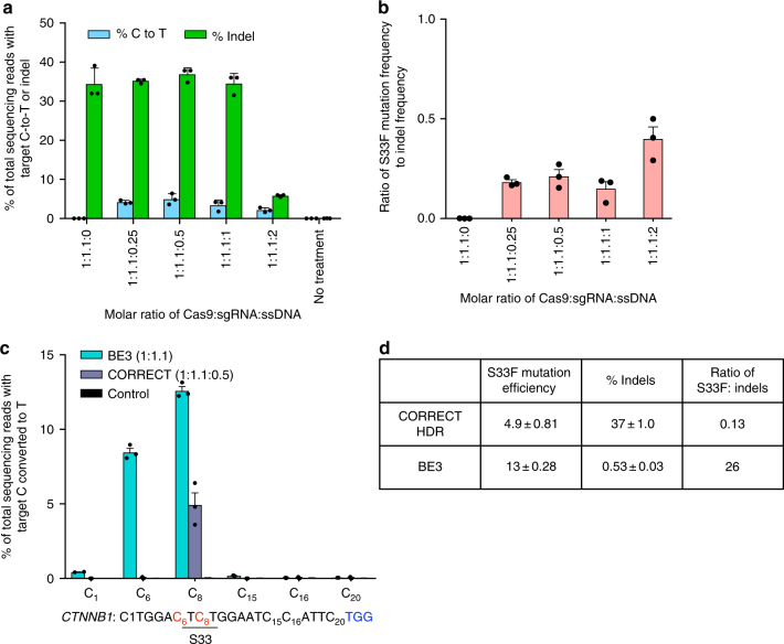
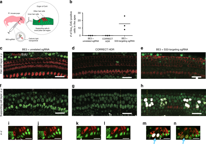
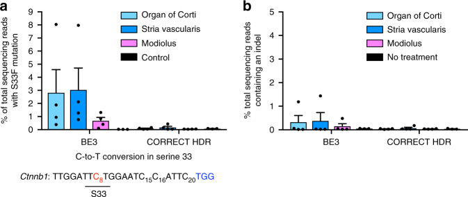
Programmable nucleases can introduce precise changes to genomic DNA through homology-directed repair (HDR). Unfortunately, HDR is largely restricted to mitotic cells, and is typically accompanied by an excess of stochastic insertions and deletions (indels). Here we present an in vivo base editing strategy that addresses these limitations. We use nuclease-free base editing to install a S33F mutation in β-catenin that blocks β-catenin phosphorylation, impedes β-catenin degradation, and upregulates Wnt signaling. In vitro, base editing installs the S33F mutation with a 200-fold higher editing:indel ratio than HDR. In post-mitotic cells in mouse inner ear, injection of base editor protein:RNA:lipid installs this mutation, resulting in Wnt activation that induces mitosis of cochlear supporting cells and cellular reprogramming. In contrast, injection of HDR agents does not induce Wnt upregulation. These results establish a strategy for modifying posttranslational states in signaling pathways, and an approach to precision editing in post-mitotic tissues.


Conflict of interest statement

D.R.L. is a consultant and co-founder of Editas Medicine, Beam Therapeutics, and Pairwise Plants, companies that are using genome editing. A.S.B.E. is a consultant and co-founder of Decibel Therapeutics. The remaining authors declare no competing interests.

Figures

Fig. 1
Fig. 1
Base editing strategy and comparison of HDR and base editing following plasmid delivery. a Schematic representation of the canonical Wnt pathway and a base editing strategy to stabilize β-catenin. In the absence of Wnt signaling, β-catenin is phosphorylated at Ser 33 by GSK-3β and degraded in a phosphorylation-dependent manner. Base editing with BE3 precisely mutates the Ser 33 codon to instead encode Phe, which cannot be phosphorylated. The resulting S33F β-catenin has an extended half-life and can activate target gene transcription by binding with TCF/LEF transcription factors. b HEK293T cells were transfected with plasmids expressing BE3 and S33-targeting sgRNA, or BE3 and an unrelated sgRNA. The percentage of total sequencing reads (with no enrichment for transfected cells) with C8 converted to T8 (resulting in the S33F mutation) was measured with high-throughput sequencing (HTS). c Plasmid delivery of Cas9 and BE3 (750 ng) with S33-targeting sgRNA (250 ng) into HEK293T cells using 1.5 µL of Lipofectamine 2000 per well of a 48-well plate. C-to-T conversion efficiency and d product selectivity ratio (desired S33F mutation: undesired indel ratio) resulting from the best-performing ratio of Cas9:sgRNA:ssDNA template and BE3. Values and error bars reflect mean ± S.E.M. of three biological replicates performed on separate days
Fig. 2
Fig. 2
Biological outcomes associated with base editing S33F in β-catenin in human cells. a HEK293T cells were transfected with plasmids encoding Topflash (β-catenin-responsive firefly luciferase reporter) or Fopflash (mutant form of Topflash that cannot be activated by β-catenin) and mutant S33F β-catenin or wild-type (Ser 33) β-catenin. Wnt signaling was measured by the ratio of Topflash:Fopflash luciferase activity. b Cytosolic and nuclear extracts of HEK293T cells treated with base editor and S33-targeting sgRNA or unrelated sgRNA were subjected to western blot analysis for total β-catenin or non-phosphorylated β-catenin (Ser33/Ser37/Thr41). Each blot represents one antibody. GAPDH was used as a loading control. c HEK293T cells were transfected with plasmids encoding the Topflash or Fopflash reporters, base editor, and S33F or unrelated control sgRNA. The Topflash:Fopflash luciferase ratio for BE3 + S33-targeting sgRNA (blue) and BE3 + unrelated sgRNA (red) are shown. d Percent C-to-T conversion at the target Ser 33 codon, which results in the S33F mutation in β-catenin, assayed by HTS. Values and error bars reflect mean ± S.E.M. of three biological replicates performed on different days. * p ≤ 0.05, ** p ≤ 0.01, *** p ≤ 0.001, and **** p ≤ 0.0001 (Students two tailed t-test)
Fig. 3
Fig. 3
Comparison of efficiency and product selectivity of HDR vs. base editing following RNP delivery. RNP delivery into HEK293T cells of 200 nM Cas9 or 200 nM BE3 pre-complexed with the S33-targeting sgRNA or an unrelated sgRNA and delivered in a cationic liposome. a Frequency of S33F mutation (blue) and indels (green) from treatment with CORRECT HDR agents (Cas9, sgRNA, and ssDNA donor template) in the ratios shown. b Product selectivity ratio, defined as the ratio of S33F modification to indel modification, resulting from treatment with the CORRECT HDR agents used in a. c Comparison of target C to T conversion efficiency following RNP delivery of BE3 (blue) or CORRECT HDR (purple) and the S33-targeting sgRNA. The control corresponds to cells treated with BE3 protein and unrelated sgRNA. d Product selectivity of base editing and CORRECT HDR. Values and error bars reflect mean ± S.E.M. of three independent biological replicates performed on different days
Fig. 4
Fig. 4
Outcomes associated with in vivo RNP-mediated base editing in post-mitotic cochlea. a The cochlea of a postnatal day 1 (P1) mouse was injected with lipid nanoparticles encapsulating BE3 + sgRNA, CORRECT HDR agents, or controls. The next day, mice received 5-ethynyl-2-deoxyuridine (EdU) by subcutaneous injection once per day for 5 days. At postnatal day 7 (P7), one day after the fifth EdU injection, the cochlea was dissected and organ of Corti was visualized by chemical staining (to visualize EdU) and immunofluorescence (Myo7a and Sox2). In the cochlea, Myo7a (red) is expressed in hair cells and Sox2 (green) is expressed in supporting cells. EdU (white) marks newly divided cells. b Quantification of Sox2 and EdU double-positive cells at the apical region of the organ of Corti from mice treated with BE3 + S33-targeting sgRNA (n = 3), CORRECT HDR agents (n = 3), or BE3 + unrelated sgRNA (n = 3). Values and error bars reflect mean and S.E.M. cn Images from the organ of Corti tissue of mice treated with BE3 + unrelated sgRNA (c, f, i, j); CORRECT HDR agents (d, g, k, l); or BE3 + S33-targeting sgRNA (e, h, m, n). The blue arrows point to triple-positive cells that had undergone proliferation and reprogramming to cells expressing Myo7a. ce xy plane of hair cell layers. fh xy plane of supporting cell layers. (i, k, m) xz plane of samples in f, g, h at the dotted yellow lines. j, k, m xz plane of samples in f, g, h at the dotted yellow lines, but with the Myo7a and Sox2 channels shown. Scale bar (white) = 25 μm
Fig. 5
Fig. 5
In vivo S33F mutation of β-catenin induced by injection of base editor RNPs. a Tissue was harvested from the cochlea of mice injected with either BE3 + S33-targeting sgRNA, or with CORRECT HDR agents. HTS of genomic DNA isolated from tissue samples revealed the frequency of the S33F mutation. Note that because tissue samples contain cells not exposed to editing agents, the observed genome modification frequency in these samples is less than the editing efficiency of treated cells. b Indel frequency at the Ser 33 locus following treatments described in a. Values and error bars reflect mean ± S.E.M. of four mice injected with BE3 and four mice injected with CORRECT HDR agents. Control samples in a, b are organ of Corti from three contralateral uninjected ears

References

    1. Cox DBT, Platt RJ, Zhang F. Therapeutic genome editing: prospects and challenges. Nat. Med. 2015;21:121–131. doi: 10.1038/nm.3793. - DOI - PMC - PubMed
    1. Doudna, J. A. & Charpentier, E. The new frontier of genome engineering with CRISPR-Cas9. Science346, 1258096 (2014). - PubMed
    1. Komor AC, Kim YB, Packer MS, Zuris JA, Liu DR. Programmable editing of a target base in genomic DNA without double-stranded DNA cleavage. Nature. 2016;533:420–424. doi: 10.1038/nature17946. - DOI - PMC - PubMed
    1. Sander JD, Joung JK. CRISPR-Cas systems for editing, regulating and targeting genomes. Nat. Biotechnol. 2014;32:347–355. doi: 10.1038/nbt.2842. - DOI - PMC - PubMed
    1. Biot-Pelletier D, Martin VJJ. Seamless site-directed mutagenesis of the Saccharomyces cerevisiae genome using CRISPR-Cas9. J. Biol. Eng. 2016;10:6. doi: 10.1186/s13036-016-0028-1. - DOI - PMC - PubMed

Publication types