. 2018 Jun 5;9(1):2181.
doi: 10.1038/s41467-018-04639-1.

Gut vagal sensory signaling regulates hippocampus function through multi-order pathways

Affiliations

Gut vagal sensory signaling regulates hippocampus function through multi-order pathways

Andrea N Suarez et al. Nat Commun. .

Abstract

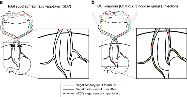
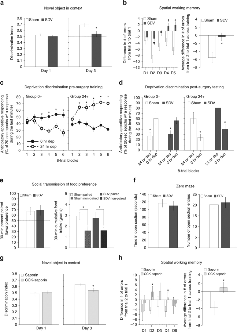
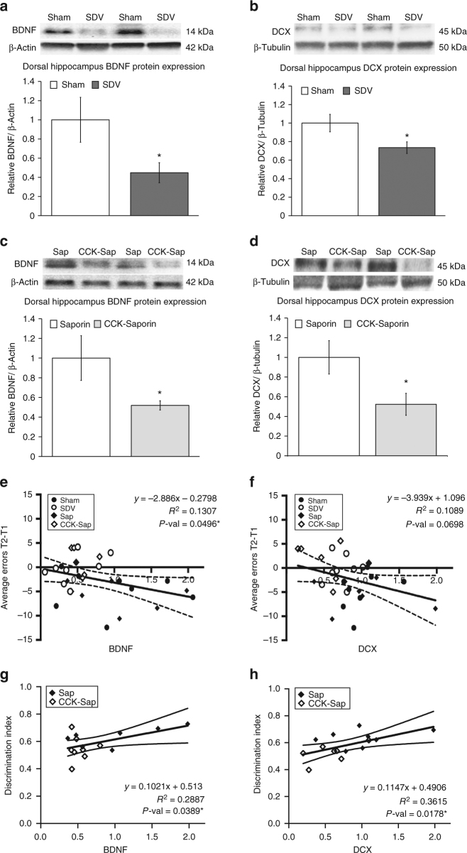
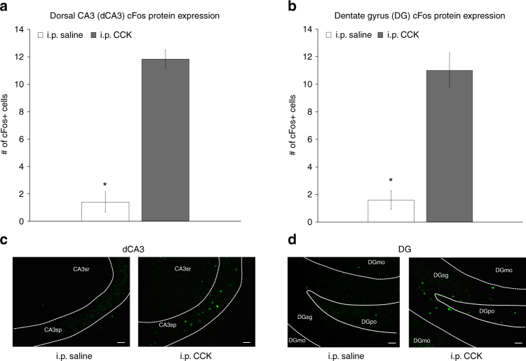
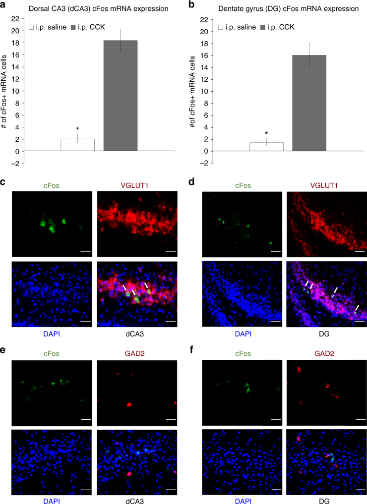
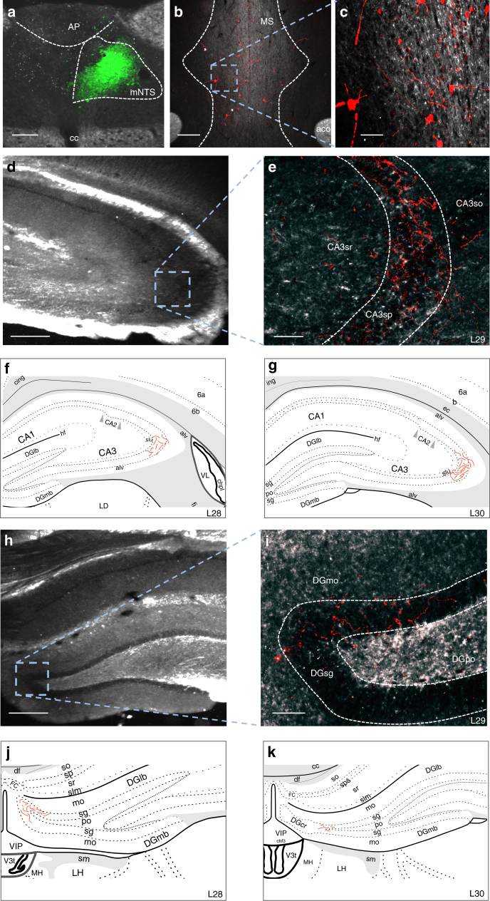
The vagus nerve is the primary means of neural communication between the gastrointestinal (GI) tract and the brain. Vagally mediated GI signals activate the hippocampus (HPC), a brain region classically linked with memory function. However, the endogenous relevance of GI-derived vagal HPC communication is unknown. Here we utilize a saporin (SAP)-based lesioning procedure to reveal that selective GI vagal sensory/afferent ablation in rats impairs HPC-dependent episodic and spatial memory, effects associated with reduced HPC neurotrophic and neurogenesis markers. To determine the neural pathways connecting the gut to the HPC, we utilize monosynaptic and multisynaptic virus-based tracing methods to identify the medial septum as a relay connecting the medial nucleus tractus solitarius (where GI vagal afferents synapse) to dorsal HPC glutamatergic neurons. We conclude that endogenous GI-derived vagal sensory signaling promotes HPC-dependent memory function via a multi-order brainstem-septal pathway, thereby identifying a previously unknown role for the gut-brain axis in memory control.


Conflict of interest statement

The authors declare no competing interests.

Figures

Fig. 1
Fig. 1
Schematic illustration of subdiaphragmatic vagus nerve ablative disconnection methods. a Classic total subdiaphragmatic vagotomy (SDV) surgical method consists of lesioning the dorsal and ventral subdiaphragmatic vagus nerve, eliminating 100% of vagal afferent (sensory) and efferent (motor) signaling below the diaphragm. b The novel CCK saporin (CCK-SAP) approach consists of nodose ganglia injections of saporin conjugated to cholecystokinin to specifically ablate ~ 80% of vagal gastrointestinal (GI)-innervating afferent signaling, while leaving 100% of vagal efferent and supradiaphragmatic vagal afferent signaling intact (see ref. ). (DMX dorsal motor nucleus of the vagus nerve, mNTS medial nucleus tractus solitarius). [Cartoon schematic made by authors based on ref. ]
Fig. 2
Fig. 2
SDV and CCK-Sap impair HPC-dependent contextual episodic and spatial working memory, but not interoceptive, social, or olfactory learning. a SDV (n = 6) impairs contextual episodic memory relative to controls (n = 9); discrimination index on day 1 (habituation) and day 3 (test day) of NOIC testing (repeated-measures ANOVA, F[1,13] = 5.564, p = 0.0347; Newman–Keuls’ post hoc, p = 0.0047). b SDV (n = 8) impairs spatial working memory relative to controls (n = 8); difference in number of errors from trial 2 (T2) to trial 1 (T1) across individual training days (left) (ANOVA, F[1,14] = 3.626, p = 0.0776 (Day 2), F[1,14] = 3.842, p = 0.0702 (Day 4), F[1,14] = 3.555, p = 0.0803 (Day 5)) and the average T2–T1 errors for each training day in the Barnes maze test (repeated-measures ANOVA, F[1,19] = 6.8565, p = 0.0169). c, d SDV does not impact deprivation intensity discrimination performance; c pre-surgery training (Group 0 + , n = 16; Group 24 + , n = 11; repeated-measures ANOVA, F[1,22] = 135.54, p < 0.0001; Newman–Keuls’ post hoc, Group 0 + block 3–6 all p < 0.0017, Group 24 + block 2–6 all p < 0.000178), d and post-surgery testing [mean percent of 20 s epochs of interval magazine entries during the last minute of test session for Group 0 + (sham, n = 8; SDV, n = 7) and 24 + (sham, n = 6; SDV, n = 5) under alternating 0 h and 24 h food restriction] (repeated-measures ANOVA, F[1,22] = 80.5115, p < 0.00001; Newman–Keuls’ post hoc, all p < 0.004). e SDV (n = 6) does not impact STFP relative to controls (n = 9); 30 min percent preference for the socially paired flavored chow and 30 min cumulative food intake (grams) in the STFP test (paired t-test, p = 0.014 (SDV), p = 0.014 (sham)). f SDV does not impact anxiety-like behavior; time spent in open arm section (seconds) and number of open section entries during zero maze test for the SDV vs. sham groups. g CCK-SAP impairs contextual episodic memory; NOIC discrimination index on days 1 and 3 in CCK-SAP (n = 9) and SAP (n = 8) control rats (repeated-measures ANOVA, F[1,15] = 6.496, p = 0.0223; Newman–Keuls’ post hoc, p = 0.0241). h CCK-SAP impairs SWM; (T2–T1 error for each individual training day (ANOVA, F[1,13] = 6.824, p = 0.0215 (Day 2)) and overall average (repeated-measures ANOVA, F[1,13] = 8.66, p = 0.0114) in CCK-SAP (n = 8) and SAP (n = 7) control rats. (*P < 0.05; ŦP < 0.08 vs. sham or SAP controls; data are mean ± SEM)
Fig. 3
Fig. 3
SDV and CCK-SAP reduce BDNF and DCX protein expression in the dorsal HPC and are functionally related to HPC-dependent memory performance. a, b SDV reduces protein expression of BDNF and DCX (expressed relative to loading control proteins) in dorsal HPC tissue in SDV (n = 8) vs. sham-operated control rats (n = 8) (ANOVA, F[1,14] = 4.609, p = 0.049 (BDNF), F[1,14] = 5.5133, p = 0.034 (DCX)). c, d CCK-SAP-mediated GI vagal afferent ablation (n = 8) reduces dorsal HPC BDNF and DCX expression relative to SAP (SAP BDNF, n = 7; SAP DCX, n = 8) controls (ANOVA, F[1,13] = 4.881, p = 0.0457 (BDNF), F[1,14] = 5.494, p = 0.034 (DCX). eh Linear regression of average number of errors from trial 2 to trial 1 (spatial working memory) and NOIC discrimination index (contextual episodic memory) against relative BDNF and DCX expression reveals a significant negative correlation for BDNF (F[1,28] = 4.211, R2 = 0.1307, p = 0.0496) (e) with a trend for DCX (F[1,29] = 3.546, R2 = 0.1089, p = 0.0698) (f). For the novel object in context (NOIC) task of contextual episodic memory, there was a positive correlation between discrimination index and protein expression of BDNF (F[1,13] = 5.277, R2 = 0.2887, p = 0.0389) (g) and DCX (F[1,13] = 7.36, R2 = 0.3615, p = 0.0178) (h). (*P < 0.05 vs. controls [sham and/or SAP]; data are mean ± SEM. BDNF brain-derived neurotrophic factor, CCK-SAP cholecystokinin–saporin, DCX doublecortin, SDV subdiaphragmatic vagotomy
Fig. 4
Fig. 4
Peripheral administration of CCK activates c-Fos protein expression in the dorsal CA3 (dCA3) and dentate gyrus (DG). Intraperitoneal injections of CCK (n = 6) (a vagally mediated gastrointestinal-derived satiation signal) increases the number of c-Fos-immunoreactive (-ir) cells (a marker for neural activation) expressed in the a dCA3 and b DG vs. saline (n = 5) treatment (ANOVA, F[1,9] = 20.236, p = 0.001492 (dCA3), F[1,9] = 37.917, p = 0.000167 (DG)). Representative images of immunohistochemical staining of c-Fos-ir protein (green) in the c dCA3 and d DG. Scale bar: 25 μm. (*P < 0.05 vs. i.p. saline controls; data are mean ± SEM.; CCK cholesystokinin, i.p. intraperional, DG dentate gyrus, dCA3 dorsal CA3, CA3sr CA3 stratum radiatum, CA3sp CA3 pyramidal layer, DGmo dentate gyrus molecular layer, DGpo DG polymorph layer, DGsg DG granule cell layer)
Fig. 5
Fig. 5
Peripheral administration of CCK activates c-Fos mRNA expression in dCA3 and DG hippocampal glutamatergic neurons. Number of c-Fos-labeled (c-Fos + ) cells for mRNA (fluorescent in situ hybridization) expressed in the a dCA3 and b DG following i.p. administration of CCK (n = 6) or saline (n = 5) (ANOVA, F[1,9] = 53.093, p = 0.000046 (dCA3), F[1,9] = 40.496, p = 0.000131 (DG)). Approximately 93% and 94% of c-Fos + cells in the dCA3 and DG were VGLUT1 + (respectively) following i.p. CCK treatment, whereas only 4% and 8% of c-Fos + cells in the dCA3 and DG were GAD2 + (respectively) following i.p. CCK. c, d Representative images show c-Fos mRNA (green) and VGLUT1 (red) or GAD2 (e, f) (red) mRNA expression in dCA3 (c, e) and DG (d, f) cell bodies following i.p. CCK (DAPI nuclear stain; blue). Scale bar: 25 μm. (Arrows, co-expression of c-Fos/VGLUT1 mRNA cells; *P < 0.05 vs i.p. saline controls; data are mean ± SEM. CCK cholesystokinin, dCA3 dorsal CA3, DG dentate gyrus, i.p. intraperional)
Fig. 6
Fig. 6
Co-injection monosynaptic neural pathway tracing strategy identifies the medial septum (MS) as a relay region connecting the mNTS to the dHPC (CA3). Schematic representative injection sites in a dCA3 and b mNTS in Swanson Atlas level 29 and 69, respectively. c Unilateral iontophoretic dCA3 injection site of the retrograde tracer, CTB-488 (green). Scale bar: 100 μm. d Ipsilateral and unilateral iontophoretic mNTS delivery of the anterograde viral tracer, AAV1-TurboRFP (red) (n = 3 double hits, n = 11 controls). Scale bar: 500 μm. e, f, g RFP-ir axons from the mNTS in apposition to CTB-488-ir cell bodies from dCA3 in the MS (images made by authors and adapted from Swanson Atlas level 15). Scale bar: 10 μm. (AP area postrema, DG dentate gyrus, mNTS medial nucleus tractus solitarius)
Fig. 7
Fig. 7
Multisynaptic viral tracing approach reveals MS neurons that receive monosynaptic input from the mNTS directly project to the dHPC (dCA3 and DG). a Unilateral iontophoretic co-injection of AAV2/1-hSyn-Cre and CTB (CTB-ir in green; to confirm injection site placement) in the mNTS (n = 3 double hits, n = 13 controls), which drives Cre expression in second-order (but not third-order) neurons based on synaptic virion release from first-order axon terminals. Scale bar: 100 μm. b, c A 200 nl pressure injection site of a Cre-dependent anterograde tracer (AAV1-CAG-FLEX-TdTomato) in the MS Scale bar: b 200 μm, c 50 μm. Axon terminal fields in the d, e dCA3 and h, i DG of MS neurons that receive direct input from mNTS. Scale bar: d, h 250 μm, e, i 50 μm. A schematic representation of dCA3 (f, g) and DG (j, k) axon terminal field distribution (Made by co-author and adapted from Swanson atlas level 28–30). (aco anterior commissure, AP area postrema, CA3sr CA3 stratum radiatum, CA3sp CA3 pyramidal layer, DGmo dentate gyrus molecular layer, DGpo DG polymorph layer, DGsg DG granule cell layer, mNTS medial nucleus tractus solitarius, MS, medial septum)

References

    1. Grill HJ, Hayes MR. Hindbrain neurons as an essential hub in the neuroanatomically distributed control of energy balance. Cell. Metab. 2012;16:296–309. doi: 10.1016/j.cmet.2012.06.015. - DOI - PMC - PubMed
    1. Kanoski SE, Grill HJ. Hippocampus contributions to food intake control: mnemonic, neuroanatomical, and endocrine mechanisms. Biol. Psychiatry. 2017;81:748–756. doi: 10.1016/j.biopsych.2015.09.011. - DOI - PMC - PubMed
    1. Min DK, Tuor UI, Chelikani PK. Gastric distention induced functional magnetic resonance signal changes in the rodent brain. Neuroscience. 2011;179:151–158. doi: 10.1016/j.neuroscience.2011.01.051. - DOI - PubMed
    1. Min DK, Tuor UI, Koopmans HS, Chelikani PK. Changes in differential functional magnetic resonance signals in the rodent brain elicited by mixed-nutrient or protein-enriched meals. Gastroenterology. 2011;141:1832–1841. doi: 10.1053/j.gastro.2011.07.034. - DOI - PubMed
    1. Wang GJ, et al. Gastric stimulation in obese subjects activates the hippocampus and other regions involved in brain reward circuitry. Proc. Natl Acad. Sci. USA. 2006;103:15641–15645. doi: 10.1073/pnas.0601977103. - DOI - PMC - PubMed

Publication types