. 2018 Jun 5;8(1):8642.
doi: 10.1038/s41598-018-26985-2.

Large-scale additive manufacturing with bioinspired cellulosic materials

Affiliations

Large-scale additive manufacturing with bioinspired cellulosic materials

Naresh D Sanandiya et al. Sci Rep. .

Abstract

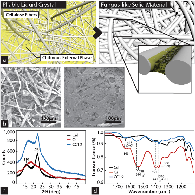
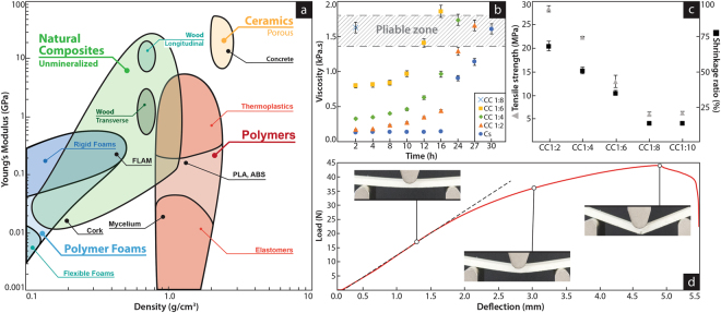
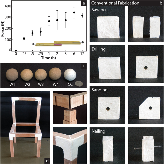
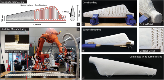
Cellulose is the most abundant and broadly distributed organic compound and industrial by-product on Earth. However, despite decades of extensive research, the bottom-up use of cellulose to fabricate 3D objects is still plagued with problems that restrict its practical applications: derivatives with vast polluting effects, use in combination with plastics, lack of scalability and high production cost. Here we demonstrate the general use of cellulose to manufacture large 3D objects. Our approach diverges from the common association of cellulose with green plants and it is inspired by the wall of the fungus-like oomycetes, which is reproduced introducing small amounts of chitin between cellulose fibers. The resulting fungal-like adhesive material(s) (FLAM) are strong, lightweight and inexpensive, and can be molded or processed using woodworking techniques. We believe this first large-scale additive manufacture with ubiquitous biological polymers will be the catalyst for the transition to environmentally benign and circular manufacturing models.


Conflict of interest statement

The authors declare no competing interests.

Figures

Figure 1
Figure 1
Supramolecular organization of fungus-like mimetic materials. (a) Cellulose fibers from plant origin are dispersed in chitosan solution. After removal of the water, chitosan crystallize in between fibers. In the process the sterically available amino groups in chitosan not involved in the crystal conformation react with the free hydroxyl groups on the surface of the cellulose fiber. As the chitosan losses intermolecular water, the polymer crystal reduced volume brings together the cellulosic fibers into a solid composite. (b) Scanning electron microscopic images of the cellulose fibers (left) and their structure in the chitinous composite (12.5% chitosan concentration). (c) X-ray diffraction pattern of the composite and their constituents. The data shows a cellulose I polymorph unaltered during the formation of the composite. A relaxation on the crystal structure, reflected in a shift of the 002 reflection, could be caused for the hydrogen bond of the cellulosic hydroxyl groups with chitosan, reducing the amount of cellulose-cellulose intermolecular hydrogen bonds. (d) FTIR fingerprint of the Chitosan-cellulose composite. The amino groups of the chitosan shifted from 1538 to 1556 cm−1 and the band associated with the hydroxyl groups of the cellulose shifted from 1640 to 1648 cm−1 indicated the interaction between amino groups of chitosan and hydroxyl groups of the cellulose.
Figure 2
Figure 2
Mechanical characteristics of fungus-like biomimetic materials. (a) Ashby plot showing the distribution of density and stiffness of natural and synthetic material commonly used in manufacture. Those relevant to this study have been highlighted, while the specific function of the fungus-like bioinspired material reported is labeled as “FLAM”. (b) Viscosity in terms of time for composites of variable amounts of chitosan. The highlighted area represents the range of viscosities suitable for manufacturing techniques, where the material can be extruded, and conform and retain a shape. (c) Determination of the optimal concentration regarding the balance between mechanical properties (tensile strength) and manufacturability (shrinking). (d) 3-point fracture test. Similar to other natural composites, the resulting material is designed for multifunctional structures, balancing strength and stiffness. The 100 × 16 × 6 mm and 3 g FLAM sample shows ductile characteristics; it holds 2.55 kg elastically, after that the internal structure starts to deform, resulting in failure when the load reaches 4.37 kg (Supplementary Movie 1).
Figure 3
Figure 3
Additive properties of fungus-like materials and their use in woodworking techniques. (a) Adhesion force with respect time following the standard test of adhesion (ASTM D5868) with respect to time. Full strength is achieved after 1 hour, from that point 21 mg of dry FLAM covering an area of 9.3 × 9.3 mm holds the equivalent to 29.02 ± 6.35 kg. This ability of the material to attach to cellulosic composites (included itself) enables its use in additive manufacturing. (b) Use of FLAM in conventional woodworking techniques. A 4 × 4 × 4 cm FLAM cube is sawn in two halves, one of the halves is then drilled and then sanded down to remove one of the corners. A nail is hammered through the other half. (Supplementary Movie 2) (c) Composites made with different sources of cellulosic materials. Samples one to four (from left) are made of wood byproducts of different qualities and sources, while the right sphere (labeled “CC”) is made of pure cellulose (Supplementary Tables 1 and 2). (d) Use of the material in combination with pieces of solid wood to produce a functional chair. All pieces are attached uniquely by the FLAM material. The fungus-like biomimetic material can be casted, 3D printed, molded but also modified using regular woodworking techniques (Supplementary Fig. 4).
Figure 4
Figure 4
Additive manufacture of fungus-like materials. (a) Design of the wind turbine blade fully made of FLAM. The inner core of the blade, built by additive manufacturing, is designed to allow ventilation and reduce weight. The outer shell is produced by coating the core with a 3 mm layer of FLAM. After drying the outer shell is sanded down to produce a smooth surface. (b) FLAM material is dispensed through a 7 mm diameter nozzle to form beads and then layers of material. The pressure is controlled by a closed loop system between a high-pressure tank (1.2 MPa) supplying material and an auger screw before the nozzle. Hot air is focused on the extruded layer just after deposition to accelerate water evaporation. The printing head is mounted on a 20 kg payload six-axis industrial robotic arm with a stationary horizontal reach radius of 1.65 m (Supplementary Fig. 6). (c) FLAM printing of the layers to support the structure of the wind turbine blade. The blade’s core was printed in two halves, each taking about 1 h and 24 h to dry. The average printing speed is of about 50 mm/s and 2.8 cm3 of FLAM per second (Supplementary Movie 3). Two halves of the inner core are assembled together using FLAM as adhesive agent. (d) The blade is coated by a layer of FLAM manually spread over the inner core. Because the ability of the material to be post processed, imperfections can be removed in post-processing stages. (e) After the outer skin is dried, it is sanded down in two steps of decreasing grit. The 1.2 m blade is estimated to be 50% hollow inside and has a weight of 5.28 kg. (Supplementary Movie 4 and Supplementary Fig. 5).

References

    1. Reiterer A, Lichtenegger H, Tschegg S, Fratzl P. Experimental evidence for a mechanical function of the cellulose microfibril angle in wood cell walls. Philosophical Magazine A. 1999;79:2173–2184. doi: 10.1080/01418619908210415. - DOI
    1. Toivonen, M. et al. Water-Resistant, Transparent Hybrid Nanopaper by Physical Cross-Linking with Chitosan. Vol. 16 (2015). - PubMed
    1. Robles E, Salaberria AM, Herrera R, Fernandes SCM, Labidi J. Self-bonded composite films based on cellulose nanofibers and chitin nanocrystals as antifungal materials. Carbohydrate Polymers. 2016;144:41–49. doi: 10.1016/j.carbpol.2016.02.024. - DOI - PubMed
    1. Bartnicki-Garcia S. Cell wall chemistry, morphogenesis, and taxonomy of fungi. Annual review of microbiology. 1968;22:87–108. doi: 10.1146/annurev.mi.22.100168.000511. - DOI - PubMed
    1. Lévesque CA. Fifty years of oomycetes—from consolidation to evolutionary and genomic exploration. Fungal Diversity. 2011;50:35. doi: 10.1007/s13225-011-0128-7. - DOI

Publication types