Review
. 2018 Oct;15(10):585-600.
doi: 10.1038/s41569-018-0036-6.

Therapeutic approaches for cardiac regeneration and repair

Affiliations
Review

Therapeutic approaches for cardiac regeneration and repair

Hisayuki Hashimoto et al. Nat Rev Cardiol. 2018 Oct.

Abstract

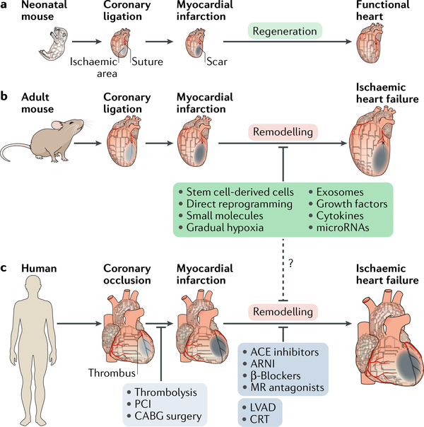
Ischaemic heart disease is a leading cause of death worldwide. Injury to the heart is followed by loss of the damaged cardiomyocytes, which are replaced with fibrotic scar tissue. Depletion of cardiomyocytes results in decreased cardiac contraction, which leads to pathological cardiac dilatation, additional cardiomyocyte loss, and mechanical dysfunction, culminating in heart failure. This sequential reaction is defined as cardiac remodelling. Many therapies have focused on preventing the progressive process of cardiac remodelling to heart failure. However, after patients have developed end-stage heart failure, intervention is limited to heart transplantation. One of the main reasons for the dramatic injurious effect of cardiomyocyte loss is that the adult human heart has minimal regenerative capacity. In the past 2 decades, several strategies to repair the injured heart and improve heart function have been pursued, including cellular and noncellular therapies. In this Review, we discuss current therapeutic approaches for cardiac repair and regeneration, describing outcomes, limitations, and future prospects of preclinical and clinical trials of heart regeneration. Substantial progress has been made towards understanding the cellular and molecular mechanisms regulating heart regeneration, offering the potential to control cardiac remodelling and redirect the adult heart to a regenerative state.


Conflict of interest statement

Competing interests

The authors declare no competing interests.

Figures

Fig. 1 |
Fig. 1 |. Response and therapeutic approaches to myocardial injury.
a | Response tomyocardial injury differs between developmental stages in mice. Neonatal mice (aged <1 week) are capable of regenerating the heart, with functional recovery after injury.This regenerative capacity is lost postnatally after the first week. b,c | In adult mice and humans, the default response to myocardial injury is fibrosis, where the infarct necrotic tissue is replaced with a fibrotic scar, causing loss of cardiac contractility. The damaged adult heart enters a negative loop of cardiac remodelling that progresses to heart failure. In humans, the current goal of clinical therapies is either to salvage the ischaemic myocardium by early revascularization (light grey box) or to prevent cardiac remodelling with drug therapy and electromechanical support (dark grey box). Accumulating evidence in preclinical studies demonstrates promising outcomes with therapeutic approaches aimed at heart regeneration (green box), although these new approaches have clinical translational problems. The dashed line indicates potential clinical therapeutic approaches. ACE, angiotensin- converting enzyme; ARNI, angiotensin receptor–neprilysin inhibitor ; CRT, cardiac resynchronization therapy; LVAD, left ventricular assist device; MR, mineralocorticoid receptor ; PCI, percutaneous coronary intervention.
Fig. 2 |
Fig. 2 |. Contributions of secretory factors to cardiac repair and regeneration.
A scheme of cardioprotective effects (green boxes) or cardiac remodelling effects (red boxes) by representative secretory factors (growth factors, microRNAs, and exosomes) is shown. Various secretory factors promote angiogenesis or cardiomyocyte proliferation, thereby promoting cardiac repair. Other secretory factors elicit cardioprotective effects by attenuating cardiac remodelling through inhibition of fibrosis, cardiomyocyte apoptosis, and oxidative stress. The dashed arrow indicates incompletely understood mechanisms. CDC, cardiosphere-derived cell; CPC, cardiac progenitor cell; FGF, fibroblast growth factor; MSC, mesenchymal stem cell; NRG1, neuregulin 1; ROS, reactive oxygen species; VEGF, vascular endothelial growth factor.
Fig. 3 |
Fig. 3 |. Direct reprogramming of fibroblasts into cardiomyocytes.
a | Forced expression of cardiac transcription factors or myogenic microRNAs directly reprogrammes mouse fibroblasts to induced cardiomyocyte-like cells (iCMs) or cardiac progenitors. As iCMs are functionally and structurally different from endogenous cardiomyocytes, studies have aimed to improve the efficiency and quality of reprogrammed iCMs by adding factors (green box) or blocking the transdifferentiation barriers (red box) in vitro. b | In humans, the cardiac reprogramming factors are different from the factors used in mouse ceüs in vitro. However, inhibition of transforming growth factor-β (TGFβ) and WNT signalling pathways enhances reprogramming in both mouse and human cells. c | The reprogramming cocktails determined in vitro can reprogramme resident cardiac fibroblasts in mice in vivo. The dashed arrows indicate differentiation potential. 9C, CHIR99021, A83–01, BIX01294, AS8351, SC1, Y27632, OAC2, SU16F, and JNJ10198409; AKT1, RAC-α serine/threonine-protein kinase; BMI1, Polycomb complex protein BMI1; ESRRγ, oestrogen-related receptor-γ; GMT, transcription factor GATA4, myocyte-specific enhancer factor 2C (MEF2C), and T-box transcription factor TBX5; HAND2, heart and neural crest derivatives-expressed protein 2; JAK, Janus kinase; MESP1, mesoderm posterior protein 1; MTGNB, MESP1, TBX5, GATA4, homeobox protein NKX2–5, and BRG1-associated factor 60C (BAF60C; also known as SMARCD3); MYOCD, myocardin; PTB, polypyrimidine tract-binding protein 1; ROCK, RHO-associated protein kinase 1.
Fig. 4 |
Fig. 4 |. Approaches to stimulate endogenous regenerative capacity for heart repair.
Approaches targeting endogenous cardiac regeneration (green boxes) involve activating the proliferation of endogenous cardiomyocytes and targeting the cardiac fibrotic response. Cardiomyocyte proliferation can be induced by overexpressing cell cycle-related genes or by inhibiting cell cycle suppressors such as the transcription factor homeobox protein MEIS1 or the Hippo signalling pathway (red boxes). Alternatively, approaches to mimic the neonatal cardiac environment by exposure to hypoxia or mechanical unloading or by providing the extracellular matrix protein agrin also evoke cardiomyocyte proliferation. Transforming growth factor-β1 (TGFβ1) and angiotensin have a pivotal role in inducing cardiac fibroblast differentiation into myofibroblasts during cardiac injury, thereby inducing cardiac fibrosis. Additionally, other cell lineages are proposed to transdifferentiate into myofibroblasts. The renin-angiotensin-aldosterone system (RAAS) and the WNT signalling pathway also contribute to cardiac fibrosis. These fibrotic processes (blue boxes) can be targeted to attenuate cardiac remodelling. The dashed arrows indicate incompletely understood mechanisms.
Fig. 5 |
Fig. 5 |. Genome editing as a therapeutic approach to heart disease.
Genome editing offers the possibility of correcting mutations postnatally in congenital muscle diseases, such as Duchenne muscular dystrophy (DMD), to restore muscle function. DMD is caused by mutations in the dystrophin gene (DMD) that lead to abnormalities in the production of dystrophin protein and are associated with premature death owing to cardiac and respiratory failure. The CRISPR-Cas9 system has been successfully used to correct Dmd mutations and restore the expression of dystrophin in a mouse model of DMD. a | The panels show heart sections from a wild-type mouse, a DMD mouse model, and a CRISPR-Cas9-edited DMD mouse model. Tissues are immunostained with an antibody for dystrophin (green). b | The panels show induced pluripotent stem cell (iPSC)-derived cardiomyocytes from a healthy person (control) and from a patient with DMD, and iPSCs from a patient with DMD that were edited with CRISPR-Cas9 to correct the DMD mutation. Cells were immunostained with antibodies for dystrophin (green) and troponin I (red). Adapted with permission from REF, AAAS.

References

    1. Roth GA et al. Global, regional, and national burden of cardiovascular diseases for 10 causes, 1990 to 2015. J. Am. Coll. Cardiol. 70, 1–25 (2017). - PMC - PubMed
    1. Cohn JN, Ferrari R & Sharpe N Cardiac remodeling — concepts and clinical implications: a consensus paper from an international forum on cardiac remodeling. J. Am. Coll. Cardiol. 35, 569–582 (2000). - PubMed
    1. Sacks CA, Jarcho JA & Curfman GD Paradigm shifts in heart-failure therapy — a timeline. N. Engl. J. Med. 371,989–991 (2014). - PubMed
    1. Packer M et al. Effect of carvedilol on survival in severe chronic heart failure. N. Engl. J. Med. 344, 1651–1658 (2001). - PubMed
    1. The SOLVD Investigators. Effect of enalapril on survival in patients with reduced left ventricular ejection fractions and congestive heart failure. N. Engl. J. Med. 325, 293–302 (1991). - PubMed

Publication types