

Identification and targeting of an FGFR fusion in a pediatric thalamic "central oligodendroglioma"
- PMID: 29872711
- PMCID: PMC5871816
- DOI: 10.1038/s41698-017-0036-8
Identification and targeting of an FGFR fusion in a pediatric thalamic "central oligodendroglioma"
Erratum in
-
Erratum: Author Correction: Identification and targeting of an FGFR fusion in a pediatric thalamic "central oligodendroglioma".NPJ Precis Oncol. 2018 Apr 25;2:12. doi: 10.1038/s41698-018-0054-1. eCollection 2018. NPJ Precis Oncol. 2018. PMID: 30202790 Free PMC article.
Abstract
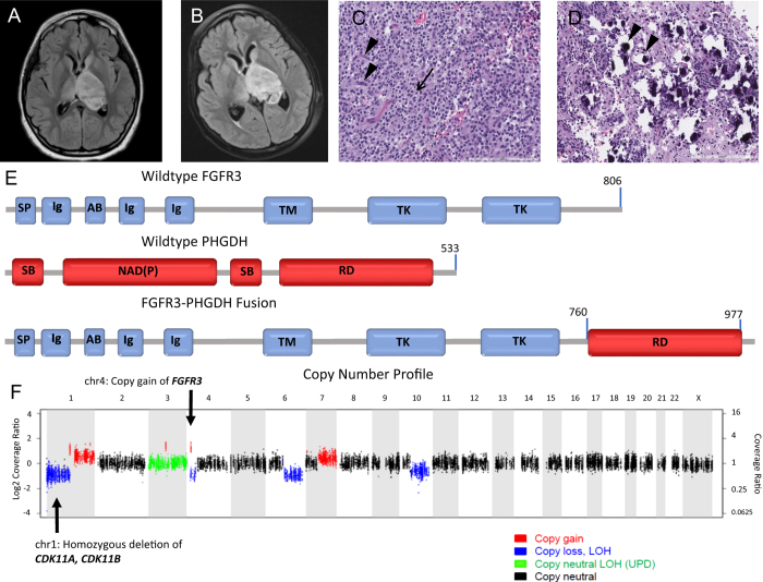
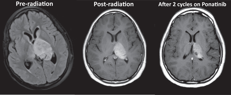
Approximately 1-5% of pediatric intracranial tumors originate in the thalamus. While great strides have been made to identify consistent molecular markers in adult oligodendrogliomas, such as the 1p/19q co-deletion, it is widely recognized that pediatric oligodendrogliomas have a vastly different molecular make-up. While pediatric thalamic or "central oligodendrogliomas" are histologically similar to peripheral pediatric oligodendrogliomas, they are behaviorally distinct and likely represent a cohesive, but entirely different entity. We describe a case of a 10-year-old girl who was diagnosed with an anaplastic glioma with features consistent with the aggressive entity often diagnosed as central or thalamic oligodendroglioma. We performed whole-exome (paired tumor and germline DNA) and transcriptome (tumor RNA) sequencing, which demonstrated an FGFR3-PHGDH fusion. We describe this fusion and our rationale for pursuing personalized, targeted therapy for the patient's tumor that may potentially play a role in the treatment of similar cases.
Conflict of interest statement
The authors declare that they have no competing financial interests.
Figures
Similar articles
-
Allelic losses at 1p36 and 19q13 in gliomas: correlation with histologic classification, definition of a 150-kb minimal deleted region on 1p36, and evaluation of CAMTA1 as a candidate tumor suppressor gene.Clin Cancer Res. 2005 Feb 1;11(3):1119-28. Clin Cancer Res. 2005. PMID: 15709179
-
Molecular characterization reveals NF1 deletions and FGFR1-activating mutations in a pediatric spinal oligodendroglioma.Pediatr Blood Cancer. 2017 Jun;64(6):10.1002/pbc.26346. doi: 10.1002/pbc.26346. Epub 2016 Nov 10. Pediatr Blood Cancer. 2017. PMID: 27862886 Free PMC article.
-
Oncogenic KIAA1549-BRAF fusion with activation of the MAPK/ERK pathway in pediatric oligodendrogliomas.Cancer Genet. 2015 Mar;208(3):91-5. doi: 10.1016/j.cancergen.2015.01.009. Epub 2015 Feb 20. Cancer Genet. 2015. PMID: 25794445
-
New treatment paradigm for patients with anaplastic oligodendroglial tumors.Anticancer Res. 2014 Apr;34(4):1587-94. Anticancer Res. 2014. PMID: 24692686 Review.
-
Oligodendroglioma: pathology, molecular mechanisms and markers.Acta Neuropathol. 2015 Jun;129(6):809-27. doi: 10.1007/s00401-015-1424-1. Epub 2015 May 6. Acta Neuropathol. 2015. PMID: 25943885 Free PMC article. Review.
Cited by
-
Clinico-pathological and epigenetic heterogeneity of diffuse gliomas with FGFR3::TACC3 fusion.Acta Neuropathol Commun. 2023 Jan 16;11(1):14. doi: 10.1186/s40478-023-01506-z. Acta Neuropathol Commun. 2023. PMID: 36647073 Free PMC article.
-
The oncogenic fusion landscape in pediatric CNS neoplasms.Acta Neuropathol. 2022 Apr;143(4):427-451. doi: 10.1007/s00401-022-02405-8. Epub 2022 Feb 15. Acta Neuropathol. 2022. PMID: 35169893 Free PMC article. Review.
-
Long-term outcomes of symptomatic optic pathway glioma: 32-year experience at a single Western Australian tertiary pediatric oncology center.Front Oncol. 2023 Jul 14;13:1157909. doi: 10.3389/fonc.2023.1157909. eCollection 2023. Front Oncol. 2023. PMID: 37519788 Free PMC article.
-
Biological and clinical implications of FGFR aberrations in paediatric and young adult cancers.Oncogene. 2023 Jun;42(23):1875-1888. doi: 10.1038/s41388-023-02705-7. Epub 2023 May 2. Oncogene. 2023. PMID: 37130917 Free PMC article. Review.
-
Molecular profiling and targeted therapy in pediatric gliomas: review and consensus recommendations.Neuro Oncol. 2019 Aug 5;21(8):968-980. doi: 10.1093/neuonc/noz022. Neuro Oncol. 2019. PMID: 30805642 Free PMC article.
References
Publication types
Grants and funding
LinkOut - more resources
Full Text Sources
Other Literature Sources
Miscellaneous
