Review
. 2018 Sep 1;120(3):1233-1246.
doi: 10.1152/jn.00848.2017. Epub 2018 Jun 6.

Cutaneous afferent innervation of the human foot sole: what can we learn from single-unit recordings?

Affiliations
Review

Cutaneous afferent innervation of the human foot sole: what can we learn from single-unit recordings?

Nicholas D J Strzalkowski et al. J Neurophysiol. .

Abstract

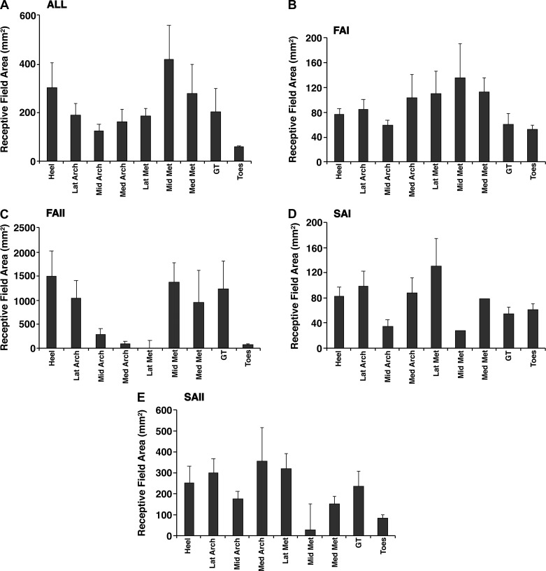
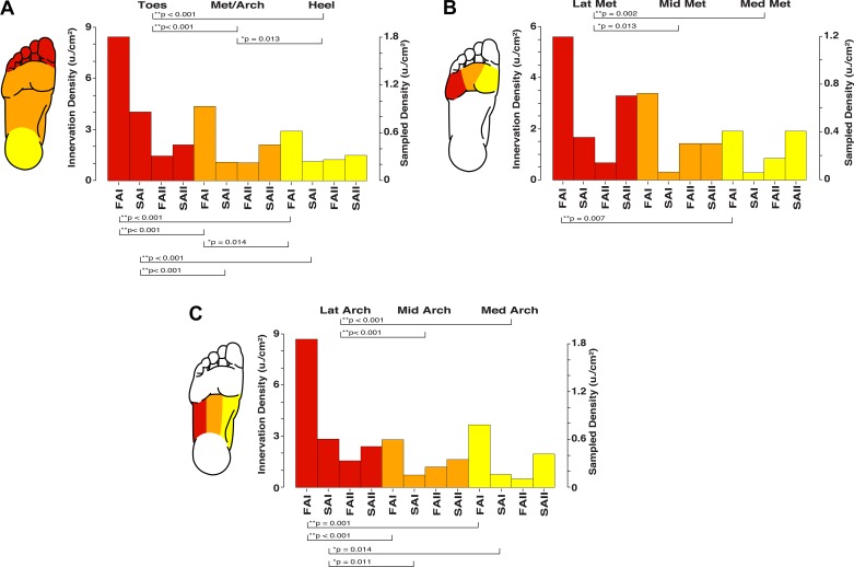
Cutaneous afferents convey exteroceptive information about the interaction of the body with the environment and proprioceptive information about body position and orientation. Four classes of low-threshold mechanoreceptor afferents innervate the foot sole and transmit feedback that facilitates the conscious and reflexive control of standing balance. Experimental manipulation of cutaneous feedback has been shown to alter the control of gait and standing balance. This has led to a growing interest in the design of intervention strategies that enhance cutaneous feedback and improve postural control. The advent of single-unit microneurography has allowed the firing and receptive field characteristics of foot sole cutaneous afferents to be investigated. In this review, we consolidate the available cutaneous afferent microneurographic recordings from the foot sole and provide an analysis of the firing threshold, and receptive field distribution and density of these cutaneous afferents. This work enhances the understanding of the foot sole as a sensory structure and provides a foundation for the continued development of sensory augmentation insoles and other tactile enhancement interventions.

Keywords: NEWS & NOTEWORTHY We present a synthesis of foot sole cutaneous afferent microneurography recordings and provide novel insights about the distribution; and firing characteristics of cutaneous afferents across the human foot sole. The foot sole is a valuable sensory structure for the control of standing balance; and our findings provide a new understanding on how the foot sole can be viewed as a sensory structure; cutaneous afferents; density; foot sole; mechanoreceptor; microneurography; tactile feedback.


Figures

Fig. 1.
Fig. 1.
An illustration of the human microneurography technique. A, top: schematic of experimental setup for recording from the tibial nerve at the level of the knee (popliteal fossa). Two tungsten microelectrodes are inserted percutaneously, with one serving as the reference electrode inserted beneath the skin near the nerve and the other serving as the active electrode, which gets inserted into the nerve. Bottom, schematic of a peripheral nerve, showing the active electrode’s placement into an individual nerve fascicle, right up next to a single axon (i.e., intrafascicular extracellular recording). B: sample recording from a fast-adapting type I (FAI) afferent showing, from top to bottom, the instantaneous firing rate (i.p.s., impulses per second), raster plot, raw neurogram (Nerve), and vibrator acceleration (Accel.) for the case of 30- and 250-Hz vibration. As expected based on the FAI bandwidth, this unit codes precisely for the 30-Hz vibration with a phase-locked 30-Hz spike train but fails to be activated by the 250-Hz stimulation. Inset left: sample of phase-locking in the FAI response with the timescale expanded. Inset right: 100 overlaid spikes (note: the double-peaked action potential morphology indicates that the microelectrode has not caused conduction blockage; see Inglis et al. 1996).
Fig. 2.
Fig. 2.
Receptive fields of the different cutaneous mechanoreceptor classes. A: foot sole maps for each afferent type showing all the receptive field locations and estimates of size in the present data set. Shaded ellipses represent individual afferent receptive fields. B: composite foot sole map showing the center of all receptive fields overlaid on the same foot template. Pie chart depicts the breakdown in terms of the percentages of each afferent type in the present data set. FAI, fast-adapting type I; FAII, fast-adapting type II; SAI, slowly adapting type I; SAII, slowly adapting type II.
Fig. 3.
Fig. 3.
Foot sole area measurement. The surface areas of 9 different individual regions were measured on the foot soles of 4 men and 4 women. At left is the largest foot encountered (male, age 25 yr, U.S. men’s size 12 shoe), and at right is the smallest (female, age 25 yr, U.S. women’s size 6 shoe). The skin regions were traced from an optical scan of each individual’s right foot sole (light green outlines), and digital area measurements were made using ImageJ software. Toes, digits 2–5; GT, great toe; LatArch, MidArch, and MedArch, lateral, middle, and medial arch; LatMet, MidMet, and MedMet, lateral, middle, and medial metatarsals; Heel, calcaneus.
Fig. 4.
Fig. 4.
Mechanical thresholds for the different cutaneous mechanoreceptor classes. Mean (SE) thresholds for evoking an action potential in the 9 different skin regions are given for all afferent types (A), fast-adapting type I (FAI; B), fast-adapting type II (FAII; C), slowly adapting type I (SAI; D), and slowly adapting type II (SAII; E). Heel, calcaneus; LatArch, MidArch, and MedArch, lateral, middle, and medial arch; LatMet, MidMet, and MedMet, lateral, middle, and medial metatarsals; GT, great toe; Toes, digits 2–5.
Fig. 5.
Fig. 5.
Receptive field sizes for the different cutaneous mechanoreceptor classes. Mean (SE) areas of receptive fields in the 9 different skin regions are given for all afferent types (A), fast-adapting type I (FAI; B), fast-adapting type II (FAII; C), slowly adapting type I (SAI; D), and slowly adapting type II (SAII; E). Heel, calcaneus; LatArch, MidArch, and MedArch, lateral, middle, and medial arch; LatMet, MidMet, and MedMet, lateral, middle, and medial metatarsals; GT, great toe; Toes, digits 2–5.
Fig. 6.
Fig. 6.
Estimates of the relative and absolute density for the different cutaneous mechanoreceptor classes across the foot sole. Significant differences are indicated by brackets and P values in A–C. A: depiction of the proximal-distal gradient in receptive field density, with greater innervation density in the toes (red) than in the metatarsals/arch (Met/Arch; orange) and heel (yellow). B: depiction of the medial-lateral gradient in receptive field density across the metatarsals, with greater innervation density in the lateral region (LatMet; red) than in the middle (MidMet; orange), and medial (MedMet; yellow) regions. C: depiction of the medial-lateral gradient in receptive field density across the arch, with greater innervation density in the lateral region (LatArch; red) than in the middle (MidArch; orange) and medial (MedArch; yellow) regions. FAI, fast-adapting type I; FAII, fast-adapting type II; SAI, slowly adapting type I; SAII, slowly adapting type II.

References

    1. Abraira VE, Ginty DD. The sensory neurons of touch. Neuron 79: 618–639, 2013. doi:10.1016/j.neuron.2013.07.051. - DOI - PMC - PubMed
    1. Ackerley R, Backlund Wasling H, Liljencrantz J, Olausson H, Johnson RD, Wessberg J. Human C-tactile afferents are tuned to the temperature of a skin-stroking caress. J Neurosci 34: 2879–2883, 2014. doi:10.1523/JNEUROSCI.2847-13.2014. - DOI - PMC - PubMed
    1. Aimonetti JM, Hospod V, Roll JP, Ribot-Ciscar E. Cutaneous afferents provide a neuronal population vector that encodes the orientation of human ankle movements. J Physiol 580: 649–658, 2007. doi:10.1113/jphysiol.2006.123075. - DOI - PMC - PubMed
    1. Auplish S, Hall S. An immunohistochemical study of palmar and plantar digital nerves. J Hand Surg [Br] 23: 6–11, 1998. doi:10.1016/S0266-7681(98)80208-8. - DOI - PubMed
    1. Bensmaïa SJ, Leung YY, Hsiao SS, Johnson KO. Vibratory adaptation of cutaneous mechanoreceptive afferents. J Neurophysiol 94: 3023–3036, 2005. doi:10.1152/jn.00002.2005. - DOI - PMC - PubMed

Publication types