. 2018 Jul 1;77(7):628-635.
doi: 10.1093/jnen/nly046.

Evaluating Circulating Tumor DNA From the Cerebrospinal Fluid of Patients With Melanoma and Leptomeningeal Disease

Affiliations

Evaluating Circulating Tumor DNA From the Cerebrospinal Fluid of Patients With Melanoma and Leptomeningeal Disease

Leomar Y Ballester et al. J Neuropathol Exp Neurol. .

Abstract

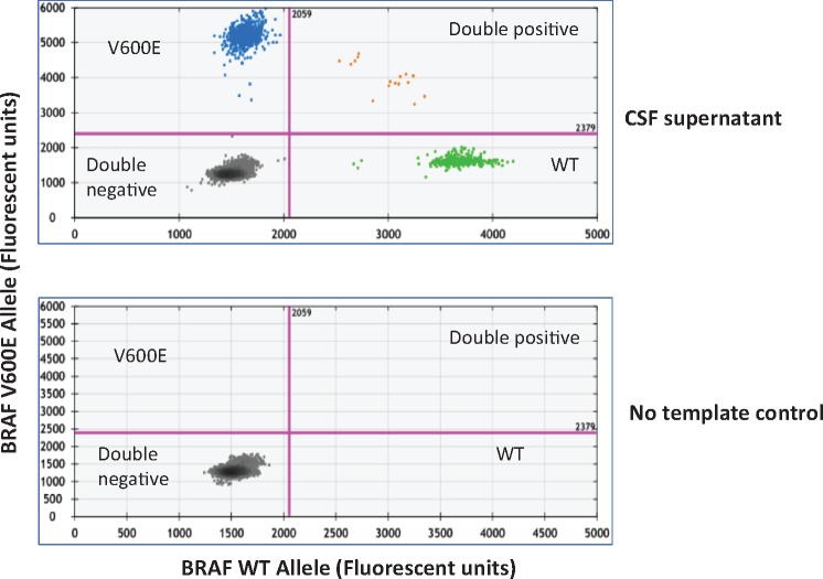
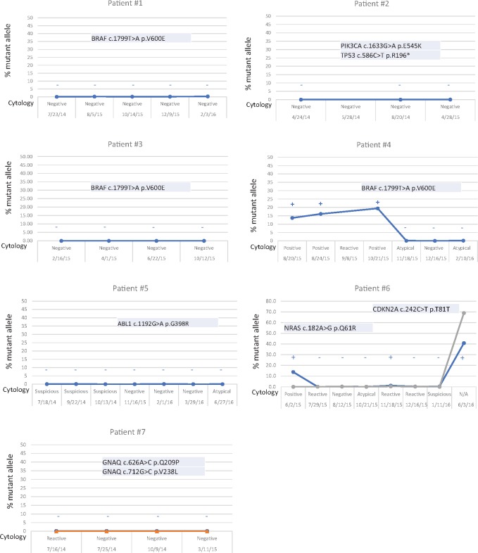
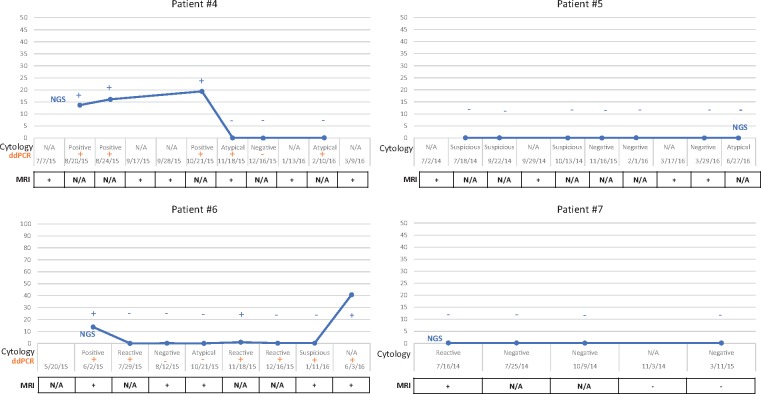
Circulating tumor DNA (ctDNA) refers to tumor-derived cell-free DNA that circulates in body fluids. Fluid samples are easier to collect than tumor tissue, and are amenable to serial collection at multiple time points during the course of a patient's illness. Studies have demonstrated the feasibility of performing mutation profiling from blood samples in cancer patients. However, detection of ctDNA in the blood of patients with brain tumors is suboptimal. Cerebrospinal fluid (CSF) can be obtained via lumbar puncture or intraventricular catheter, and may be a suitable fluid to assess ctDNA in patients with brain tumors. We detected melanoma-associated mutations by droplet-digital PCR (ddPCR) and next-generation sequencing in ctDNA obtained from the CSF (CSF-ctDNA) of melanoma patients with leptomeningeal disease. There is a strong correlation between mutation detection by ddPCR, the presence of circulating tumor cells in CSF and abnormalities in the MRI. However, approximately 30% of CSF samples that were negative or indeterminate for the presence of tumor cells by microscopic examination were positive for CSF-ctDNA by ddPCR. Our results demonstrate that CSF is a suitable fluid for evaluating ctDNA and ddPCR is superior to CSF-cytology for analysis of CSF in melanoma patients with leptomeningeal disease.


Figures

FIGURE 1.
FIGURE 1.
Mutation detection in CSF-ctDNA by ddPCR. Representative 2D plots of ddPCR droplet reads. Bottom plot represents droplet reads from a no-template control sample. All droplets are double negative in the no-template control. The top panel represents the droplet reads from the CSF-cfDNA from Patient 4. Note the presence of WT-DNA in the X-axis (green dots) and BRAF p.V600E mutant DNA in the Y-axis (blue dots). A few droplets contained both WT and mutant DNA and fell in the double positive category (orange dots).
FIGURE 2.
FIGURE 2.
NGS versus CSF-cytology. MAFs and CSF-cytology results at different time points. MAFs are determined from NGS data. If negative, only the MAFs for the expected mutations that were present in the solid mass (intra- or extracranial mass) are shown. Also, any mutation detected in any of 50 genes tested by the NGS panel in the CSF-ctDNA is shown. Note the presence of the BRAF p.V600E mutation in Patient 4. Also, note the presence of the NRAS p.Q61R in the first and last samples tested in Patient 6. Interestingly, a silent change not present initially (CDKN2A p.T81T) is detected in the last sample tested, when the CSF-ctDNA levels increase suggesting tumor evolution. No false positive results were detected in any of the samples tested. There is a good correlation between the results of CSF-cytology (shown in the X-axis) and CSF-ctDNA. Samples positive for CSF-cytology are positive for CSF-ctDNA and samples negative for CSF-cytology are negative for CSF-ctDNA. Interestingly, several samples interpreted as atypical (indeterminate by CSF-cytology) are negative for CSF-ctDNA. These suggest that CSF-ctDNA might provide more definitive results when trying to evaluate the presence of tumor in the CSF.
FIGURE 3.
FIGURE 3.
MRI, CSF-ctyology and CSF-ctDNA. No signal abnormalities were detected in the MRI for Patients 1–3 (not shown), which correlates with the absence of tumor cells or mutant DNA. In contrast, signal abnormalities in the MRI were identified in Patients 4–7. Minimal signal abnormalities were noted in the imaging studies for Patients 5 and 7, even in the absence of detectable levels of mutant DNA in the CSF by NGS.

References

    1. Remon J, Le Rhun E, Besse B.. Leptomeningeal carcinomatosis in non-small cell lung cancer patients: A continuing challenge in the personalized treatment era. Cancer Treatment Rev 2017;53:128–37 - PubMed
    1. Davies MA, Liu P, McIntyre S et al. , . Prognostic factors for survival in melanoma patients with brain metastases. Cancer 2011;117:1687–96 - PubMed
    1. Scott BJ, Oberheim-Bush NA, Kesari S.. Leptomeningeal metastasis in breast cancer - a systematic review. Oncotarget 2016;7:3740–7 - PMC - PubMed
    1. Le Rhun E, Taillibert S, Chamberlain MC.. Carcinomatous meningitis: Leptomeningeal metastases in solid tumors. Surg Neurol Int 2013;4:S265–88 - PMC - PubMed
    1. Chamberlain MC. Leptomeningeal metastasis. Curr Opin Oncol 2010;22:627–35 - PubMed

Publication types