Review
. 2018 Jun 5;27(6):1176-1199.
doi: 10.1016/j.cmet.2018.05.011.

Hallmarks of Brain Aging: Adaptive and Pathological Modification by Metabolic States

Affiliations
Review

Hallmarks of Brain Aging: Adaptive and Pathological Modification by Metabolic States

Mark P Mattson et al. Cell Metab. .

Abstract

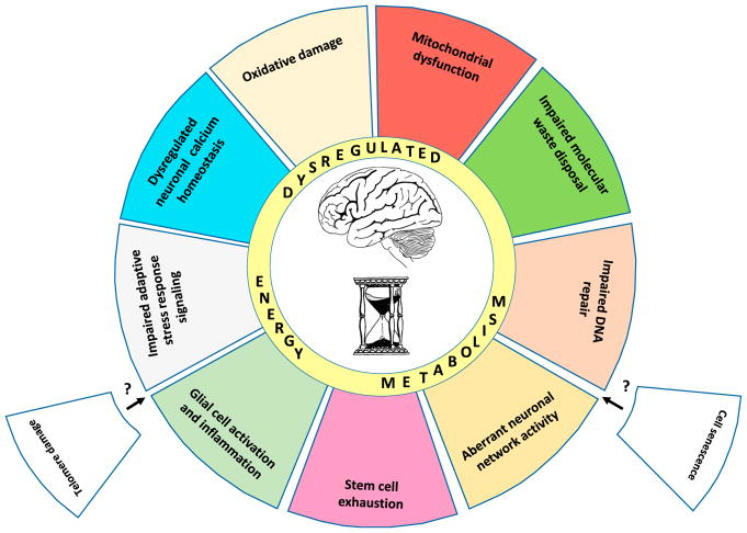
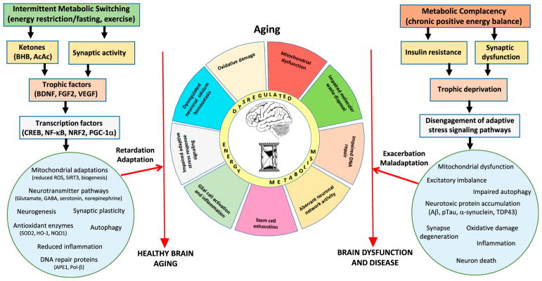
During aging, the cellular milieu of the brain exhibits tell-tale signs of compromised bioenergetics, impaired adaptive neuroplasticity and resilience, aberrant neuronal network activity, dysregulation of neuronal Ca2+ homeostasis, the accrual of oxidatively modified molecules and organelles, and inflammation. These alterations render the aging brain vulnerable to Alzheimer's and Parkinson's diseases and stroke. Emerging findings are revealing mechanisms by which sedentary overindulgent lifestyles accelerate brain aging, whereas lifestyles that include intermittent bioenergetic challenges (exercise, fasting, and intellectual challenges) foster healthy brain aging. Here we provide an overview of the cellular and molecular biology of brain aging, how those processes interface with disease-specific neurodegenerative pathways, and how metabolic states influence brain health.

Keywords: Alzheimer’s disease; Parkinson’s disease; aging; amyloid; autophagic; hippocampus; ketones; mitochondrial dysfunction; synaptic dysfunction; synuclein.


Figures

Figure 1
Figure 1. Hallmarks of Brain Aging
There are ten established hallmarks of brain aging. The illustration depicts nine of the hallmarks as colored slices of pie interacting prominently with “dysregulated energy metabolism,” which is shown as an inner ring of the aging wheel. Also shown are two slices (telomere damage and cell senescence) that are considered hallmarks of aging in proliferative peripheral tissues, but have not yet been established as hallmarks of brain aging.
Figure 2
Figure 2. Core Neuronal Circuitry and Glial Cells of the Mammalian Brain
Excitatory neurons deploy the neurotransmitter glutamate and typically elaborate long axons that project relatively long distances within and between brain regions. Ca2+ is the principal second messenger mediating both presynaptic and postsynaptic plasticity at excitatory synapses. The major inhibitory neurons within brain regions are GABAergic and function to constrain excitatory neuronal circuits within physiological limits. Glutamatergic neurons also receive inputs from neuromodulatory transmitters including norepinephrine, serotonin, and acetylcholine. Astrocytes are the most abundant type of glial cell in the brain; they remove glutamate from the extracellular milieu and produce neurotrophic factors, lactate, and ketones to support neuronal growth and bioenergetics. Oligo-dendrocytes are glia that myelinate axons to increase the speed of action potential propagation along the axon. Microglia are the major innate immune cell in the brain; they produce reactive oxygen species (ROS) and cytokines and remove apoptotic cells and extracellular debris.
Figure 3
Figure 3. Examples of Roles for Aging Processes, Acting Both Upstream and Downstream of Disease-Defining Molecular Lesions, in the Pathogenesis of AD, PD, and Stroke
There is considerable evidence that the indicated hallmarks of brain aging act upstream of the disease-defining Aβ plaques and pTau neurofibrillary tangles. On the other hand, aggregating Aβ and pTau can cause oxidative stress, dysregulation of Ca2+ homeostasis, mitochondrial dysfunction, and other hallmarks of aging in neurons. In PD, core aging processes result in the intracellular accumulation of neurotoxic forms of α-synuclein, and conversely, the accumulation of α-synuclein exacerbates aging processes resulting in neuronal dysfunction and death. Aging renders the brain vulnerable to stroke by promoting atherosclerosis and by compromising the ability of neurons to withstand and recover from the ischemic stress.
Figure 4
Figure 4. Working Model for How Intermittent Metabolic Challenges Bolster Brain Health during Aging, Whereas a Chronic Positive Energy Balance Hastens Brain Aging and Associated Brain Diseases
Left: eating and lifestyle patterns that result in intermittent depletion of liver glycogen stores and mobilization of fatty acids to generate ketones (fasting and exercise) also typically increase neuronal network activity in many brain regions. Signaling pathways are activated in brain cells that upregulate the expression of trophic factors and activate transcription factors that induce the expression of genes encoding proteins that enhance neural plasticity and resilience during aging. These adaptations to intermittent metabolic switching include mitochondrial biogenesis and stress resistance; adaptive modifications of neurotransmitter signaling pathways; upregulation of autophagy, antioxidant defenses, and DNA repair; stimulation of neurogenesis; and suppression of inflammation. In these ways intermittent metabolic switching counteracts core brain aging mechanisms, thereby slowing age-related declines in neurological function and reducing the risk of AD, PD, and stroke. Right: sedentary overindulgent lifestyles accelerate brain aging and increase the risk of AD, PD, and stroke. A chronic positive energy balance results in metabolic morbidity (insulin resistance and dyslipidemia) and reduced activation of signaling pathways that promote synaptic plasticity and cellular stress resistance. As a consequence, neurons suffer: impaired mitochondrial function, autophagy, and DNA repair; excessive oxidative stress; dysregulated neuronal network activity and Ca2+ homeostasis; the accumulation of potentially toxic protein aggregates; and inflammation. In these ways, metabolic complacency accelerates age-related decrements in brain function and increases the risk of AD, PD, and stroke.

References

    1. Aarsland D, Creese B, Politis M, Chaudhuri KR, Ffytche DH, Weintraub D, Ballard C. Cognitive decline in Parkinson disease. Nat Rev Neurol. 2017;13:217–231. - PMC - PubMed
    1. Adamczak SE, de Rivero Vaccari JP, Dale G, Brand FJ, 3rd, Nonner D, Bullock MR, Dahl GP, Dietrich WD, Keane RW. Pyroptotic neuronal cell death mediated by the AIM2 inflammasome. J Cereb Blood Flow Metab. 2014;34:621–629. - PMC - PubMed
    1. Alexander GE, Ryan L, Bowers D, Foster TC, Bizon JL, Geldmacher DS, Glisky EL. Characterizing cognitive aging in humans with links to animal models. Front Aging Neurosci. 2012;4:21. - PMC - PubMed
    1. Alirezaei M, Kemball CC, Flynn CT, Wood MR, Whitton JL, Kiosses WB. Short-term fasting induces profound neuronal autophagy. Autophagy. 2010;6:702–710. - PMC - PubMed
    1. Alzheimer’s Association. 2016 Alzheimer’s disease facts and figures. Alzheimers Dement. 2016;12:459–509. - PubMed