. 2018 Jun 5;23(10):2967-2975.
doi: 10.1016/j.celrep.2018.05.011.

Excess Synaptojanin 1 Contributes to Place Cell Dysfunction and Memory Deficits in the Aging Hippocampus in Three Types of Alzheimer's Disease

Affiliations

Excess Synaptojanin 1 Contributes to Place Cell Dysfunction and Memory Deficits in the Aging Hippocampus in Three Types of Alzheimer's Disease

Andre M Miranda et al. Cell Rep. .

Abstract

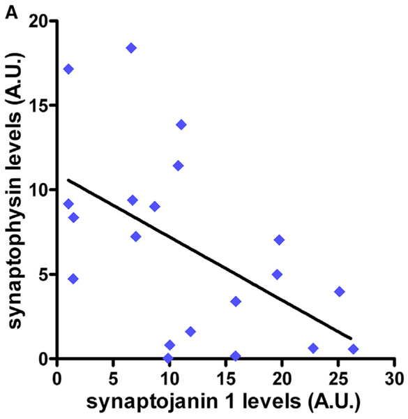
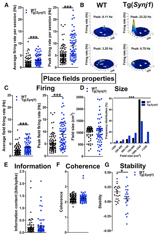
The phosphoinositide phosphatase synaptojanin 1 (SYNJ1) is a key regulator of synaptic function. We first tested whether SYNJ1 contributes to phenotypic variations in familial Alzheimer's disease (FAD) and show that SYNJ1 polymorphisms are associated with age of onset in both early- and late-onset human FAD cohorts. We then interrogated whether SYNJ1 levels could directly affect memory. We show that increased SYNJ1 levels in autopsy brains from adults with Down syndrome (DS/AD) are inversely correlated with synaptophysin levels, a direct readout of synaptic integrity. We further report age-dependent cognitive decline in a mouse model overexpressing murine Synj1 to the levels observed in human sporadic AD, triggered through hippocampal hyperexcitability and defects in the spatial reproducibility of place fields. Taken together, our findings suggest that SYNJ1 contributes to memory deficits in the aging hippocampus in all forms of AD.

Keywords: Long-term memory; SYNJ1; hyperexcitability; in vivo electrophysiology; neurodegenerative disorders; single nucleotide polymorphisms; synaptic dysfunction.


Figures

Figure 1.
Figure 1.. Elevated SYNJ1 Levels Are Associated with Synaptic Deficits in DS/AD
Western blot analysis of SYNJ1 and synaptophysin in human post-mortem brain samples from the mid-frontal cortex (BA46) of individuals with DS, aged 40–52 years (Martin et al., 2014) (n = 20). The line represents the linear regression (R2 = 0.29, p = 0.015). See also Figure S3 for additional information.
Figure 2.
Figure 2.. Overexpression of Synj1 Drives Hippocampal-Dependent Cognitive Deficits in an Age-Dependent Manner
(A) Western blot analysis of Synj1 in 19-month-old WT and Tg(Synj1) mice (n = 4). Tubulin was used as an equal loading marker. Synj1 protein levels were 76% higher in Tg(Synj1) (1.76 ± 0.11) than in WT (1.00 ± 0.05) mice. ***p < 0.001 in unpaired Student’s t test. (B) Performance of WT and Tg(Synj1) mice at 9 (n = 8 WT and 7 Tg[Synj1] mice) and 19 (n = 9 mice for both genotypes) months in the radial arm water maze (RAWM). Mice were administered 30 trials over a 2-day period, and the number of errors was averaged over three trials. Two-way ANOVA revealed an interaction between genotype and trial block at 19 months but not at 9 months. ns, p > 0.05, and **p < 0.01 for the overall effect of genotype in two-way ANOVA. In trial 6, ***p < 0.001 for the effect of genotype in two-way ANOVA with Bonferroni post-test. (C) Age-dependent modification of RAWM performance of WT and Tg(Synj1) mice. Age-dependent cognitive deficits were more severe in Tg(Synj1) mice (188 ± 25%, n = 9) than in WT mice (100 ± 20%, n = 9). *p < 0.05 in unpaired Student’s t test. (D and E) Freezing response in the contextual and cued FC paradigm in (D) 9-month-old WT (n = 10) and Tg(Synj1) (n = 9) mice and in (E) 19-month-old WT (n = 11) and Tg(Synj1) (n = 7) mice. *p < 0.05 in unpaired Student’s t test. Data are represented as mean ± SEM. See also Figure S4 for additional information.
Figure 3.
Figure 3.. Overexpression of Synj1 Results in Hippocampal Hyperexcitability and Decreased Place Field Stability
(A) Left: average firing rate of hippocampal excitatory (pyramidal) neurons in 24-month-old Tg(Synj1) mice (3.2 ± 0.2 Hz, n = 72 neurons from six animals) and controls (2.0 ± 0.2 Hz, n = 98 neurons from five animals). Right: peak firing rate of pyramidal neurons in Tg(Synj1) mice (9.3 ± 0.7 Hz) and controls (5.6 ± 0.4 Hz). ***p < 0.001 in Mann Whitney test. (B) Representative examples of firing rate maps showing place fields obtained after WT and Tg(Synj1) mice explored a 50-cm-diameter cylindrical arena for 20 min. The firing rate is represented by a heatmap ranging from blue (no firing) to red (peak firing). White spaces indicate locations not visited by the animal. (C) Left: average field firing rate in Tg(Synj1) mice (3.8 ± 0.3 Hz, n = 72 neurons) and controls (2.2 ± 0.2 Hz, n = 98 neurons). Right: peak field firing rate in Tg(Synj1) mice (9.2 ± 0.7 Hz) and controls (5.5 ± 0.4 Hz). ***p < 0.001 in Mann-Whitney test. (D) Size of place fields in Tg(Synj1) and WT mice. The average size (left) of place fields was comparable (p > 0.05 in Mann-Whitney test) between Tg(Synj1) (1,066 ± 58 cm2, n = 72 neurons) and WT (1,043 ± 24 cm2, n = 98 neurons) mice, although the distribution (right) of place field sizes was significantly different (***p < 0.001 in chi-square test). (E) Comparable (p > 0.05, Mann-Whitney test) information content between Tg(Synj1) (0.29 ± 0.05 bits/spike, n = 72) and WT (0.19 ± 0.03 bits/spike, n = 98) neurons. (F) Similar spatial coherence (p > 0.05, Mann-Whitney test) between Tg(Synj1) (2.25 ± 0.03, n = 72) and WT (2.21 ± 0.02, n = 98) neurons. (G) Place field stability at 18–24 hr was significantly decreased (*p < 0.05 in unpaired Student’s t test) in Tg(Synj1) mice (0.05 ± 0.04, n = 20 neurons) compared with controls (0.17 ± 0.03, n = 24 neurons).

References

    1. Alharby E, Albalawi AM, Nasir A, Alhijji SA, Mahmood A, Ramzan K, Abdusamad F, Aljohani A, Abdelsalam O, Eldardear A, et al. (2017). A homozygous potentially pathogenic variant in the PAXBP1 gene in a large family with global developmental delay and myopathic hypotonia. Clin. Genet 92, 579–586. - PubMed
    1. Antonarakis SE (2017). Down syndrome and the complexity of genome dosage imbalance. Nat. Rev. Genet 18, 147–163. - PubMed
    1. Arai Y, Ijuin T, Takenawa T, Becker LE, and Takashima S (2002). Excessive expression of synaptojanin in brains with Down syndrome. Brain Dev 24, 67–72. - PubMed
    1. Athan ES, Williamson J, Ciappa A, Santana V, Romas SN, Lee JH, Rondon H, Lantigua RA, Medrano M, Torres M, et al. (2001). A founder mutation in presenilin 1 causing early-onset Alzheimer disease in unrelated Caribbean Hispanic families. JAMA 286, 2257–2263. - PubMed
    1. Austin-Tse C, Halbritter J, Zariwala MA, Gilberti RM, Gee HY, Hellman N, Pathak N, Liu Y, Panizzi JR, Patel-King RS, et al. (2013). Zebrafish ciliopathy screen plus human mutational analysis identifies C21orf59 and CCDC65 defects as causing primary ciliary dyskinesia. Am. J. Hum. Genet 93, 672–686. - PMC - PubMed

Publication types

MeSH terms