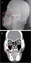
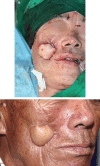
Multiple foreign bodies causing an orocutaneous fistula of the cheek
- PMID: 29874906
- PMCID: PMC6057133
- DOI: 10.7181/acfs.2018.00017
Multiple foreign bodies causing an orocutaneous fistula of the cheek
Abstract
Foreign bodies impacted in the maxillofacial region are often a diagnostic challenge. They can be a source of chronic inflammatory reactions and infections leading to the formation of an orocutaneous fistula. Such orocutaneous fistulas cause significant morbidity in most patients, eventually requiring surgery. Recently, we encountered a very rare case of an orocutaneous fistula caused by multiple foreign bodies in the cheek. Precise removal of the foreign bodies was required, and a double-sided anterolateral thigh free flap was used to reconstruct the defect. Surgeons should be aware of the complications of multiple foreign bodies and should be able to diagnose these on careful clinical examination.
Keywords: Flap; Foreign body; Orocutaneous fistula.
Conflict of interest statement
No potential conflict of interest relevant to this article was reported.
Figures


Similar articles
-
Bilateral Orocutaneous Fistula Secondary to Pericoronal Infection of Mandibular Third Molars: A Rare Case Report.Clin Case Rep. 2025 Jan 14;13(1):e70017. doi: 10.1002/ccr3.70017. eCollection 2025 Jan. Clin Case Rep. 2025. PMID: 39816707 Free PMC article.
-
A linguoverted impacted tooth with orocutaneous fistula - a rare case report.Clujul Med. 2018 Oct;91(4):479-483. doi: 10.15386/cjmed-945. Epub 2018 Oct 30. Clujul Med. 2018. PMID: 30564028 Free PMC article.
-
Awake intubation in a patient with huge orocutaneous fistula: a case report.J Dent Anesth Pain Med. 2017 Dec;17(4):313-316. doi: 10.17245/jdapm.2017.17.4.313. Epub 2017 Dec 28. J Dent Anesth Pain Med. 2017. PMID: 29349354 Free PMC article.
-
Review of Ingested and Aspirated Foreign Bodies in Children and Their Clinical Significance for Radiologists.Radiographics. 2015 Sep-Oct;35(5):1528-38. doi: 10.1148/rg.2015140287. Epub 2015 Aug 21. Radiographics. 2015. PMID: 26295734 Review.
-
Pediatric foreign bodies and their management.Curr Gastroenterol Rep. 2005 Jun;7(3):212-8. doi: 10.1007/s11894-005-0037-6. Curr Gastroenterol Rep. 2005. PMID: 15913481 Review.
Cited by
-
Residual foreign body inflammation caused by a lumber beam penetrating the facial region: a case report.Arch Craniofac Surg. 2023 Feb;24(1):37-40. doi: 10.7181/acfs.2022.01018. Epub 2023 Feb 20. Arch Craniofac Surg. 2023. PMID: 36858360 Free PMC article.
-
Management of Maxillofacial Trauma in Attempt Suicide Patients During COVID-19 Pandemic.J Craniofac Surg. 2021 Jun 1;32(4):e394-e396. doi: 10.1097/SCS.0000000000007428. J Craniofac Surg. 2021. PMID: 33427781 Free PMC article.
-
Factors Associated with Treatment Outcomes and Pathological Features in Patients with Osteoradionecrosis: A Retrospective Study.Int J Environ Res Public Health. 2022 May 27;19(11):6565. doi: 10.3390/ijerph19116565. Int J Environ Res Public Health. 2022. PMID: 35682149 Free PMC article.
References
-
- Bede SY, Ahmed FT. Management of retained foreign bodies in missile injuries of the maxillofacial region. J Craniofac Surg. 2011;22:1440–4. - PubMed
-
- Auluck A, Behanan AG, Pai KM, Shetty C. Recurrent sinus of the cheek due to a retained foreign body: report of an unusual case. Br Dent J. 2005;198:337–9. - PubMed
-
- Raman R, Ariayanayagam C. Closure of orocutaneous and pharyngocutaneous fistulas. Plast Reconstr Surg. 1987;79:310. - PubMed
-
- Naude GP, Bongard FS, Demetriades D. Trauma secrets. Philadelphia: Hanley & Belfus; 2003.
-
- McLean JN, Nicholas C, Duggal P, Chen A, Grist WG, Losken A, et al. Surgical management of pharyngocutaneous fistula after total laryngectomy. Ann Plast Surg. 2012;68:442–5. - PubMed
LinkOut - more resources
Full Text Sources
Other Literature Sources

