Review
. 2018;25(35):4616-4637.
doi: 10.2174/0929867325666180605122633.

Emerging Technologies for Cancer Research: Towards Personalized Medicine with Microfluidic Platforms and 3D Tumor Models

Affiliations
Review

Emerging Technologies for Cancer Research: Towards Personalized Medicine with Microfluidic Platforms and 3D Tumor Models

Matteo Turetta et al. Curr Med Chem. 2018.

Abstract

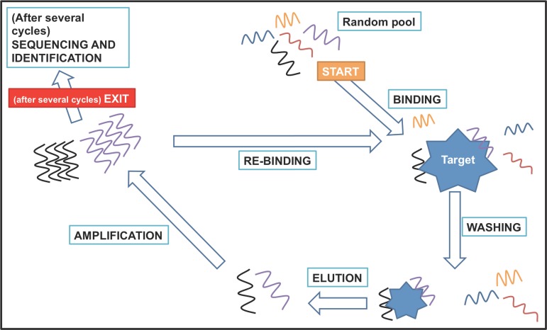
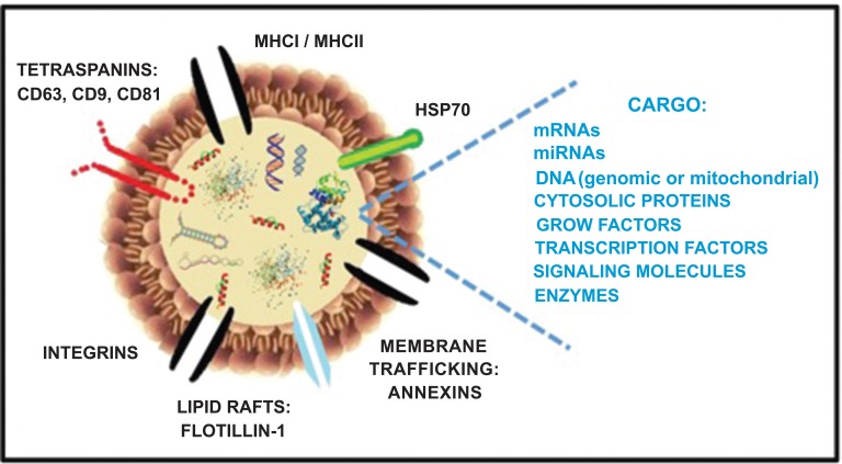
In the present review, we describe three hot topics in cancer research such as circulating tumor cells, exosomes, and 3D environment models. The first section is dedicated to microfluidic platforms for detecting circulating tumor cells, including both affinity-based methods that take advantage of antibodies and aptamers, and "label-free" approaches, exploiting cancer cells physical features and, more recently, abnormal cancer metabolism. In the second section, we briefly describe the biology of exosomes and their role in cancer, as well as conventional techniques for their isolation and innovative microfluidic platforms. In the third section, the importance of tumor microenvironment is highlighted, along with techniques for modeling it in vitro. Finally, we discuss limitations of two-dimensional monolayer methods and describe advantages and disadvantages of different three-dimensional tumor systems for cell-cell interaction analysis and their potential applications in cancer management.

Keywords: 3D cell cultures; Cancer; circulating tumor cells; exosomes; microfluidic Platforms; tumor microenvironment..


Figures

Fig. (1)
Fig. (1)
Schematic view of technologies for CTCs isolation. Methods based on biological properties, described in 2.2.1 and on physical properties, described in 2.2.2.
Fig. (2)
Fig. (2)
Schematic view of the SELEX technology. Starting point is a random pool of synthetic DNA oligonucleotides. The SELEX procedure consists of several cycles of the following steps. At first, the random pool and the target molecules are incubated for binding. Unbound oligonucleotides are then removed by washing. The target-bound oligonucleotides are eluted and amplified by PCR or RT-PCR. A new pool of oligonucleotides is thus generated and is then used for the next selection round. In general, 5 to 20 complete cycles are needed for the selection of specific aptamers. The process can be interrupted and selected oligonucleotides can be characterized by sequencing.
Fig. (3)
Fig. (3)
Schematic representation of metabolism-based method steps. Step 1: Starting from a cell suspension in which both white blood cells (WBCs) and cancer cells (CTCs) are present, the sample, containing a fluorescent pH probe (e.g., SNARF), is emulsified in millions of picoliter droplets at a microfluidic flow-focusing junction using fluorinated oil and surfactant to avoid droplet fusion. Encapsulation of single cells can be seen at bottom-left corner. Then, the emulsion is incubated for a variable amount of time at 37°C to activate metabolic processes. Step 2: Droplets are reinjected and fluorescence is screened with a laser slit to measure pH. In the bottom-right corner a sample track is displayed, showing the altered emission ratio between two fluorescent channels (SNARF 580 and SNARF 630) indicating an alteration of physiological pH [62].
Fig. (4)
Fig. (4)
Exosome structure and content. Exosomes are small membrane bound vesicles characterized by a phospholipid bilayer and by the presence of proteins involved in membrane trafficking (e.g. annexins and flotillin), cell targeting (e.g. tetraspanins and integrins) as well as other proteins involved in exosomal biogenesis (Alix and TSG101, not shown). Additionally, depending on the cell of origin, exsosomes contain a molecular cargo constituted by cytosolic proteins, growth factors, cytokines, DNA, RNA and miRNA, which can be delivered to target cells. Abbreviations: HSP, heat shock protein; MHC, major histocompatibility complex.
Fig. (5)
Fig. (5)
Methods for exosome enrichment from biological fluids. Representative pictures of (a) differential ultracentrifugation protocol; (b) size-based separation chromatography; (c) immunoaffinity approach for antibody-specific capture; (d) polymer-based precipitation technique.
Fig. (6)
Fig. (6)
Atomic force microscopy topographic height images of A172-derived exosomes precipitated by Exoquick. The exosomes appear as circular structures with an average diameter of about 70 nm. Courtesy of F. Caponnetto.
Fig. (7)
Fig. (7)
Schematic illustration of a) spheroids, b) hydrogel, and c) fibrous-based scaffold. Each system can support growth and rearrangement of more than one cell type and allows for exchange of oxygen, nutrients and bioactive molecules.

References

    1. Stewart B. W., Wild C. P. World Cancer Report 2014 . 2014.
    1. Li H., Fan X., Houghton J. Tumor microenvironment: The role of the tumor stroma in cancer. J. Cell. Biochem. 2007:805–815. - PubMed
    1. Gu L., Mooney D.J. Biomaterials and emerging anticancer therapeutics: Engineering the microenvironment. Nat. Rev. Cancer. 2015;16(1):56–66. - PMC - PubMed
    1. Wistuba I.I., Gelovani J.G., Jacoby J.J., Davis S.E., Herbst R.S. Methodological and practical challenges for personalized cancer therapies. Nat. Rev. Clin. Oncol. 2011;8(3):135–141. - PubMed
    1. Schilsky R.L. Personalized medicine in oncology: The future is now. Nat. Rev. Drug Discov. 2010;9:363–366. - PubMed