Review
. 2018 Jun 7;13(1):12.
doi: 10.1186/s13064-018-0109-6.

Assembly and maintenance of GABAergic and Glycinergic circuits in the mammalian nervous system

Affiliations
Review

Assembly and maintenance of GABAergic and Glycinergic circuits in the mammalian nervous system

Clare R Gamlin et al. Neural Dev. .

Abstract

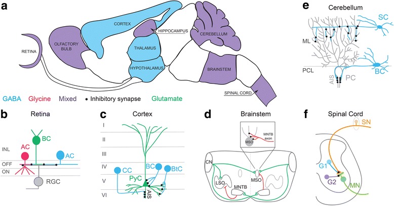
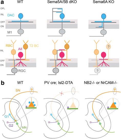
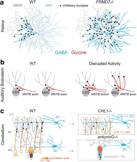
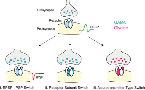
Inhibition in the central nervous systems (CNS) is mediated by two neurotransmitters: gamma-aminobutyric acid (GABA) and glycine. Inhibitory synapses are generally GABAergic or glycinergic, although there are synapses that co-release both neurotransmitter types. Compared to excitatory circuits, much less is known about the cellular and molecular mechanisms that regulate synaptic partner selection and wiring patterns of inhibitory circuits. Recent work, however, has begun to fill this gap in knowledge, providing deeper insight into whether GABAergic and glycinergic circuit assembly and maintenance rely on common or distinct mechanisms. Here we summarize and contrast the developmental mechanisms that regulate the selection of synaptic partners, and that promote the formation, refinement, maturation and maintenance of GABAergic and glycinergic synapses and their respective wiring patterns. We highlight how some parts of the CNS demonstrate developmental changes in the type of inhibitory transmitter or receptor composition at their inhibitory synapses. We also consider how perturbation of the development or maintenance of one type of inhibitory connection affects other inhibitory synapse types in the same circuit. Mechanistic insight into the development and maintenance of GABAergic and glycinergic inputs, and inputs that co-release both these neurotransmitters could help formulate comprehensive therapeutic strategies for treating disorders of synaptic inhibition.

Keywords: Circuit refinement; Inhibition; Synapse formation; Synapse maturation; Synaptic targeting.


Conflict of interest statement

Ethics approval and consent to participate

Not applicable.

Competing interests

The authors declare that they have no competing interests.

Publisher’s Note

Springer Nature remains neutral with regard to jurisdictional claims in published maps and institutional affiliations.

Figures

Fig. 1
Fig. 1
Types of inhibitory circuits across the CNS. a Modulation of neuronal activity in many CNS regions relies primarily on GABAergic inhibition (blue regions), whereas other regions engage both GABAergic and glycinergic inhibition (purple regions; mixed). In regions such as the retina, GABA and glycine are often released by separate populations of inhibitory neurons. However, inhibitory neurons in the spinal cord can co-release both transmitter types. Sagittal outline of the mouse brain adapted from the GENSAT brain atlas [153]. b-f Schematics showing outline of inhibitory circuits discussed in the review. Some circuits show laminar organization (b, c, e), and interneurons typically target specific subcellular compartments of their postsynaptic partners (b-f). b Schematic of the retina showing glycinergic and GABAergic amacrine cells (AC) contacting glutamatergic bipolar cells (BC) and retinal ganglion cells (RGC) in OFF and ON subdivisions of the inner nuclear layer (INL). [–15]. (c) In the primary cortex, multiple types of GABAergic interneurons (blue) synapse onto glutamatergic pyramidal cells (PyC, green), each interneuron targeting a specific subcellular location on the PyC. For example, chandelier cells (CC) form synapses onto PyC axon initial segments (AIS). Cortical basket cells (BC) and bitufted cells (BtC) form synapses onto the soma and distal dendrites of PyCs, respectively. Summarized from: [27, 141]. (d) Schematic of circuits between mammalian brainstem nuclei. Dotted grey line represents the midline of the cross-section through the brainstem. The lateral superior olive (LSO) neurons receive excitatory input from the ipsilateral cochlear nucleus (CN) and inhibitory glycinergic input from the ipsilateral medial nucleus of the trapezoid body (MNTB), which is driven by the contralateral CN. Medial superior olive (MSO) neurons receive excitatory input from both ipsi- and contralateral CN, as well as inhibitory glycinergic input from the ipsilateral MNTB. MNTB axons provide glycinergic inhibition onto the soma of MSO neurons. Summarized from [29]. (e) Cerebellar inhibitory circuits. In the cerebellum, GABAergic stellate cells (SC) and basket cells (BC) target distinct subcellular compartments of Purkinje cells (PC). Summarized from [33, 36]. ML: Molecular layer, PCL: Purkinje cell layer, AIS: Axon initial segment. (f) Schematic of a spinal cord inhibitory circuit. Distinct inhibitory interneurons (G1 and G2), which are GABAergic and/or mixed GABA/glycinergic, target sensory afferents (SN) and motor neurons (MN) in the spinal cord, respectively. Summarized from [21, 22]
Fig. 2
Fig. 2
Molecular cues guide partner selection of inhibitory neurons. a Schematic showing the lamination of GABAergic-dopaminergic amacrine cells (DACs) and glycinergic AII amacrine cells together with their synaptic partners in wildtype (WT), Sema5A/6A double knockout mutants (dKO) and Sema6A knockouts (KO). T2 BC: Type 2 bipolar cell, M1: melanopsin-expressing retinal ganglion cell, RBC: rod bipolar cell, RGC: retinal ganglion cell, ON: inner sublamina of the retinal plexiform layer, OFF: outer sublamina of the retinal plexiform layer, INL: inner nuclear layer, OPL: outer plexiform layer. Summarized from [18, 19]. Question mark indicates non-examined synaptic partners. b Organization of inhibitory connections in the spinal cord sensory-motor circuit. Distinct populations of inhibitory neurons (G1 and G2) target sensory afferent terminals (SN) and motor neurons (MN), respectively, in WT mice. When sensory afferents are eliminated in PV cre/Isl2-DTA mice, G1 neurons do not form aberrant connections with motor neurons. Inhibitory synapses from G2 to motor neurons are still present in these mutants. In NB2−/− or NrCAM−/− mice, the number of inhibitory synapses from G1 to sensory neurons is significantly reduced but G2 interneuronal contacts onto motor neurons remain unaffected. G1: GABAergic neurons; G2: GABAergic and/or glycinergic neurons. Summarized from [21, 22]
Fig. 3
Fig. 3
Mechanisms that regulate pre- and postsynaptic subcellular targeting of inhibitory connections. a In wildtype (WT) mouse retina, only a specific quadrant of the arbor of GABAergic starburst amacrine cells (SACs) form inhibitory synapses onto direction-selective retinal ganglion cells (DSGCs). In FRMD7−/− mice, this pattern of connectivity between SACs and DSGCs that prefer horizontal movement is disrupted. Summarized from [25]. b During normal development, excess MNTB axon targeting individual LSO neurons are eliminated. In the gerbil auditory brainstem, MNTB neurons initially provide inhibition to MSO neurons across their soma and dendritic arbor, but during development, dendritic synapses are eliminated after the onset of binaural input. Disrupted activity, such as loss of glutamate release or disrupted binaural input, prevents synapse elimination during development. Summarized from: [, , –137]. c In the cerebellum, GABAergic stellate cells (SC) and basket cells (BC) utilize distinct cellular mechanisms to target distal dendrites and axon initial segments (AIS) of Purkinje cells (PC). In WT mice, ankyrinG binds to neurofascin and both are highly expressed in the AISs of PCs. Accordingly, in ankyrinG−/− mice the expression pattern of neurofascin is disrupted and basket cell processes erroneously target PC soma and distal processes, following the perturbed neurofascin expression pattern. The number of inhibitory synapses from basket cell to PC AISs is also reduced. In wildtype mice, stellate cells follow processes of Bergmann glia (BG) to make contact with distal dendrites of PCs. Both SCs and BGs express the cell surface molecule (CHL1). Consequently, in CHL1−/− mice stellate cells cannot recognize processes of BG and the number of SC synapses onto PC distal dendrites is reduced. Summarized from [33, 36]. ML: Molecular layer; PCL: Purkinje cell layer
Fig. 4
Fig. 4
Maturational ‘switches’ at inhibitory synapses. i) GABAergic and glycinergic transmission is initially depolarizing early in development due to the high intracellular chloride concentration within the postsynaptic cell. Reversal of the chloride gradient with maturation leads to hyperpolarization upon activation of GABA and glycine receptors [104]. EPSP: excitatory postsynaptic potential, IPSP: inhibitory postsynaptic potential. ii) During maturation, the composition of GABA and glycine receptor pentamers switches to incorporate different subunits, typically resulting in faster synaptic transmission [122, 123]. iii) Inhibitory circuits can also undergo a neurotransmitter type switch accompanied by a change in postsynaptic receptor expression. The transition from GABA-releasing to glycine-releasing is more common, but the reverse has also been documented [112, 114], see text for more details
Fig. 5
Fig. 5
Cross-talk between inhibitory neurotransmitter circuits. In some circuits, perturbing either GABAergic or glycinergic signaling leads to potentially compensatory postsynaptic changes. In both the spinal cord and retina, there are conditions in which there is cross-talk between inhibitory neurotransmitter circuits. In the spinal cord, oscillator mice carry a mutation that results in non-functional glycine receptors (non-α1 subunit containing glycine receptors, faded) and spastic mice carry a mutation that results in a dramatic reduction of glycine receptors at the synapse (dotted lines). Both mutations result in decreased glycinergic inhibitory postsynaptic currents (IPSCs, red traces). However, in the spastic mice there is an increase in extrasynaptic GABAA receptors and in the amplitude of GABAergic IPSCs (blue trace) [149, 150]. In the wildtype retina, Neuroligin 2 (NL2) is found at GABAergic synapses, and NL4 is localized at glycinergic synapses. In the retina of a NL4 knockout (KO) animal, α1-containing glycine receptors are lost, but there is no change in the expression of other NLs. However, in the NL2 KO retina, GABAAα3 and GABAAγ2-containing synapses are down-regulated, and there is an up-regulation of NL4 [69, 70]

References

    1. Asari H, Meister M. Divergence of visual channels in the inner retina. Nat Neurosci. 2012;15(11):1581–1589. doi: 10.1038/nn.3241. - DOI - PMC - PubMed
    1. Dumoulin A, Triller A, Dieudonné S. IPSC kinetics at identified GABAergic and mixed GABAergic and glycinergic synapses onto cerebellar Golgi cells. J Neurosci. 2001;21(16):6045–6057. doi: 10.1523/JNEUROSCI.21-16-06045.2001. - DOI - PMC - PubMed
    1. Huang ZJ, Di Cristo G, Ango F. Development of GABA innervation in the cerebral and cerebellar cortices. Nat Rev Neurosci. 2007;8(9):673–686. doi: 10.1038/nrn2188. - DOI - PubMed
    1. Decavel C, Van Den Pol AN. GABA: a dominant neurotransmitter in the hypothalamus. J Comp Neurol. 1990;302(4):1019–1037. doi: 10.1002/cne.903020423. - DOI - PubMed
    1. Halassa MM, Acsády L. Thalamic inhibition: diverse sources, diverse scales. Trends Neurosci. 2016;39(10):680–693. doi: 10.1016/j.tins.2016.08.001. - DOI - PMC - PubMed

Publication types

LinkOut - more resources