. 2018 Jul 15;201(2):337-342.
doi: 10.4049/jimmunol.1800279. Epub 2018 Jun 6.

Cutting Edge: Evidence for Nonvascular Route of Visceral Organ Immunosurveillance by T Cells

Affiliations

Cutting Edge: Evidence for Nonvascular Route of Visceral Organ Immunosurveillance by T Cells

Elizabeth M Steinert et al. J Immunol. .

Abstract

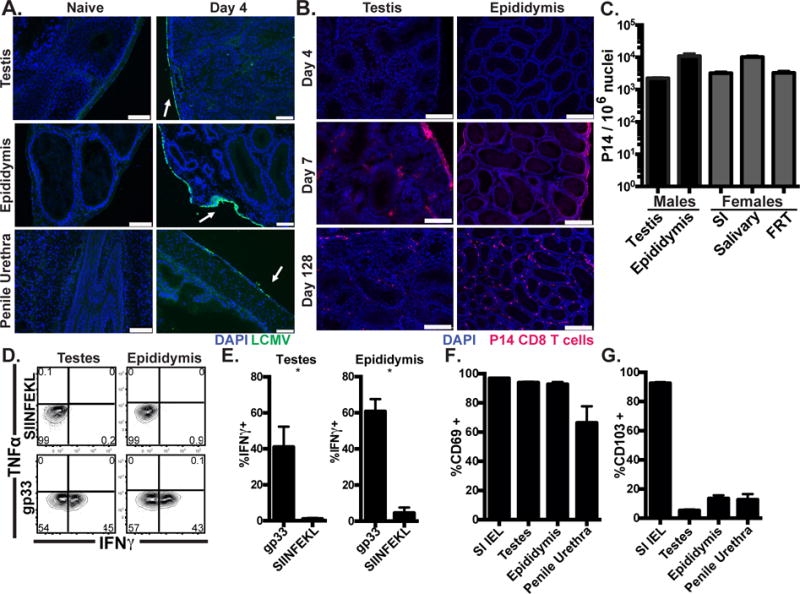
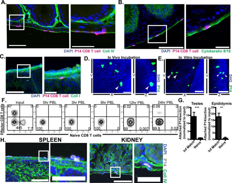
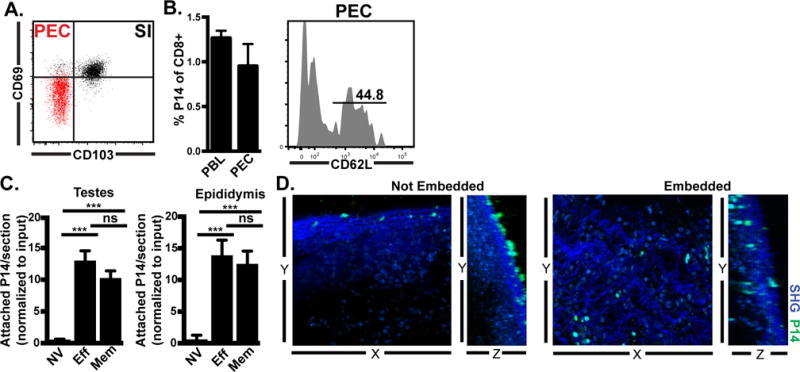
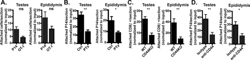
Lymphocytes enter tissues from blood vessels through a well-characterized three-step process of extravasation. To our knowledge, nonvascular routes of lymphocyte entry have not been described. In this article, we report that Ag-experienced CD8 T cells in mice recirculate from blood through the peritoneal cavity. In the event of infection, Ag-experienced CD8 T cell subsets adhered to visceral organs, indicating potential transcapsular immunosurveillance. Focusing on the male genital tract (MGT), we observed Ag-experienced CD8 T cell migration from the peritoneal cavity directly to the infected MGT across the capsule, which was dependent on the extracellular matrix receptor CD44. We also observed that, following clearance of infection, the MGT retained functional resident memory CD8 T cells. These data suggest that recirculation through body cavities may provide T cells with opportunities for broad immunosurveillance and potential nonvascular mechanisms of entry.


Figures

FIGURE 1
FIGURE 1. Resident-phenotype memory CD8 T cells persist in male genital tract
(A) C57BL/6 mice were infected with LCMV i.p. and stained with anti-LCMV nucleoprotein antibody (Green), and DAPI (Blue). Scale bar=100μm. n=8 from 3 experiments. (B) Representative images 4, 7 or 128 days after Thy1.1+ P14 cell transfer and LCMV infection, Thy1.1+ P14 (red), DAPI (Blue), scale bar=250μm, n=6-8 from 2 or 3 experiments per time point. (C) Enumeration of Thy1.1+ P14 in tissues from male (black) or age-matched female (grey) mice 115-160 days after LCMV infection, n=6, representative of 4 experiments. (D-E) 120-160 days after LCMV infection, 50μg of gp33 (cognate) or SIINFEKL (irrelevant) peptide was transurethrally instilled. 12h later, P14 were isolated and cytokine expressing cells were assessed by flow cytometry. n=6, representative of 3 experiments. (F-G) CD69 and CD103 expression on P14 memory cells, n=12 from 3 experiments. Graphs show mean and SEM, *p 0.0357, Mann-Whitney.
FIGURE 2
FIGURE 2. Evidence that effector CD8 T cells enter visceral tissues via direct transcapsular migration
0.5-1×10⁶ CD45.1+ naïve P14 CD8 T cells and 3-4×10⁶ Thy1.1+ effector P14 from mice infected with LCMV 6 days prior (D6 effector) were mixed and co-transferred i.p. into day 6 LCMV infection-matched recipients. 3h after injection tissues were harvested and analyzed for migration of transferred cells. (A-C) Representative images of adhered and migrating effector P14 CD8 T cells (in magenta) on epididymis, DAPI (in blue) indicates nuclei. Part of the section on the left (white rectangle) is shown in higher magnification in the right. Scale bars=100μm (left panels) and 25μm (right panels). Sections were also stained for stromal markers (in green) including (A) collagen IV, (B) cytokeratin 8 and 18, and (C) collagen I. (D) 3-4×10⁶ LCMV D6 effector P14-gfp were i.p. transferred to infection-matched recipients. 3h after injection, the MGT was harvested and P14-gfp attachment was evaluated using 2-photon microscopy. Still images from 2-photon microscopy video show a P14 CD8 T cell (white arrowhead) under the collagen capsule layer, third dimension is flattened. Scale bar=30μm (left, in vivo panel). (E) 4×10⁶ D6 effector P14-gfp cells were incubated in vitro with infection-matched tissue for 1-3h before 2-photon microscopy. Still images from video with third dimension flattened show P14 cells (white arrowhead) under the collagen capsule layer, Scale bar=50μm. (F) Congenically marked D6 effector (11×106) and naïve (1.61×10⁶) P14 CD8 T cells were mixed and co-transferred i.p. into infection matched recipients. PBL was taken at indicated time points after injection. (G) D6 effector P14 were transferred i.p. into D6 LCMV infection matched or naïve recipients. 3h after injection, tissues were harvested and analyzed for attachment of transferred cells. (H) Representative images of D6 effector P14 cells adhered to spleen and kidney, scale bar=100μm(left) or 50μm (right). Graph shows mean and SEM, ***p<0.0001, **p 0.0012, Mann-Whitney test. n= at least 6 from 2-3 separate experiments.
FIGURE 3
FIGURE 3. Recirculation through peritoneal cavity by memory CD8 T cells allows adherence and transcapsular migration to infected tissues
(A) CD69 and CD103 on gated memory P14 CD8 T cells isolated from small intestine epithelium (SI, black) or peritoneal exudate cells (PEC, red). (B) Memory P14 CD8 T cells isolated from lymphoid organs were transferred i.v. into naïve mice. Donor cells were assessed in recipient blood (PBL) and peritoneal lavage (PEC) 18h later. (C) Naïve (NV), effector (Eff, 6 days after LCMV infection) or memory (Mem, 30 days after LCMV infection) congenically distinct P14 populations were isolated from spleen and co-injected i.p. (4-7.5×10⁶ of each cell type) into day 6 LCMV recipients. 3h later, attached P14 were quantified by immunofluorescence. Graph shows mean and SEM and results of Wilcoxon matched pairs statistical analysis, ***p 0.0005, ***p 0.001. (D) Memory P14 CD8 T cells were co-cultured for 2-3h with MGT freshly isolated from LCMV D6 mice. Live imaging was captured by 2-photon microscopy (representative still images shown). n=3 from 2 separate experiments.
FIGURE 4
FIGURE 4. Antigen-independent, CD44-dependent mechanism of visceral immunosurveillance
(A) Congenically marked effector P14 (4×10⁶) and OT-I (7×10⁶) CD8 T cells from day 6 post infection with LCMV and VSV-OVA immune chimeras, respectively, were co-injected i.p. into LCMV infection-matched recipients. 3h after i.p. injection, adhered P14 and OT-I were quantified by immunofluorescence. n=4, representative of n=9 from 2 independent experiments. (B) Effector P14 cells from day 6 LCMV immune chimeras were untreated (Ctrl) or treated with pertussis toxin (PTx) in vitro then injected (4-12×106 cells) i.p. into day 6 LCMV infection-matched hosts. 3h later, adhered P14 cells were quantified by immunofluorescence. n=6 from 2 independent experiments. Graphs show mean and SEM. Mann-Whitney statistical analysis, **p 0.0043, *p 0.0260. (C) 6 days after LCMV infection, lymphocytes from WT or CD44KO mice were injected i.p. into WT infection matched recipients. 3h after i.p. injection, adhered CD8β+ transferred cells were quantified by immunofluorescence. n=8 representative of 2 separate experiments. Graphs show mean and SEM. Wilcoxon statistical analysis, **p 0.0043. (D) 4-9×10⁶ day 6 effector P14 were untreated or treated with anti-CD44 antibody in vitro for 1h prior to i.p. injection into untreated or hyaluronidase treated LCMV infection-matched recipients, respectively. 3h later, adhered P14 were quantified by immunofluorescence. n=6 from 2 separate experiments. Graphs show mean and SEM. Mann-Whitney statistical analysis, **p 0.0027, *p 0.0116.

References

    1. von Andrian UH, Mackay CR. T-Cell Function and Migration — Two Sides of the Same Coin. N Engl J Med. 2000;343:1020–1034. - PubMed
    1. Butcher EC, Picker LJ. Lymphocyte homing and homeostasis. Science. 1996;272:60. - PubMed
    1. Masopust D, Schenkel JM. The integration of T cell migration, differentiation and function. Nat Rev Immunol. 2013;13:309–320. - PubMed
    1. Mueller SN, Gebhardt T, Carbone FR, Heath WR. Memory T cell subsets, migration patterns, and tissue residence. Annu Rev Immunol. 2013;31:137–161. - PubMed
    1. Steinert EM, Schenkel JM, Fraser KA, Beura LK, Manlove LS, Igyártó BZ, Southern PJ, Masopust D. Quantifying memory CD8 T cells reveals regionalization of immunosurveillance. Cell. 2015;161:737–749. - PMC - PubMed

Publication types

Substances