. 2018 Jul 31;92(16):e00325-18.
doi: 10.1128/JVI.00325-18. Print 2018 Aug 15.

Ulk1 Governs Nerve Growth Factor/TrkA Signaling by Mediating Rab5 GTPase Activation in Porcine Hemagglutinating Encephalomyelitis Virus-Induced Neurodegenerative Disorders

Affiliations

Ulk1 Governs Nerve Growth Factor/TrkA Signaling by Mediating Rab5 GTPase Activation in Porcine Hemagglutinating Encephalomyelitis Virus-Induced Neurodegenerative Disorders

Zi Li et al. J Virol. .

Abstract

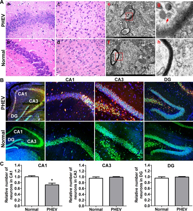
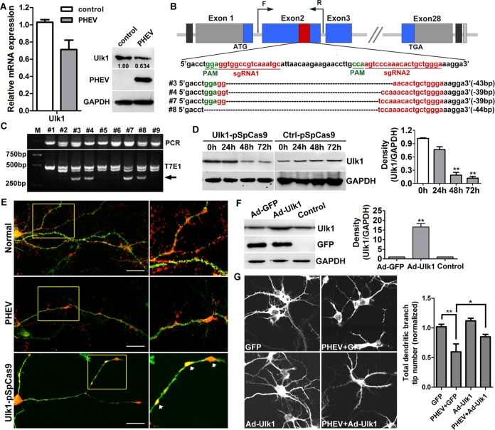
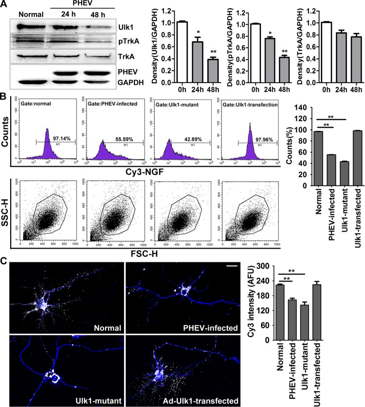
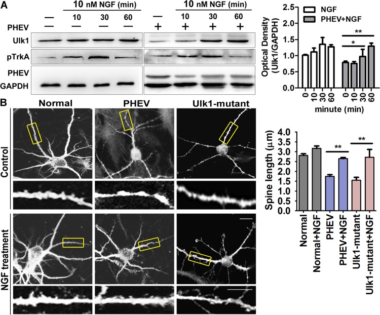
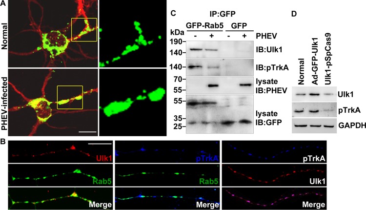
Porcine hemagglutinating encephalomyelitis virus (PHEV) is a highly neurovirulent coronavirus and causes neurological dysfunction in the central nervous system (CNS), but the neuropathological mechanism of PHEV remains poorly understood. We report that Unc51-like kinase 1 (Ulk1/Unc51.1) is a pivotal regulator of PHEV-induced neurological disorders and functions to selectively control the initiation of nerve growth factor (NGF)/TrkA endosome trafficking. We first identified the function of Ulk1 by histopathologic evaluation in a PHEV-infected mouse model in which neuronal loss was accompanied by the suppression of Ulk1 expression. Morphogenesis assessments in the primary cortical neurons revealed that overexpression or mutations of Ulk1 modulated neurite outgrowth, collateral sprouting, and endosomal transport. Likewise, Ulk1 expression was decreased following PHEV infection, suggesting that there was a correlation between the neurodegeneration and functional Ulk1 deficiency. We then showed that Ulk1 forms a multiprotein complex with TrkA and the early endosome marker Rab5 and that Ulk1 defects lead to either blocking of NGF/TrkA endocytosis or premature degradation of pTrkA via constitutive activation of the Rab5 GTPase. Further investigation determined that the ectopic expression of Rab5 mutants induces aberrant endosomal accumulation of activated pTrkA, proving that targeting of Ulk1-TrkA-NGF signaling to the retrograde transport route in the neurodegenerative process that underlies PHEV infection is dependent on Rab5 GTPase activity. Therefore, we described a long-distance signaling mechanism of PHEV-driven deficits in neurons and suggested that such Ulk1 repression may result in limited NGF/TrkA retrograde signaling within activated Rab5 endosomes, explaining the progressive failure of neurite outgrowth and survival.IMPORTANCE Porcine hemagglutinating encephalomyelitis virus (PHEV) is a neurotropic coronavirus and targets neurons in the nervous system for proliferation, frequently leaving behind grievous neurodegeneration. Structural plasticity disorders occur in the axons, dendrites, and dendritic spines of PHEV-infected neurons, and dysfunction of this neural process may contribute to neurologic pathologies, but the mechanisms remain undetermined. Further understanding of the neurological manifestations underlying PHEV infection in the CNS may provide insights into both neurodevelopmental and neurodegenerative diseases that may be conducive to targeted approaches for treatment. The significance of our research is in identifying an Ulk1-related neurodegenerative mechanism, focusing on the regulatory functions of Ulk1 in the transport of long-distance trophic signaling endosomes, thereby explaining the progressive failure of neurite outgrowth and survival associated with PHEV aggression. This is the first report to define a mechanistic link between alterations in signaling from endocytic pathways and the neuropathogenesis of PHEV-induced CNS disease.

Keywords: NGF; Rab5; Ulk1; neurodegeneration; neurovirulent coronavirus; porcine hemagglutinating encephalomyelitis virus.


Figures

FIG 1
FIG 1
Decreased Ulk1 expression in the CNS of PHEV-infected mice. (A) Mice that were intranasally inoculated with 100 μl PHEV (104.45 TCID50/0.1 ml) showed typical neurological symptoms at 3 dpi, such as abnormal gait, tremors, listlessness, standing upright, raised forelimbs, and arched waists. Mice in the control group survived normally. (B) Survival curves of PHEV-infected mice (n = 10 mice per group; data from three independent experiments). (C) Body weight curves of PHEV-infected and control mice from 0 to 6 dpi. From 4 dpi, PHEV-infected mice exhibited a significantly lesser body weight than that observed in the control mice. (D) qRT-PCR analysis of Ulk1 mRNA levels in mouse brain after PHEV infection. Data are presented as the means ± standard errors of the means (SEM) (n = 6). (E) In situ hybridization of the mouse brain for Ulk1 mRNA (green) with an antisense probe. Quantitative analysis is summarized in the histograms on the right. The y axis is the average Ulk1 mRNA intensity in arbitrary fluorescence units.
FIG 2
FIG 2
Degeneration of neurons in the CNS of PHEV-infected mice. (A) Serial mouse brain sections stained with H&E demonstrated the loss of pyramidal neurons in the PHEV-infected mice (a to d). Ultrathin sections from mouse brain that were stained with uranyl acetate and lead citrate showed myelin degeneration (e to h). The arrow indicates a PHEV particle. (B) Representative images of serial brain sections (hippocampal areas CA1, CA3, and dentate gyrus [DG]) stained with DAPI (blue) and antibodies against the neuronal markers NeuN (green) and PHEV (red). (C) Average number of neurons (normalized to that in uninfected controls) ± SEM in the indicated area (n = 3 mice). *, P < 0.05 (Student's t test).
FIG 3
FIG 3
Ulk1 deficiency in cortical neurons is associated with PHEV-induced degeneration. (A) The suppression of Ulk1 in the cortical neurons that were preincubated with PHEV was verified by qRT-PCR and Western blotting. The levels of Ulk1 expression were normalized to the uninfected controls. Anti-Ulk1 antibodies were used to determine the level of Ulk1 and mouse anti-GAPDH antibody to determine protein loading. *, P < 0.05 (Student's t test). (B) CRISPR/Cas9-mediated gene targeting of Ulk1 in cortical neurons. Schematic diagram of sgRNA targeting the Ulk1 gene loci (top panel) and Sanger sequencing of the modified Ulk1 alleles (bottom panel). (C) The target fragments were amplified by PCR from genomic DNA that was extracted from independent transgenic neurons, and detection of target mutations was performed by T7E1 cleavage assay. M, DL2000; #1 to #9 represent 9 independent transgenic Ulk1 sgRNAs neurons. Arrows indicate the fragments digested by T7E1. (D) Cortical neurons at 4 DIV were transfected with Ctrl-pSpCas9 or Ulk1-pSpCas9. Western blot analysis confirmed that Ulk1 protein was significantly decreased in these knockdown neurons. Relative protein levels were analyzed by using ImageJ software. (E) In the transgenic group, cortical neurons at 4 DIV were transfected with Ulk1-pSpCas9, followed by puromycin screening for a total of 48 h. In the infected group, cortical neurons at 5 DIV were incubated with PHEV for 24 h. The expression and localization of Ulk1 were determined by antibody staining using antibodies against Ulk1 (1:100, red) and MAP2 (1:100, green). The boxed insets are shown at a greater magnification in the panels on the right. Neuritic beading or focal beadlike swellings in the dendrites and axons are indicated by arrows. Bars, 50 μm. (F) The primary cortical neurons were transfected with Ad-GFP-Ulk1 or Ad-GFP at an MOI of 100. Forty-eight hours after transfection, the cells were harvested for analysis of Western blots. Relative protein levels were analyzed by using ImageJ software. (G) Cortical neurons at 5 DIV were incubated with PHEV for 24 h and were then transfected with Ad-GFP or Ad-GFP-Ulk1 for 48 h. Representative images are shown. Histograms show the normalized total dendritic branch tip number. The mean values from the Ad-GFP-transfected neurons were set at 1.0. Data are shown as the means ± SEM from the three experiments. The numbers in the histograms indicate the neuron numbers from the three experiments. *, P < 0.05; **, P < 0.01 (Student's t test).
FIG 4
FIG 4
PHEV infection suppresses the Ulk1-mediated endocytosis of NGF/TrkA complexes. (A) Cortical neurons were incubated with PHEV as indicated, and cell lysates were analyzed by Western blotting with antibodies against Ulk1, TrkA, pTrkA, PHEV, and GAPDH. Ulk1 and pTrkA expression were inhibited by PHEV, although the total expression of TrkA was not significantly affected. Densitometric analysis was performed; Ulk1, pTrkA, and TrkA intensities were normalized against the amount of GAPDH. (B) Cy3-NGF internalization assay: Cy3-NGF in the Ad-GFP-Ulk1-transfected, Ulk1 mutant, PHEV-infected, and normal cortical neurons was detected by FACS, and the data were normalized to internalization rates in normal neurons. Representative FACS profiles obtained during the analysis are shown, and Cy3 signals were gated in M1 and expressed as a percentage of the total number of cells analyzed. SSC-H, side scatter height; FSC-H, forward scatter height. (C) Representative images of Cy3-NGF (white) internalization in the MAP2-marked neurons (blue). Quantitative analyses revealed that the Cy3-NGF internalization was markedly suppressed in the PHEV-infected or Ulk1 mutant neurons. The y axis represents the average Cy3 intensity using arbitrary fluorescence units. Bars, 20 μm. *, P < 0.05; **, P < 0.01 (Student's t test).
FIG 5
FIG 5
NGF internalization is required for Ulk1-dependent production of neurite morphogenesis. (A) Time course of NGF-stimulated phosphorylation of TrkA. Neurons were stimulated with 10 nM NGF for different amounts of time (0, 10, 30, and 60 min) and lysed. Then, Western blotting was used to check for Ulk1, pTrkA, and GAPDH expression. TrkA was maximally phosphorylated within 30 min after adding NGF. Densitometric analysis was performed, and Ulk1 intensities were normalized against GAPDH. (B) Cortical neurons were preincubated with PHEV at 5 DIV for 24 h or pretransfected with Ulk1-pSpCas9 at 4 DIV for 48 h, followed by being treated with 10 nM NGF. After 24 h, these neurons were stained for MAP2; representative micrographs are shown. The bottom panels (insets of boxed areas) show high-magnification images of shorter spines. Bars, 20 μm (top images) or 5 μm (high-magnification images). *, P < 0.05; **, P < 0.01 (Student's t test).
FIG 6
FIG 6
Ulk1 is associated with TrkA within Rab5 endosomes. (A) Cortical neurons were transfected with GFP-Rab5 at 4 DIV for 24 h and incubated with PHEV at 5 DIV for 24 h and then fixed and subjected to immunocytochemistry with an antibody against MAP2 (red). Typical images of GFP-Rab5-expressing neurons are shown in the left panels. Boxed regions are shown at a higher magnification in the right panels. Bar, 20 μm. (B) Cortical neurons (4 DIV) were transfected with GFP-Rab5, fixed, and subjected to immunocytochemistry with antibodies against Ulk1 (red) and pTrkA (blue) at 6 DIV. The representative micrographs showed that the Ulk1 and pTrkA dots were localized along the growing axon within Rab5 endosomes. Bar, 5 μm. (C) GFP-Rab5- or GFP-expressing neurons were infected with PHEV for 24 h, and the cell lysates were immunoprecipitated (IP) by anti-GFP antibody-conjugated Sepharose beads, followed by SDS-PAGE and immunoblot (IB) analysis with the indicated antibodies. (D) With pretreatment of Ad-GFP-Ulk1 transfection or CRISPR/Cas9-mediated Ulk1 mutant in the cortical neurons, the cell lysates were subjected to Western blot assay to determine the levels of Ulk1 and pTrkA.
FIG 7
FIG 7
Rab5 GTPase activation blocks Ulk1-mediated NGF/TrkA endocytosis. (A) Ad-GFP-Ulk1-overexpressing or Ulk1 mutant cortical neurons were infected with PHEV for 24 h, and then the cell lysates were subjected to Rab5 activity assays and Western blotting using the indicated antibodies. Immunoprecipitation was performed with the anti-active Rab5 monoclonal antibody (1:500). Immunoblotting was performed with an anti-Rab5 polyclonal antibody (1:1,000). (B) Densitometric analysis of blots in panel A was performed; Rab5 intensities were normalized against GAPDH, and pTrkA intensities were normalized against total TrkA. (C) Differential treatments of the cortical neurons as described for panel A were cultured with standard culture medium containing 10 nM Cy3-NGF (red) for 30 min and then fixed and stained with MAP2 (blue). Representative images were acquired using a confocal microscope. Bars, 20 μm. (D) Cortical neurons were transfected with plasmids encoding Rab5:wt, Rab5:Q79L, or Rab5:S34N at 4 DIV. After 24 h of expression, neurons were infected with PHEV for 24 h and were then harvested for Western blot and Rab5 activity assays using the indicated antibodies. (E) Representative images obtained after GFP-Rab5:wt-, Rab5:Q79L-, or Rab5:S34N-expressing neurons were fixed and stained for pTrkA (red). Bar, 20 μm. Quantification analysis revealed that the pTrkA trafficking was markedly suppressed in the Rab5:Q79L-expressing neurons. The y axis represents the average pTrkA intensity per 5 mm of dendrite, and the dendritic field is represented as the sum of the lengths of all the dendrites on one neuron. *, P < 0.05; **, P < 0.01 (Student's t test).

Similar articles

Cited by

References

    1. Greig AS, Mitchell D, Corner AH, Bannister GL, Meads EB, Julian RJ. 1962. A hemagglutinating virus producing encephalomyelitis in baby pigs. Can J Comp Med Vet Sci 26:49–56. - PMC - PubMed
    1. Vijgen L, Keyaerts E, Lemey P, Maes P, Van Reeth K, Nauwynck H, Pensaert M, Van Ranst M. 2006. Evolutionary history of the closely related group 2 coronaviruses: porcine hemagglutinating encephalomyelitis virus, bovine coronavirus, and human coronavirus OC43. J Virol 80:7270–7274. doi:10.1128/JVI.02675-05. - DOI - PMC - PubMed
    1. Alsop JE. 2006. A presumptive case of vomiting and wasting disease in a swine nucleus herd. J Swine Health Production 14:97–100.
    1. Quiroga MA, Cappuccio J, Pineyro P, Basso W, More G, Kienast M, Schonfeld S, Cancer JL, Arauz S, Pintos ME, Nanni M, Machuca M, Hirano N, Perfumo CJ. 2008. Hemagglutinating encephalomyelitis coronavirus infection in pigs, Argentina. Emerg Infect Dis 14:484–486. doi:10.3201/eid1403.070825. - DOI - PMC - PubMed
    1. Mengeling WL. 1975. Incidence of antibody for hemagglutinating encephalomyelitis virus in serums from swine in the United States. Am J Vet Res 36:821–823. - PubMed

Publication types

MeSH terms

LinkOut - more resources