. 2018 Jul 31;92(16):e00690-18.
doi: 10.1128/JVI.00690-18. Print 2018 Aug 15.

Mamu-B*17+ Rhesus Macaques Vaccinated with env, vif, and nef Manifest Early Control of SIVmac239 Replication

Affiliations

Mamu-B*17+ Rhesus Macaques Vaccinated with env, vif, and nef Manifest Early Control of SIVmac239 Replication

Mauricio A Martins et al. J Virol. .

Abstract

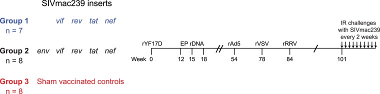
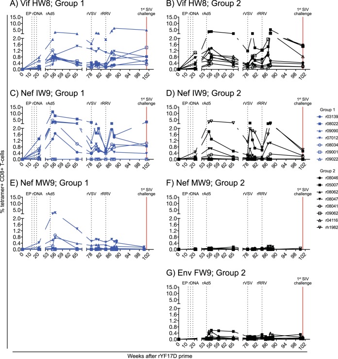
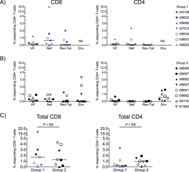
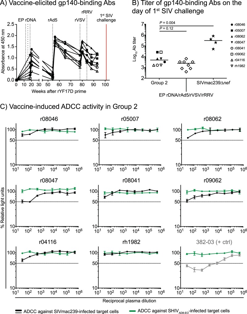
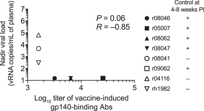
Certain major histocompatibility complex class I (MHC-I) alleles are associated with spontaneous control of viral replication in human immunodeficiency virus (HIV)-infected people and simian immunodeficiency virus (SIV)-infected rhesus macaques (RMs). These cases of "elite" control of HIV/SIV replication are often immune-mediated, thereby providing a framework for studying anti-lentiviral immunity. In this study, we examined how vaccination impacts SIV replication in RMs expressing the MHC-I allele Mamu-B*17 Approximately 21% of Mamu-B*17+ and 50% of Mamu-B*08+ RMs control chronic-phase viremia after SIVmac239 infection. Because CD8+ T cells targeting Mamu-B*08-restricted SIV epitopes have been implicated in virologic suppression in Mamu-B*08+ RMs, we investigated whether this might also be true for Mamu-B*17+ RMs. Two groups of Mamu-B*17+ RMs were vaccinated with genes encoding Mamu-B*17-restricted epitopes in Vif and Nef. These genes were delivered by themselves (group 1) or together with env (group 2). Group 3 included MHC-I-matched RMs and served as the control group. Surprisingly, the group 1 vaccine regimen had little effect on viral replication compared to group 3, suggesting that unlike Mamu-B*08+ RMs, preexisting SIV-specific CD8+ T cells alone do not facilitate long-term virologic suppression in Mamu-B*17+ RMs. Remarkably, however, 5/8 group 2 vaccinees controlled viremia to <15 viral RNA copies/ml soon after infection. No serological neutralizing activity against SIVmac239 was detected in group 2, although vaccine-elicited gp140-binding antibodies correlated inversely with nadir viral loads. Collectively, these data shed new light on the unique mechanism of elite control in Mamu-B*17+ RMs and implicate vaccine-induced, nonneutralizing anti-Env antibodies in the containment of immunodeficiency virus infection.IMPORTANCE A better understanding of the immune correlates of protection against HIV might facilitate the development of a prophylactic vaccine. Therefore, we investigated simian immunodeficiency virus (SIV) infection outcomes in rhesus macaques expressing the major histocompatibility complex class I allele Mamu-B*17 Approximately 21% of Mamu-B*17+ macaques spontaneously controlled chronic phase viremia after SIV infection, an effect that may involve CD8+ T cells targeting Mamu-B*17-restricted SIV epitopes. We vaccinated Mamu-B*17+ macaques with genes encoding immunodominant epitopes in Vif and Nef alone (group 1) or together with env (group 2). Although neither vaccine regimen prevented SIV infection, 5/8 group 2 vaccinees controlled viremia to below detection limits shortly after infection. This outcome, which was not observed in group 1, was associated with vaccine-induced, nonneutralizing Env-binding antibodies. Together, these findings suggest a limited contribution of Vif- and Nef-specific CD8+ T cells for virologic control in Mamu-B*17+ macaques and implicate anti-Env antibodies in containment of SIV infection.

Keywords: AIDS; human immunodeficiency virus; simian immunodeficiency virus; vaccines.


Figures

FIG 1
FIG 1
Experimental design. Twenty-three Mamu-B*17+ RMs were vaccinated with an rYF17D/EP rDNA/rAd5/rVSV/rRRV regimen and divided into three groups depending on which vaccine inserts they received. Animals in group 1 (n = 7) were vaccinated with vif and nef sequences. Animals in group 2 (n = 8) were vaccinated with the same SIV inserts plus env. RMs in group 3 were sham vaccinated with vectors lacking SIV genes or expressing irrelevant inserts and served as the control group for this experiment. At study week 101, vaccine efficacy was assessed by subjecting all animals to repeated intrarectal (i.r.) challenges with a marginal dose of SIVmac239 (200 TCID50) every 2 weeks.
FIG 2
FIG 2
Development of vaccine-induced CD8+ T-cell responses against Mamu-B*17-restricted SIV epitopes in groups 1 and 2. Fluorochrome-labeled Mamu-B*17 tetramers folded with peptides corresponding to SIV epitopes were used to track vaccine-elicited SIV-specific CD8+ T cells in PBMC from RMs in group 1 (A, C, and E) and group 2 (B, D, F, and G). The percentages of live tetramer+ CD8+ T cells specific for Vif HW8 (A and B), Nef IW9 (C and D), Nef MW9 (E and F), and Env FW9 (G) are shown at multiple time points throughout the vaccine phase. No data for Env FW9-specific CD8+ T cells are available for the group 1 RMs because those animals were not vaccinated with env. The times of each vaccination (black vertical dotted lines) and the day of the first i.r. SIV challenge (red vertical solid line) are indicated in each graph. RMs in groups 1 and 2 are color-coded in blue and black, respectively.
FIG 3
FIG 3
Vaccine-induced SIV-specific CD4+ and CD8+ T-cell responses in groups 1 and 2 at the time of the first SIV challenge. CD8+ and CD4+ T-cell responses were measured in PBMC by ICS using pools of peptides (15-mers overlapping by 11 amino acids) spanning the appropriate SIVmac239 proteins. Peptides covering the Rev and Tat proteins were tested together. The percentages of responding CD4+ or CD8+ T cells displayed in all panels were calculated by adding the frequencies of positive responses producing any combination of three immunological functions (IFN-γ, TNF-α, and CD107a). The magnitude and specificity of vaccine-induced CD8+ (left panels) and CD4+ (right panels) T-cell responses are shown for group 1 (A) and group 2 (B). (C) Comparison of the total magnitude of vaccine-elicited SIV-specific CD8+ (left) and CD4+ (right) between groups 1 and 2. The Mann-Whitney U test was used for these comparisons, and no statistically significant difference was found. RMs in groups 1 and 2 are color-coded in blue and black, respectively, and each symbol corresponds to one vaccinee. Lines represent medians. NA, not applicable; NS, not significant.
FIG 4
FIG 4
Vaccine-induced Env-specific humoral immune responses in group 2. (A) Env-binding antibodies (Abs) were measured by ELISA using plate-bound gp140 at multiple time points during the vaccine phase. Straight 1:200 dilutions of plasma from each RM in group 2 were used for this analysis. (B) Log-transformed endpoint titers of vaccine-induced gp140-binding Abs in sera from the group 2 vaccinees collected at the time of the first SIV challenge. As a reference, these values were plotted alongside the endpoint titers of gp140-binding Abs in four RMs that had been infected with SIVmac239Δnef for 28 weeks as part of a previous experiment (70). The endpoint titers of gp140-binding Abs in macaques vaccinated with an EP rDNA/rAd5/rVSV/rRRV regimen encoding env, gag, vif, tat, rev, and nef are also shown (27). (C) Ab-dependent cellular cytotoxicity (ADCC) activity was screened against SIVmac239-infected target cells using plasma collected from the group 2 vaccinees at the time of the first i.r. SIV challenge (black lines). SHIVAD8-EO-infected target cells were used as internal controls (green lines) for nonspecific killing. The decrease in relative light units (RLU) indicates the loss of virus-infected cells in the presence of an NK cell line during the duration of the assay. Dotted lines indicate 50% activity. Plasma from an SIV-infected RM (382-03) with a defined ADCC titer against SIVmac239-infected cells was used as a positive control for these measurements.
FIG 5
FIG 5
Kaplan-Meier rate of infection after repeated i.r. challenges with SIVmac239. Macaques in groups 1 to 3 were inoculated intrarectally with 200 TCID50 of SIVmac239 every other week. The rate of SIV infection in groups 1 and 2 was not significantly different than that of group 3. The P value for the comparison of group 1 versus group 3 was 0.96. The P value for the comparison of group 2 versus group 3 was 0.28.
FIG 6
FIG 6
Plasma viral RNA levels after SIVmac239 infection. Viral load (VL) traces for individual animals in group 1 (A), group 2 (B), and group 3 (C). VLs were log transformed and correspond to the number of vRNA copies/milliliter of plasma. The dotted lines in all the graphs are for reference only and indicate a VL of 103 vRNA copies/ml. The dashed lines are also for reference only and denote a VL of 106 vRNA copies/ml. The pink rectangle in each graph frames the interval during which five group 2 vaccinees controlled viremia to <15 vRNA copies/ml. Groups 1, 2, and 3 are color-coded in blue, black, and red, respectively.
FIG 7
FIG 7
SIV plasma viral load comparisons among groups 1, 2, and 3. (A) Plasma VLs measured on day 6 postinfection (p.i.). (B) Peak VLs. (C) Nadir VLs. (D) Setpoint VLs, calculated as the geometric mean of VLs measured between week 8 p.i. and the last chronic phase time point available. (E) Time to peak viremia, determined as the week p.i. when each animal experienced its peak VL. The dotted lines in panels A to D are for reference only and indicate a VL of 103 vRNA copies/ml. The dashed lines in panels A to D are also for reference only and denote a VL of 106 vRNA copies/ml. Lines represent medians and P values were calculated using the Mann-Whitney U test. Groups 1, 2, and 3 are color-coded in blue, black, and red, respectively, and each symbol corresponds to one vaccinee.
FIG 8
FIG 8
Viral loads in the five group 2 controllers compared to those in RMs historically infected with SIVmac239. The outcome of SIVmac239 infection in the five group 2 vaccinees that manifested early control of viral replication was compared to those of 197 RMs that were rectally infected with SIVmac239 as part of eight SIV vaccine trials conducted by our group. The historical VLs for each independent SIV vaccine trial are plotted in panels A to H and include both vaccinated (gray lines) and control (salmon lines) RMs. VLs from RMs that expressed MHC-I alleles associated with elite control of SIV infection (i.e., Mamu-B*08 and Mamu-B*17) are shown in dashed lines. VLs from RMs that did not express these protective MHC-I alleles are shown in solid lines. To better visualize the early control of viral replication manifested by the five group 2 vaccinees, their VLs (black lines) were also plotted in each panel. Additionally, only the first 20 weeks of SIV infection are shown in each graph. (A) In trial 1, 24 RMs were vaccinated with env, gag, vif, rev, tat, and nef delivered by either rRRV alone or rRRV followed by two boosts with rAd5 and rVSV. Eight RMs served as the controls for the challenge phase (M. A. Martins, D. C. Tully, N. Pedreño-Lopez, B. von Bredow, M. G. Pauthner, Y. C. Shin, M. Yuan, N. S. Lima, D. J. Bean, L. Gonzalez-Nieto, A. Domingues, M. J. Gutman, H. S. Maxwell, D. M. Magnani, M. J. Ricciardi, V. K. Bailey, J. D. Altman, D. R. Burton, K. Ejima, D. B. Allison, D. T. Evans, E. G. Rakasz, C. L. Parks, M. C. Bonaldo, S. Capuano III, J. D. Lifson, R. C. Desrosiers, T. M. Allen, D. I. Watkins, unpublished data). (B) The details of this experiment were published recently (27). Briefly, 32 RMs were vaccinated with an EP rDNA/rAd5/rVSV/rRRV regimen encoding four different sets of SIV inserts. Eight RMs served as the controls for this experiment. One vaccinee did not get infected, so VL traces for 39 RMs are shown in the graph. (C) The details of this experiment were published recently (71). Briefly, four different mixed-modality vaccine regimens were used to deliver minigenes of SIV gag, vif, and nef to 32 RMs. Eight RMs served as the controls for this experiment. (D) Ten Mamu-B*08+ RMs were vaccinated with a rAd5/rVSV/rRRV regimen including vif, rev, tat, and nef, and six MHC-I-matched RMs served as the controls for the challenge phase (Martins et al., unpublished). Three vaccinees did not acquire SIV infection in this experiment, so VL traces for 13 RMs are shown in the graph. (E) The details of this experiment were published recently (72). Briefly, 16 Mamu-B*08+ RMs were vaccinated with an EP rDNA/rYF17D/rAd5 regimen containing nef, and two MHC-I-matched macaques served as the controls for the challenge phase. (F) The details of this experiment were published recently (30). Four RMs, two of which were Mamu-B*08+, were vaccinated with an EP rDNA/rYF17D/rRRV regimen containing either gag or nef. For unknown reasons, the animal highlighted in pink harbored a nef deletion SIV variant as early as week 2 p.i. The replicative fitness cost imposed by this nef deletion likely underlies the stringent control of viral replication manifested by this animal. (G) Twenty RMs were vaccinated with an EP rDNA/recombinant vaccinia/rVSV/rAd5/rRRV regimen encoding vif only. Twenty MHC-I-matched RMs served as the controls for the challenge phase. Eight vaccinees and nine control RMs expressed either Mamu-B*08 or Mamu-B*17 (Martins et al., unpublished). One vaccinee did not acquire SIV infection, so VL traces for 39 RMs are shown in the graph. (H) The details of this experiment were published elsewhere (21). Briefly, 16 Mamu-B*08+ RMs were vaccinated with an rYF17D/rAd5 regimen including either vif and nef minigenes containing Mamu-B*08-restricted epitopes or regions of the SIV proteome lacking epitopes restricted by Mamu-B*08. Four MHC-I-matched RMs served as the controls for the challenge phase.
FIG 9
FIG 9
nef sequence diversity in acute-phase plasma from four group 2 controllers. Heat maps illustrate the levels of sequence diversity at each codon in the nef open reading frame. The range of amino acids covered by each row is indicated on the right-hand side of the figure and should be used only as a reference because both synonymous and nonsynonymous mutations are considered in these heat maps. The codon corresponding to the first amino acid in Nef is located in the top left corner of each grid. Each row spans 17 amino acids of the Nef protein, except for the bottom row, which covers 9 amino acids and includes the last codon at position 264. The images on the left correspond to plasma samples collected at week 2 postinfection. The images on the right correspond to plasma samples collected at week 3 p.i., or week 4 p.i. in the case of r08046. Data from r08046, r05007, r08062, and r08047 are shown in panels A, B, C, and D, respectively.
FIG 10
FIG 10
env sequence diversity in acute-phase plasma from four group 2 controllers. Heat maps illustrate the levels of sequence diversity at each codon in the env open reading frame. The range of amino acids covered by each row is indicated on the right-hand side of the figure only as a reference because both synonymous and nonsynonymous mutations are considered in these heat maps. When applicable, the frequency and location of amino acid substitutions are described in the text and in Fig. 11. The codon corresponding to the first amino acid in Env is located in the top left corner of each grid. Each row spans 30 amino acids of the Env protein, except for the bottom row, which covers 9 amino acids and includes the last residue at position 879. The images on the left correspond to plasma samples collected at week 2 postinfection. The images on the right correspond to plasma samples collected at week 3 p.i., or week 4 p.i. in the case of r08046. Data from r08046, r05007, r08062, and r08047 are shown in panels A, B, C, and D, respectively.
FIG 11
FIG 11
Summary of amino acid substitutions in Env present at ≥10% frequencies in acute phase samples from four group 2 controllers. Each substitution is enclosed by a square, and its details are listed in a text box outside the Env main sequence. The details provided for each amino acid substitution include the animal identifier (ID), time point postinfection, position in the Env protein, and the frequency of sequence reads displaying the wild-type (boldface type) and mutant residues. As a reference, several topological features of the SIV Env protein are shown, including the five variable loops (V) in gp120, the gp120/gp41 cleavage site, the membrane spanning domain, and the highly immunogenic region of gp41. The location of the Mamu-B*17-restricted Env FW9 epitope is also indicated in the carboxyl terminus of gp41.
FIG 12
FIG 12
Association between titers of vaccine-induced gp140-binding Abs at the time of the 1st SIV challenge and nadir viral loads in group 2. This is a graphical representation of the comparison between the log10 endpoint titers of vaccine-induced gp140-binding antibodies at the time of the first i.r. SIV challenge and nadir viral loads in group 2 listed in Table 2. The symbol for macaque r09062 (open black square) is masked by the symbol of r05007 (filled black square) because both animals had the same titer of gp140-binding antibodies (25,600) and nadir VL (15 vRNA copies/ml of plasma). The Bonferroni-corrected P value is shown. Each symbol corresponds to one group 2 vaccinee.

Similar articles

Cited by

References

    1. UNAIDS. 2016. Global AIDS update. UNAIDS, Geneva, Switzerland.
    1. Buchbinder SP, Mehrotra DV, Duerr A, Fitzgerald DW, Mogg R, Li D, Gilbert PB, Lama JR, Marmor M, Del Rio C, McElrath MJ, Casimiro DR, Gottesdiener KM, Chodakewitz JA, Corey L, Robertson MN. 2008. Efficacy assessment of a cell-mediated immunity HIV-1 vaccine (the Step Study): a double-blind, randomised, placebo-controlled, test-of-concept trial. Lancet 372:1881–1893. doi:10.1016/S0140-6736(08)61591-3. - DOI - PMC - PubMed
    1. Flynn NM, Forthal DN, Harro CD, Judson FN, Mayer KH, Para MF. 2005. Placebo-controlled phase 3 trial of a recombinant glycoprotein 120 vaccine to prevent HIV-1 infection. J Infect Dis 191:654–665. doi:10.1086/428404. - DOI - PubMed
    1. Gray GE, Allen M, Moodie Z, Churchyard G, Bekker LG, Nchabeleng M, Mlisana K, Metch B, de Bruyn G, Latka MH, Roux S, Mathebula M, Naicker N, Ducar C, Carter DK, Puren A, Eaton N, McElrath MJ, Robertson M, Corey L, Kublin JG. 2011. Safety and efficacy of the HVTN 503/Phambili study of a clade-B-based HIV-1 vaccine in South Africa: a double-blind, randomised, placebo-controlled test-of-concept phase 2b study. Lancet Infect Dis 11:507–515. doi:10.1016/S1473-3099(11)70098-6. - DOI - PMC - PubMed
    1. Hammer SM, Sobieszczyk ME, Janes H, Karuna ST, Mulligan MJ, Grove D, Koblin BA, Buchbinder SP, Keefer MC, Tomaras GD, Frahm N, Hural J, Anude C, Graham BS, Enama ME, Adams E, DeJesus E, Novak RM, Frank I, Bentley C, Ramirez S, Fu R, Koup RA, Mascola JR, Nabel GJ, Montefiori DC, Kublin J, McElrath MJ, Corey L, Gilbert PB. 2013. Efficacy trial of a DNA/rAd5 HIV-1 preventive vaccine. N Engl J Med 369:2083–2092. doi:10.1056/NEJMoa1310566. - DOI - PMC - PubMed

Publication types

MeSH terms

LinkOut - more resources