. 2018 Jun 7;3(11):e96492.
doi: 10.1172/jci.insight.96492.

Intracapillary immune complexes recruit and activate slan-expressing CD16+ monocytes in human lupus nephritis

Affiliations

Intracapillary immune complexes recruit and activate slan-expressing CD16+ monocytes in human lupus nephritis

Florina Olaru et al. JCI Insight. .

Abstract

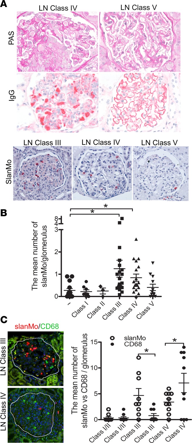
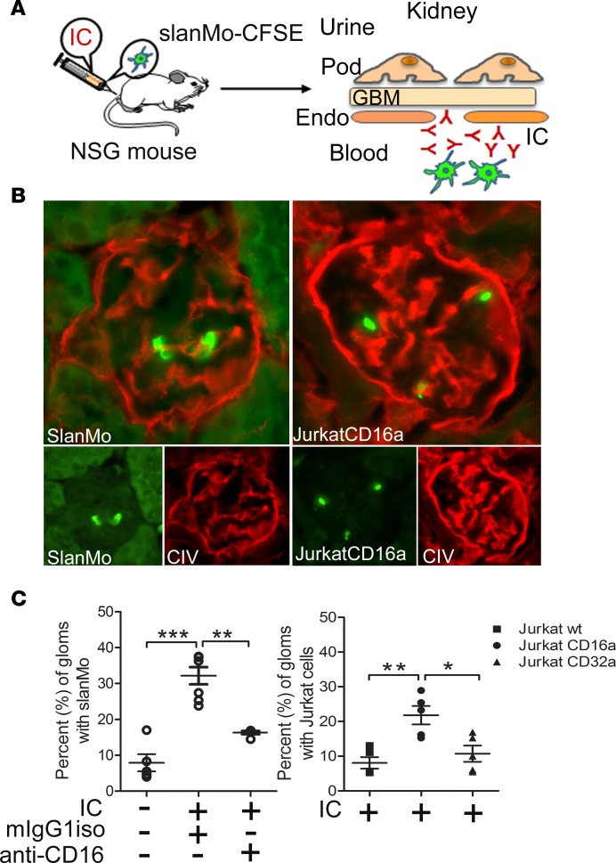
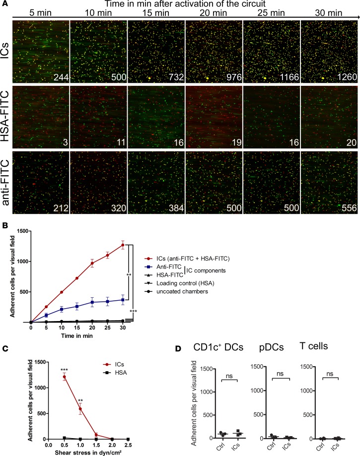
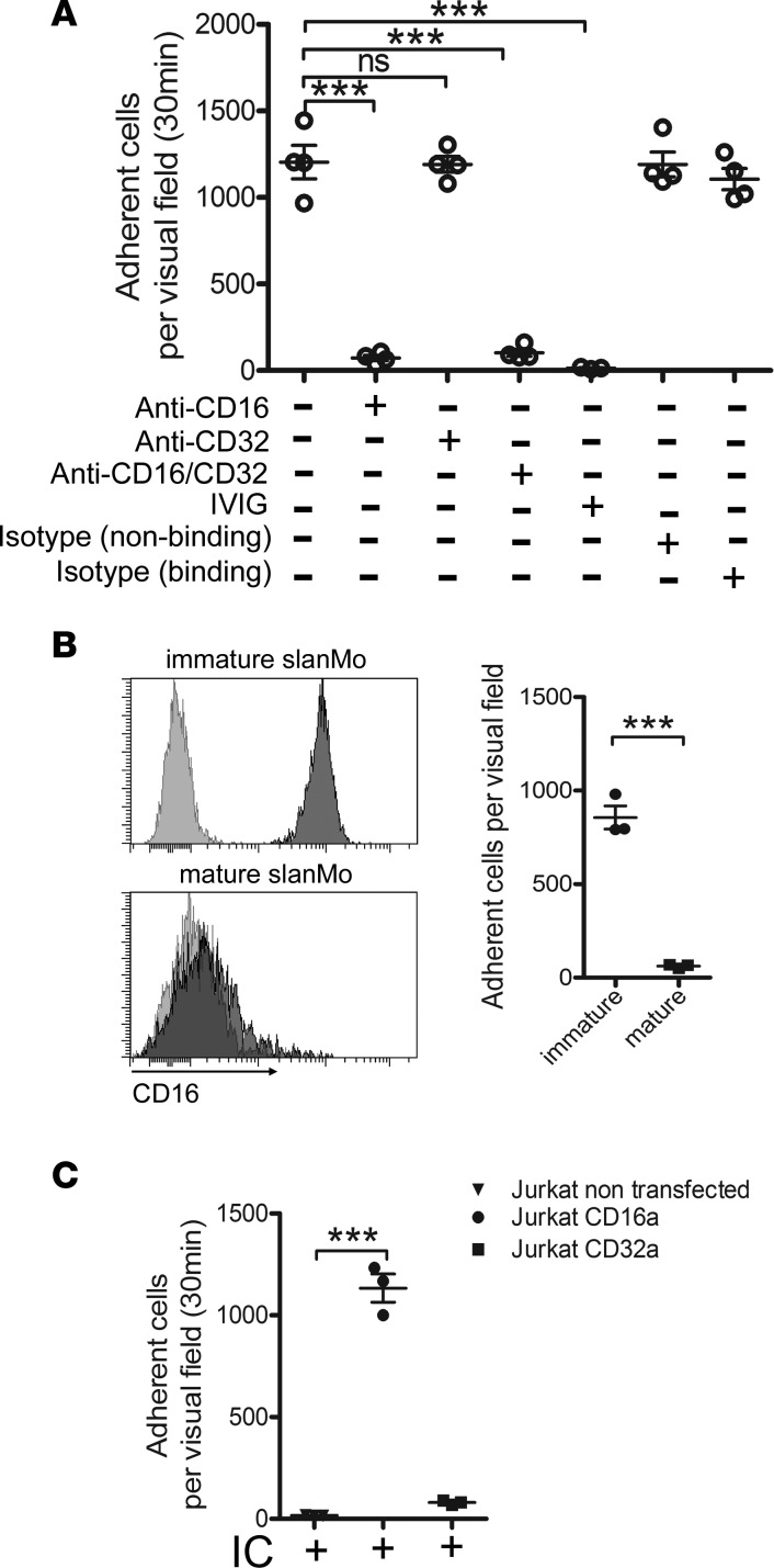
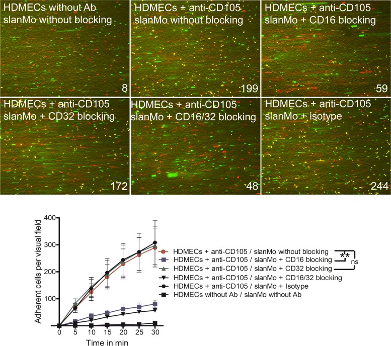
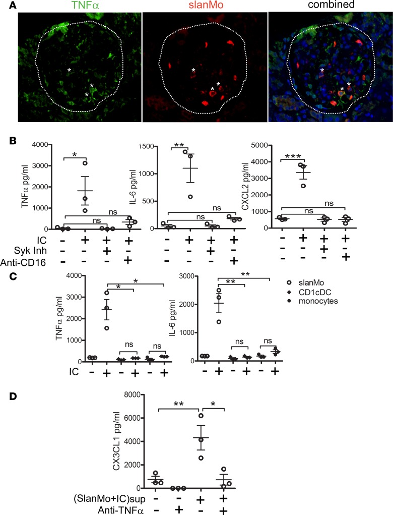
Lupus nephritis is a major cause of morbidity in patients with systemic lupus erythematosus. Among the different types of lupus nephritis, intracapillary immune complex (IC) deposition and accumulation of monocytes are hallmarks of lupus nephritis class III and IV. The relevance of intracapillary ICs in terms of monocyte recruitment and activation, as well as the nature and function of these monocytes are not well understood. For the early focal form of lupus nephritis (class III) we demonstrate a selective accumulation of the proinflammatory population of 6-sulfo LacNAc+ (slan) monocytes (slanMo), which locally expressed TNF-α. Immobilized ICs induced a direct recruitment of slanMo from the microcirculation via interaction with Fc γ receptor IIIA (CD16). Interestingly, intravenous immunoglobulins blocked CD16 and prevented cell recruitment. Engagement of immobilized ICs by slanMo induced the production of neutrophil-attracting chemokine CXCL2 as well as TNF-α, which in a forward feedback loop stimulated endothelial cells to produce the slanMo-recruiting chemokine CX3CL1 (fractalkine). In conclusion, we observed that expression of CD16 equips slanMo with a unique capacity to orchestrate early IC-induced inflammatory responses in glomeruli and identified slanMo as a pathogenic proinflammatory cell type in lupus nephritis.

Keywords: Autoimmunity; Immunology; Monocytes.


Conflict of interest statement

Conflict of interest: The authors have declared that no conflict of interest exists.

Figures

Figure 1
Figure 1. Preferential glomerular accumulation of slanMo in type III and IV lupus nephritis.
(A) Histological images of lupus nephritis patients (class IV and V) stained with periodic acid–Schiff (PAS) and for IgG deposits and hyaline thrombi. The presence of slanMo was shown using the slan-specific antibody DD2. Original magnification, ×40 (PAS and IgG) and ×20 (SlanMo). (B) The mean number of immunohistochemically identified slanMo in glomeruli found in kidney samples of patients with lupus nephritis: healthy (n = 14), class I (n = 8), class II (n = 4), III (n = 22), IV (n = 18) or V (n = 12). One-way ANOVA (nonparametric) followed by Dunn’s multiple-comparison test. Mean values are shown ± SEM. *P < 0.05; comparison between healthy kidney samples and classes I–V of patients with lupus nephritis. (C) Costaining of slanMo and CD68-expressing cells (PG-M1 mAb). Original magnification, ×20. The mean number of slanMo and CD68-expressing cells in glomeruli of lupus nephritis patients (n = 8–10). One-way ANOVA (nonparametric) followed by Newman-Keuls multiple-comparison test. Mean values are shown ± SEM. *P < 0.05; comparison between slanMo and CD68-expressing cells in glomeruli.
Figure 2
Figure 2. Recruitment of slanMo by ICs in vivo.
(A) Schematic representation of the mouse model. Preformed immune complexes (ICs) were injected i.v. into immunodeficient NSG mice followed by the injection of either fluorescently labeled freshly isolated slanMo or Jurkat cells transfected with either CD16 or CD32. (B) Immunofluorescence staining of collagen IV (CIV) on kidney sections (red) of experimental animals and localization of fluorescently labeled cells (slanMo and Jurkat CD16a) (green) in the glomeruli (n = 5 mice per group). Original magnification, ×40. (C) Percentage of glomeruli (gloms) with slanMo and Jurkat cells, respectively, recruited by ICs in the presence or absence of blocking mAbs for CD16 (3G8). Mean values are shown ± SEM (n = 5 mice per group). *P < 0.05, **P < 0.01, ***P < 0.001 by 1-way ANOVA followed by Bonferroni’s multiple-comparison test.
Figure 3
Figure 3. Immobilized ICs recruit slanMo from shear flow conditions.
(A and B) Capture of slanMo by immobilized immune complexes (ICs) from the flow. Freshly isolated slanMo were labeled for fluorescence microscopic detection and subsequently perfused over immobilized ICs for 30 minutes at a shear stress of 0.5 dyn/cm2 in a flow chamber assay. Adherent cells appear in yellow after merge of consecutive frames (slanMo in red or green) at intervals of 5 frames in the off-line analysis at the respective time points after the initiation of shear flow. The images are a representative example of 4 experiments. Original magnification, ×20. ICs were preformed by incubating FITC-labeled human serum albumin (HSA, 1 mg/ml) (HSA-FITC) with polyclonal rabbit anti-FITC IgG (1 mg/ml) (anti-FITC) at 1:6 for 1 hour at 4°C. (C) Capture of slanMo at increasing levels of surface sheer stress (n = 3). (D) Capture of purified CD1c+ DCs, pDCs, and CD4+ T cells by immobilized ICs under the same conditions. n = 3 for CD1c+ DCs and pDCs and n = 4 for CD4+ T cells. Mean values are shown ± SEM. **P < 0.01, ***P < 0.001 by 1-way ANOVA followed by Tukey’s post hoc test.
Figure 4
Figure 4. Recruitment of slanMo from the flow requires expression of CD16.
(A) CD16-specific capture of slanMo by immobilized immune complexes (ICs). Freshly isolated slanMo were labeled for fluorescence microscopic detection and incubated with specific blocking mAbs for CD16 (3G8), CD32 (AT10), or both as well as a binding (DX17) and a nonbinding (MOPC-21) isotype control. Additionally, intravenous immunoglobulin (IVIG) was used for blocking FcγR. SlanMo subsequently were perfused over immobilized ICs for 30 minutes with an applied surface shear stress of 0.5 dyn/cm2 (n = 4). (B) Freshly isolated and CD16-negative mature slanMo of the same donor run over immobilized ICs for 30 minutes at a shear stress of 0.5 dyn/cm2 (n = 3). The histograms give an example of the CD16 expression of freshly isolated and mature slanMo of the same donor. (C) CD16a-transfected, CD32a-transfected, and nontransfected Jurkat cells were fluorescently labeled and subsequently run over immobilized ICs for 30 minutes at a shear stress of 0.5 dyn/cm2 (n = 3). Mean values are shown ± SEM. ***P < 0.001. ns, not significant by 1-way ANOVA followed by Tukey’s post hoc test.
Figure 5
Figure 5. SlanMo are captured by antibodies deposited on endothelial cells.
Antibody-dependent capture of slanMo on endothelial cells under shear-flow conditions. Human microvascular endothelial cells (HDMECs) were seeded into flow chamber slides and grown to 100% confluence. Immediately before the perfusion assay, HDMECs were treated with an antibody against CD105. Subsequently, slanMo that were either untreated or treated with specific blocking mAbs for CD16 (3G8), CD32 (AT10), or both as well as an isotype control (MOPC-21) were perfused for 30 minutes at a shear stress of 0.5 dyn/cm2 over the HDMECs. Adherent cells appear in yellow after merge of consecutive frames (slanMo in red or green) at intervals of 5 frames in the off-line analysis at the respective time points after the initiation of shear flow (n = 3). Original magnification, ×32. **P < 0.01. ns, not significant by 1-way ANOVA followed by Tukey’s post hoc test.
Figure 6
Figure 6. IC-slanMo interaction mediates inflammatory responses.
(A) Single-color images and combined colors for the detection of TNF-α (green), slanMo (red), and cell nuclei (DAPI; blue) in lupus nephritis glomeruli class III. Original magnification, ×20. Results are representative of multiple sections from 3 independent donors (n = 3). Asterisks indicate double-positive cells. (B) Cytokine induction by immobilized immune complexes (ICs). Freshly isolated slanMo were treated or not with Syk inhibitor or specific blocking mAbs for CD16 (3G8) and incubated with immobilized ICs for 20 hours. TNF-α, IL-6, and CXCL2 protein levels in the culture medium were determined by ELISA (n = 3). (C) TNF-α and IL-6 levels in the culture medium of CD1c+ DCs and monocytes in the presence or absence of immobilized ICs (n = 3). (D) HDMECs were cultured for 20 hours with supernatants from slanMo previously incubated with immobilized ICs and in the presence or absence of a neutralizing TNF-α mAb (10 μg/ml). CX3CL1 protein levels were determined by ELISA (n = 3). Mean values are shown ± SEM. *P < 0.05, **P < 0.01, ***P < 0.001 by 1-way ANOVA followed by Bonferroni’s multiple-comparison test.

Similar articles

Cited by

References

    1. Cohen SD, Kimmel PL. Immune complex renal disease and human immunodeficiency virus infection. Semin Nephrol. 2008;28(6):535–544. doi: 10.1016/j.semnephrol.2008.08.006. - DOI - PubMed
    1. de Zubiria Salgado A, Herrera-Diaz C. Lupus nephritis: an overview of recent findings. Autoimmune Dis. 2012;2012:849684. - PMC - PubMed
    1. Gross WL, Trabandt A, Reinhold-Keller E. Diagnosis and evaluation of vasculitis. Rheumatology (Oxford) 2000;39(3):245–252. doi: 10.1093/rheumatology/39.3.245. - DOI - PubMed
    1. Weening JJ, et al. The classification of glomerulonephritis in systemic lupus erythematosus revisited. Kidney Int. 2004;65(2):521–530. doi: 10.1111/j.1523-1755.2004.00443.x. - DOI - PubMed
    1. Means TK, Latz E, Hayashi F, Murali MR, Golenbock DT, Luster AD. Human lupus autoantibody-DNA complexes activate DCs through cooperation of CD32 and TLR9. J Clin Invest. 2005;115(2):407–417. doi: 10.1172/JCI23025. - DOI - PMC - PubMed

Publication types

MeSH terms