

Phosphoinositide 3-kinase δ inhibition promotes antitumor responses but antagonizes checkpoint inhibitors
- PMID: 29875319
- PMCID: PMC6124416
- DOI: 10.1172/jci.insight.120626
Phosphoinositide 3-kinase δ inhibition promotes antitumor responses but antagonizes checkpoint inhibitors
Abstract
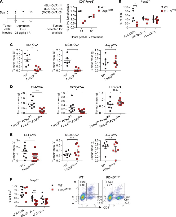
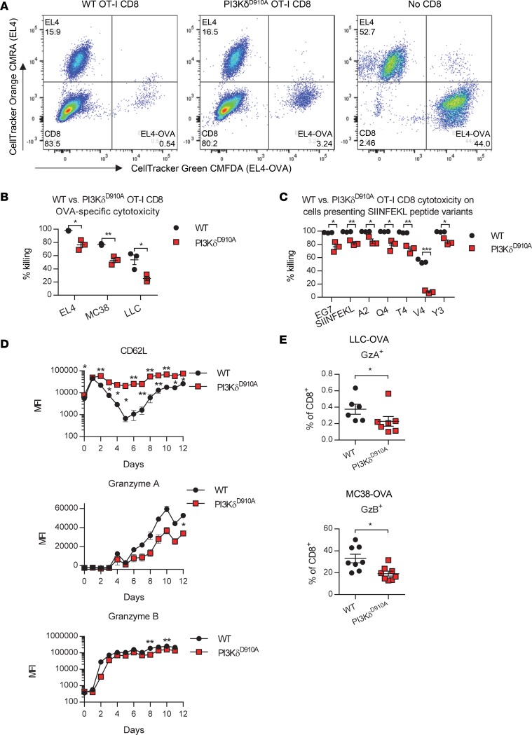
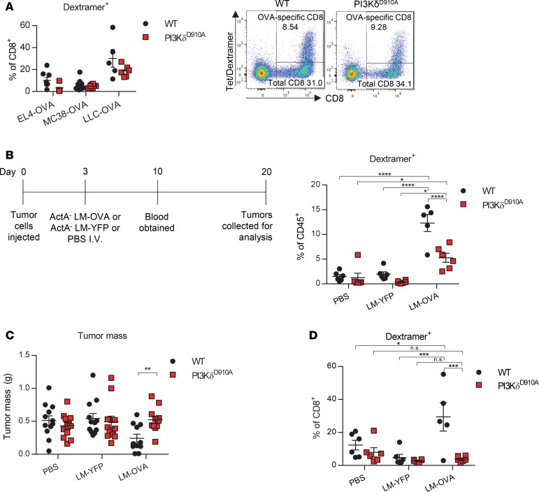
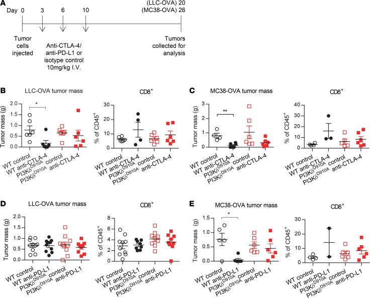
Multiple modes of immunosuppression restrain immune function within tumors. We previously reported that phosphoinositide 3-kinase δ (PI3Kδ) inactivation in mice confers resistance to a range of tumor models by disrupting immunosuppression mediated by regulatory T cells (Tregs). The PI3Kδ inhibitor idelalisib has proven highly effective in the clinical treatment of chronic lymphocytic leukemia and the potential to extend the use of PI3Kδ inhibitors to nonhematological cancers is being evaluated. In this work, we demonstrate that the antitumor effect of PI3Kδ inactivation is primarily mediated through the disruption of Treg function, and correlates with tumor dependence on Treg immunosuppression. Compared with Treg-specific PI3Kδ deletion, systemic PI3Kδ inactivation is less effective at conferring resistance to tumors. We show that PI3Kδ deficiency impairs the maturation and reduces the capacity of CD8+ cytotoxic T lymphocytes (CTLs) to kill tumor cells in vitro, and to respond to tumor antigen-specific immunization in vivo. PI3Kδ inactivation antagonized the antitumor effects of tumor vaccines and checkpoint blockade therapies intended to boost the CD8+ T cell response. These findings provide insights into mechanisms by which PI3Kδ inhibition promotes antitumor immunity and demonstrate that the mechanism is distinct from that mediated by immune checkpoint blockade.
Keywords: Adaptive immunity; Cancer immunotherapy; Immunology; Oncology; Signal transduction.
Conflict of interest statement
Figures
Similar articles
-
Compensation between CSF1R+ macrophages and Foxp3+ Treg cells drives resistance to tumor immunotherapy.JCI Insight. 2018 Jun 7;3(11):e120631. doi: 10.1172/jci.insight.120631. eCollection 2018 Jun 7. JCI Insight. 2018. PMID: 29875321 Free PMC article.
-
PI3Kδ inhibition modulates regulatory and effector T-cell differentiation and function in chronic lymphocytic leukemia.Leukemia. 2019 Jun;33(6):1427-1438. doi: 10.1038/s41375-018-0318-3. Epub 2018 Dec 20. Leukemia. 2019. PMID: 30573773
-
The PI3K p110δ Isoform Inhibitor Idelalisib Preferentially Inhibits Human Regulatory T Cell Function.J Immunol. 2019 Mar 1;202(5):1397-1405. doi: 10.4049/jimmunol.1701703. Epub 2019 Jan 28. J Immunol. 2019. PMID: 30692213
-
CD8+ cytotoxic T lymphocytes in cancer immunotherapy: A review.J Cell Physiol. 2019 Jun;234(6):8509-8521. doi: 10.1002/jcp.27782. Epub 2018 Nov 22. J Cell Physiol. 2019. PMID: 30520029 Review.
-
Phosphoinositide 3-kinase δ is a regulatory T-cell target in cancer immunotherapy.Immunology. 2019 Jul;157(3):210-218. doi: 10.1111/imm.13082. Immunology. 2019. PMID: 31107985 Free PMC article. Review.
Cited by
-
Signaling pathways and targeted therapies in lung squamous cell carcinoma: mechanisms and clinical trials.Signal Transduct Target Ther. 2022 Oct 5;7(1):353. doi: 10.1038/s41392-022-01200-x. Signal Transduct Target Ther. 2022. PMID: 36198685 Free PMC article. Review.
-
PI3-Kinase p110α Deficiency Modulates T Cell Homeostasis and Function and Attenuates Experimental Allergic Encephalitis in Mature Mice.Int J Mol Sci. 2021 Aug 13;22(16):8698. doi: 10.3390/ijms22168698. Int J Mol Sci. 2021. PMID: 34445401 Free PMC article.
-
PI3Kδ as a Novel Therapeutic Target for Aggressive Prostate Cancer.Cancers (Basel). 2025 May 9;17(10):1610. doi: 10.3390/cancers17101610. Cancers (Basel). 2025. PMID: 40427108 Free PMC article. Review.
-
Beyond Anti-PD-1/PD-L1: Improving Immune Checkpoint Inhibitor Responses in Triple-Negative Breast Cancer.Cancers (Basel). 2024 Jun 11;16(12):2189. doi: 10.3390/cancers16122189. Cancers (Basel). 2024. PMID: 38927895 Free PMC article. Review.
-
The Next Generation of Immunotherapy for Cancer: Small Molecules Could Make Big Waves.J Immunol. 2019 Jan 1;202(1):11-19. doi: 10.4049/jimmunol.1800991. J Immunol. 2019. PMID: 30587569 Free PMC article. Review.
References
Publication types
MeSH terms
Substances
Grants and funding
- 095198/Z/10/Z/WT_/Wellcome Trust/United Kingdom
- BBS/E/B/000C0427/BB_/Biotechnology and Biological Sciences Research Council/United Kingdom
- 105663/Z/14/Z/WT_/Wellcome Trust/United Kingdom
- 22597/CRUK_/Cancer Research UK/United Kingdom
- BB/N007794/1/BB_/Biotechnology and Biological Sciences Research Council/United Kingdom
- BBS/E/B/000C0428/BB_/Biotechnology and Biological Sciences Research Council/United Kingdom
- BBS/E/B/000C0407/BB_/Biotechnology and Biological Sciences Research Council/United Kingdom
- BB/E009867/1/BB_/Biotechnology and Biological Sciences Research Council/United Kingdom
- BBS/E/B/000C0409/BB_/Biotechnology and Biological Sciences Research Council/United Kingdom
- 095691/WT_/Wellcome Trust/United Kingdom
LinkOut - more resources
Full Text Sources
Other Literature Sources
Research Materials
