. 2018 Jun 7;3(11):e120137.
doi: 10.1172/jci.insight.120137.

Modulation of subsets of cardiac B lymphocytes improves cardiac function after acute injury

Affiliations

Modulation of subsets of cardiac B lymphocytes improves cardiac function after acute injury

Luigi Adamo et al. JCI Insight. .

Abstract

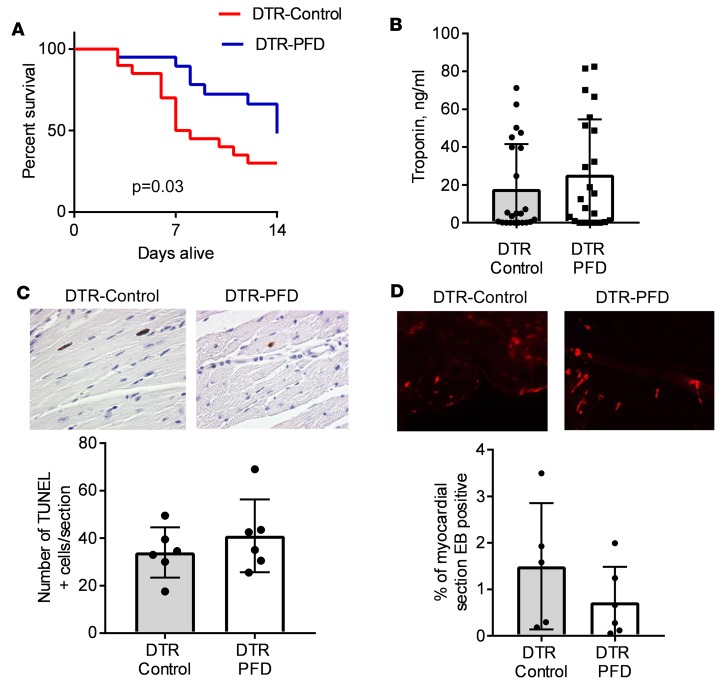
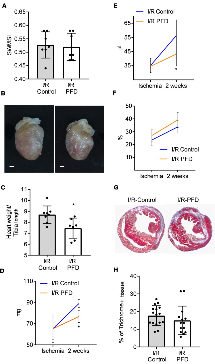
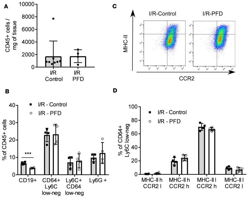
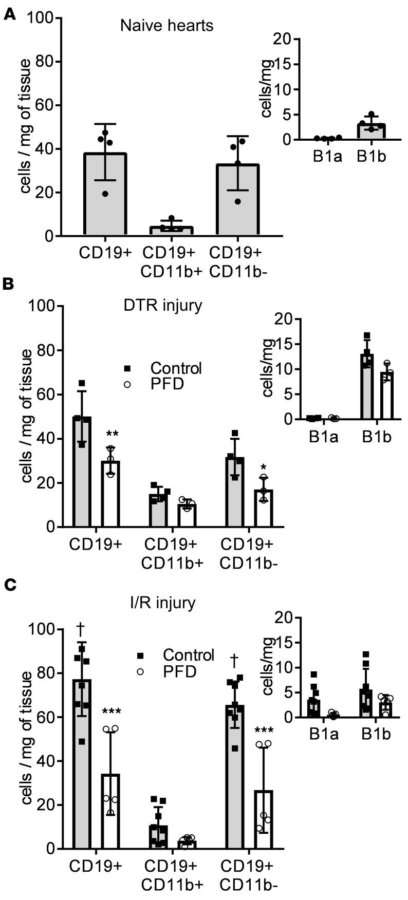
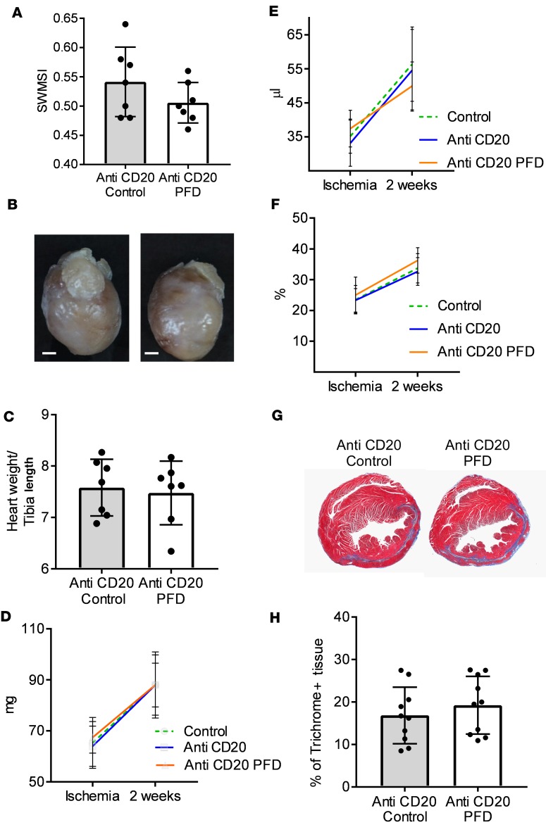
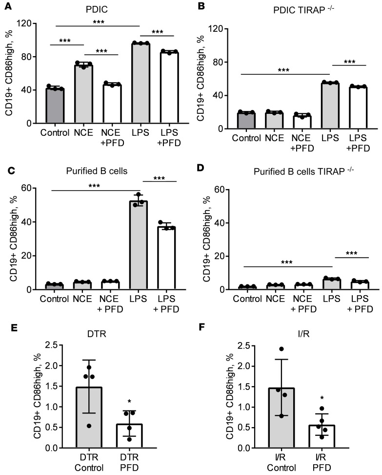
Despite the long-standing recognition that the immune response to acute myocardial injury contributes to adverse left ventricular (LV) remodeling, it has not been possible to effectively target this clinically. Using 2 different in vivo models of acute myocardial injury, we show that pirfenidone confers beneficial effects in the murine heart through an unexpected mechanism that depends on cardiac B lymphocytes. Naive hearts contained a large population of CD19+CD11b-CD23-CD21-IgD+IgMlo lymphocytes, and 2 smaller populations of CD19+CD11b+ B1a and B1b cells. In response to tissue injury, there was an increase in neutrophils, monocytes, macrophages, as well as an increase in CD19+ CD11b- B lymphocytes. Treatment with pirfenidone had no effect on the number of neutrophils, monocytes, or macrophages, but decreased CD19+CD11b- lymphocytes. B cell depletion abrogated the beneficial effects of pirfenidone. In vitro studies demonstrated that stimulation with lipopolysaccharide and extracts from necrotic cells activated CD19+ lymphocytes through a TIRAP-dependent pathway. Treatment with pirfenidone attenuated this activation of B cells. These findings reveal a previously unappreciated complexity of myocardial B lymphocytes within the inflammatory infiltrate triggered by cardiac injury and suggest that pirfenidone exerts beneficial effects in the heart through a unique mechanism that involves modulation of cardiac B lymphocytes.

Keywords: B cells; Cardiology; Heart failure.


Conflict of interest statement

Conflict of interest: The authors have declared that no conflict of interest exists.

Figures

Figure 1
Figure 1. Effect of pirfenidone on mortality and cardiac myocyte cell death after DT treatment.
Mice expressing the diphtheria toxin receptor (DTR) in the myocardium were exposed to diphtheria toxin (DT) and fed either chow enriched with pirfenidone (DTR-PFD) or regular chow (DTR-control). (A) Kaplan-Meier survival curves of DTR control and DTR-PFD mice (n = 20 per group). (B) Serum troponin levels measured at day 4 after DT treatment in DTR-PFD and DTR-control animals (n = 23/group). (C) Cardiac myocyte apoptosis measured at day 4 after treatment with DT. Upper panels are representative histological sections of myocardium from DTR-control and DTR-PFD mice at ×40 magnification. Lower panel summarizes the group data (n = 6 mice/group, 4 sections per animal analyzed). (D) Evans blue (EB) dye uptake at day 4 after DT treatment in DTR-control and DTR-PFD animals; upper panels are representative fluorescence microscopy images at ×10 magnification; lower panel summarizes the group data (n = 5 control; n = 6 mice with pirfenidone; 4 sections per animal analyzed). Bars represent the mean, and error bars represent standard deviation. P values were calculated with the Gehan-Breslow-Wilcoxon method for panel A and with Student’s t test for panels BD.
Figure 2
Figure 2. Effect of pirfenidone on myocardial inflammation (day 4) after DT treatment.
Mice expressing the diphtheria toxin receptor (DTR) in the myocardium were exposed to diphtheria toxin (DT) and fed either chow enriched with pirfenidone (DTR-PFD) or regular chow (DTR-control). Mice were sacrificed at day 4 after DT injection and the heart was collected for analysis via flow cytometry. (A) Total number of CD45+ cells/mg heart tissue (n = 17 control, n = 19 pirfenidone). (B) Leukocyte subsets in the myocardium (percentage of total: CD19+, n = 14 control, n = 16 pirfenidone; Ly6g+, n = 6/group, Ly6C+CD64lo/–, n = 10 control, n = 12 pirfenidone; CD64+Ly6Clo/–, n = 10 control, n = 12 pirfenidone). (C) Representative FACS analysis of MHC-II and CCR-2 macrophages and monocytes. (D) Macrophage/monocyte subsets in the myocardium as defined by expression of CCR2, low (l) or high (h) and MHC-II expression, low (l) or high (h). Percentage of total, n = 10 control, n = 12 pirfenidone.*P < 0.05. Bars represent the mean, and error bars represent standard deviation. P values were calculated with Student’s t test.
Figure 3
Figure 3. Effect of pirfenidone on LV structure and function after closed-chest I/R injury.
Wild-type mice were subjected to 90 minutes of closed-chest ischemia followed by 2 weeks of reperfusion (I/R injury). Mice were fed either chow enriched with pirfenidone or regular chow. (A) Area at risk during closed-chest ischemia as determined by the simplified segmental wall motion score index (SWMSI) at time of ischemia. (B) Representative pictures of hearts harvested from control mice (left) and pirfenidone-treated animals (right) 2 weeks after I/R injury. Scale bar: 1 mm. (C) Gravimetric analysis of hearts harvested from control mice (I/R-control) and pirfenidone-treated animals (I/R-PFD). n = 8 control, n = 7 pirfenidone. (DF) Echocardiographic assessment of myocardial function at the time of ischemia and 2 weeks after I/R injury. n = 8 I/R-control, n = 7 I/R-PFD. (D) Left ventricular (LV) mass (LVM) by 2-D echocardiography. (E) LV end-diastolic volume (LVEDV). (F) LV ejection fraction (LVEF). (G) Representative trichrome staining of histological sections of hearts from control animals (left panel) and pirfenidone-treated animals (right panel). Original magnification, ×1.25. (H) Quantitative assessment of the percentage trichrome-positive staining, 2 sections analyzed per heart. n = 16 sections I/R-control, n = 14 sections I/R-PFD. *P < 0.05. Bars represent the mean, and error bars represent standard deviation. P values were calculated with Student’s t test.
Figure 4
Figure 4. Effect of pirfenidone on myocardial inflammation (day 4) after I/R injury.
Wild-type mice were subjected to 90 minutes closed-chest ischemia followed by reperfusion (I/R injury). Mice were fed either chow enriched with pirfenidone (PFD) or regular chow. Mice were sacrificed at day 4 after I/R injury and the heart was collected for analysis via flow cytometry. n = 8 I/R-control, n = 4 I/R-PFD. (A) Total number of CD45+ cells/mg heart tissue. (B) Leukocyte subsets in the myocardium (percentage of total: CD19+, Ly6g+, Ly6C+CD64lo/–, CD64+Ly6Clo; n = 4/group). (C) Representative FACS analysis of MHC-II and CCR-2 macrophages and monocytes. (D) Macrophage/monocyte subsets in the myocardium as defined by expression of CCR 2, low (l) or high (h) and MHC-II expression, low (l) or high (h). Percentage of total, n = 4/group.***P < 0.001. Bars represent the mean, and error bars represent standard deviation. P values were calculated with Student’s t test.
Figure 5
Figure 5. Characterization of subsets of myocardial B lymphocytes at baseline and after DT-induced injury and I/R injury.
(A) Analysis of subsets of myocardial CD19+ B lymphocytes in naive hearts (n = 4). (B) Mice expressing the diphtheria toxin receptor (DTR) in the myocardium were exposed to diphtheria toxin (DT) and fed either regular chow (control, gray bars) or chow enriched with pirfenidone (PFD, white bars). Mice were sacrificed at day 4 after DT injection and the heart was collected for analysis of myocardial CD19+ B lymphocytes via flow cytometry (n = 4 control, n = 3 pirfenidone). (C) Wild-type mice were subjected to 90 minutes closed-chest ischemia followed by reperfusion (I/R injury). Mice were fed either regular chow (control, gray bars) or chow enriched with pirfenidone (PFD, white bars). Mice were sacrificed at day 4 after I/R injury and the heart was collected for analysis of myocardial CD19+ B lymphocytes via flow cytometry (n = 8 controls, n = 5 pirfenidone). *P < 0.05, **P < 0.01, ***P < 0.001 versus control; P < 0.001 versus naive hearts. Bars represent the mean, and error bars represent standard deviation. P values were calculated with 2-way ANOVA followed by Tukey’s test for multiple comparisons.
Figure 6
Figure 6. Effect of B cell depletion on pirfenidone cardioprotective effect after I/R injury.
Wild-type mice were B cell depleted via injection of anti-CD20 antibody. Seven days after injection of anti-CD20 antibody, B cell–depleted mice were subjected to 90 minutes of closed-chest ischemia followed by reperfusion (I/R injury). Mice were fed either chow enriched with pirfenidone (anti-CD20 PFD) or regular chow (anti-CD20 control). (A) Area at risk during closed chest-ischemia as determined by the simplified segmental wall motion score index (SWMSI) at time of ischemia. (B) Representative pictures of hearts harvested from anti-CD20–treated mice (left) and anti-CD20 + pirfenidone–treated animals (right). Scale bar: 1 mm. (C) Gravimetric analysis of hearts harvested from pirfenidone-treated animals (anti-CD20 PFD) and untreated controls (anti-CD20 control), n = 7/group. (DF) Echocardiographic assessment of myocardial function at the time of ischemia and 2 weeks after I/R injury, n = 7/group. Data from non–anti-CD20–treated animals already reported in Figure 3, D–F are shown again for comparison only (control). (D) Left ventricular (LV) mass (LVM) by 2-D echocardiography. (E) LV end-diastolic volume (LVEDV). (F) LV ejection fraction (LVEF). (G) Representative trichrome staining of histological sections of hearts from control animals (left panel) and pirfenidone-treated animals (right panel). Original magnification, ×1.25. (H) Quantitative assessment of the percentage of trichrome-positive staining, 2 sections analyzed per heart, n = 10 sections/group. Bars represent the mean, and error bars represent standard deviation. P values were calculated with Student’s t test.
Figure 7
Figure 7. Analysis of B cell activation in wild-type and TIRAP-deficient immune cells.
(A and B) Unfractionated peritoneum-derived inflammatory cells (PDICs) were collected on day 4 after intraperitoneal injection of thioglycollate and placed in culture. Cells were cultured in media alone (control), in the presence of necrotic cell extracts from H9c2 cells (NCEs), in the presence of NCEs and pirfenidone (NCE-PFD), in the presence of LPS (LPS), or in the presence of LPS and pirfenidone (LPS-PFD). After 24 hours of culture, cells were collected for flow cytometric analysis and the prevalence of CD19+CD86hi cells was quantified. Three biological replicates per experiment are reported. (A) Results from inflammatory cells collected from wild-type mice. (B) Results from inflammatory cells collected from TIRAP–/– animals. (C and D) B lymphocytes were purified from the spleen and cultured for 24 hours under the same conditions described for panels A and B. After 24 hours of culture, cells were collected for flow cytometric analysis and the prevalence of CD19+CD86hi cells was quantified. Three biological replicates per experiment are reported. Panel C shows results from splenic B cells purified from wild-type mice, and panel D shows results from B lymphocytes collected from TIRAP–/– animals. (E and F) Mice were subjected to acute myocardial injury either by exposure to diphtheria toxin (DT) (E) or through 90 minutes of closed-chest ischemia followed by reperfusion (I/R) (F). Mice were fed regular chow (control) or chow enriched with pirfenidone (PFD). On day 4 after injury, the myocardium was collected and analyzed via flow cytometry to assess the number of myocardial CD19+CD86hi cells. In E, n = 4 per/group; in F, n = 4 control, n = 5 PFD. *P < 0.05, ***P < 0.001. Bars represent the mean, and error bars represent standard deviation. P values were calculated with 1-way ANOVA followed by Tukey’s test for multiple comparisons in panels AD and with Student’s t test in panels E and F.

References

    1. Mann DL. The emerging role of innate immunity in the heart and vascular system: for whom the cell tolls. Circ Res. 2011;108(9):1133–1145. doi: 10.1161/CIRCRESAHA.110.226936. - DOI - PMC - PubMed
    1. Rymer JA, Newby LK. Failure to launch: Targeting inflammation in acute coronary syndromes. JACC: Basic to Translational Science. 2017;2(4):484–497. doi: 10.1016/j.jacbts.2017.07.001. - DOI - PMC - PubMed
    1. Carter NJ. Pirfenidone: in idiopathic pulmonary fibrosis. Drugs. 2011;71(13):1721–1732. doi: 10.2165/11207710-000000000-00000. - DOI - PubMed
    1. Wang Y, Wu Y, Chen J, Zhao S, Li H. Pirfenidone attenuates cardiac fibrosis in a mouse model of TAC-induced left ventricular remodeling by suppressing NLRP3 inflammasome formation. Cardiology. 2013;126(1):1–11. doi: 10.1159/000351179. - DOI - PubMed
    1. Yamagami K, et al. Pirfenidone exhibits cardioprotective effects by regulating myocardial fibrosis and vascular permeability in pressure-overloaded hearts. Am J Physiol Heart Circ Physiol. 2015;309(3):H512–H522. doi: 10.1152/ajpheart.00137.2015. - DOI - PubMed

Publication types

MeSH terms