

Effects of Post-Treatment Hydrogen Gas Inhalation on Uveitis Induced by Endotoxin in Rats
- PMID: 29875353
- PMCID: PMC6020745
- DOI: 10.12659/MSM.907269
Effects of Post-Treatment Hydrogen Gas Inhalation on Uveitis Induced by Endotoxin in Rats
Abstract
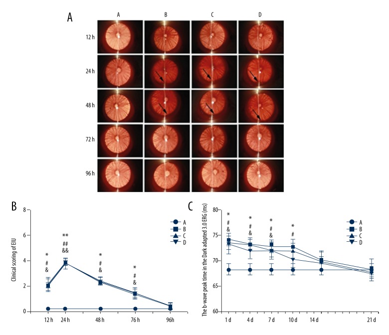
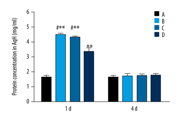
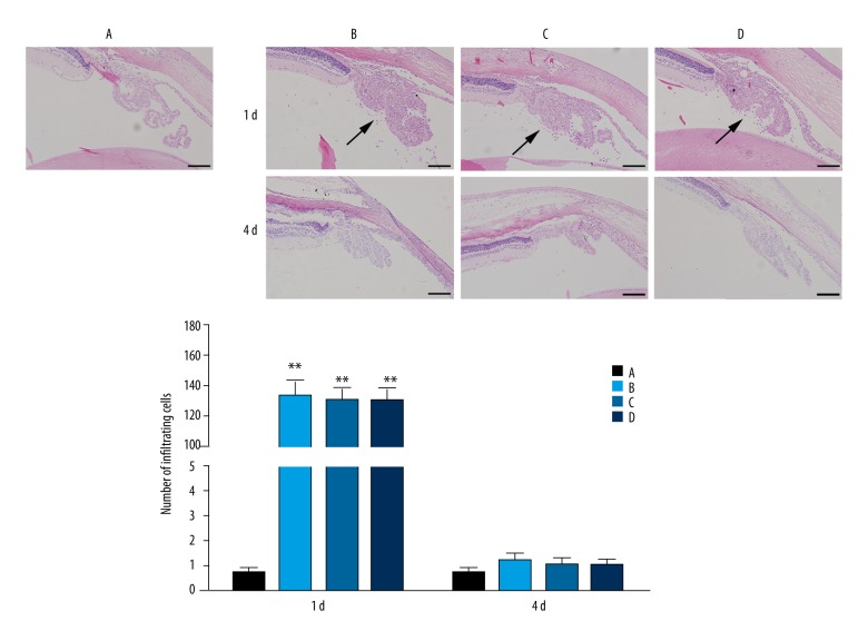
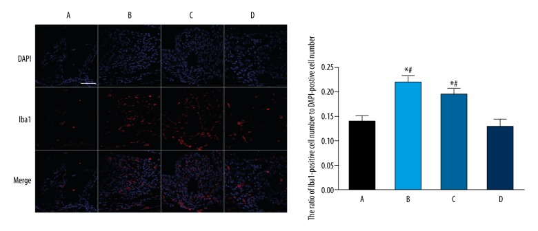
BACKGROUND Molecular hydrogen (H2) has been widely reported to have benefiicial effects in diverse animal models and human disease through reduction of oxidative stress and inflammation. The aim of this study was to investigate whether hydrogen gas could ameliorate endotoxin-induced uveitis (EIU) in rats. MATERIAL AND METHODS Male Sprague-Dawley rats were divided into a normal group, a model group, a nitrogen-oxygen (N-O) group, and a hydrogen-oxygen (H-O) group. EIU was induced in rats of the latter 3 groups by injection of lipopolysaccharide (LPS). After that, rats in the N-O group inhaled a gas mixture of 67% N2 and 33% O2, while those in the H-O group inhaled a gas mixture of 67% H2 and 33% O2. All rats were graded according to the signs of uveitis after electroretinography (ERG) examination. Protein concentration in the aqueous humor (AqH) was measured. Furthermore, hematoxylin-eosin staining and immunostaining of anti-ionized calcium-binding adapter molecule 1 (Iba1) in the iris and ciliary body (ICB) were carried out. RESULTS No statistically significant differences existed in the graded score of uveitis and the b-wave peak time in the Dark-adapted 3.0 ERG among the model, N-O, and H-O groups (P>0.05), while rats of the H-O group showed a lower concentration of AqH protein than that of the model or N-O group (P<0.05). The number of the infiltrating cells in the ICB of rats from the H-O group was not significantly different from that of the model or N-O group (P>0.05), while the activation of microglia cells in the H-O group was somewhat reduced (P<0.05). CONCLUSIONS Post-treatment hydrogen gas inhalation did not ameliorate the clinical signs, or reduce the infiltrating cells of EIU. However, it inhibited the elevation of protein in the AqH and reduced the microglia activation.
Conflict of interest statement
None.
Figures
References
-
- Seve P, Cacoub P, Bodaghi B, et al. Uveitis: Diagnostic work-up. A literature review and recommendations from an expert committee. Autoimmun Rev. 2017;16:1254–64. - PubMed
-
- Prete M, Dammacco R, Fatone MC, Racanelli V. Autoimmune uveitis: Clinical, pathogenetic, and therapeutic features. Clin Exp Med. 2016;16:125–36. - PubMed
-
- Dick AD, Tundia N, Sorg R, et al. Risk of ocular complications in patients with noninfectious intermediate uveitis, posterior uveitis, or panuveitis. Ophthalmology. 2016;123:655–62. - PubMed
-
- Turk A, Aykut M, Akyol N, et al. Serum anti-carbonic anhydrase antibodies and oxidant-antioxidant balance in patients with acute anterior uveitis. Ocul Immunol Inflamm. 2014;22:127–32. - PubMed
-
- Rao NA, Wu GS. Free radical mediated photoreceptor damage in uveitis. Prog Retin Eye Res. 2000;19:41–68. - PubMed
MeSH terms
Substances
LinkOut - more resources
Full Text Sources
Other Literature Sources
Molecular Biology Databases
