

MAdCAM costimulation through Integrin-α4β7 promotes HIV replication
- PMID: 29875402
- PMCID: PMC6160318
- DOI: 10.1038/s41385-018-0044-1
MAdCAM costimulation through Integrin-α4β7 promotes HIV replication
Abstract
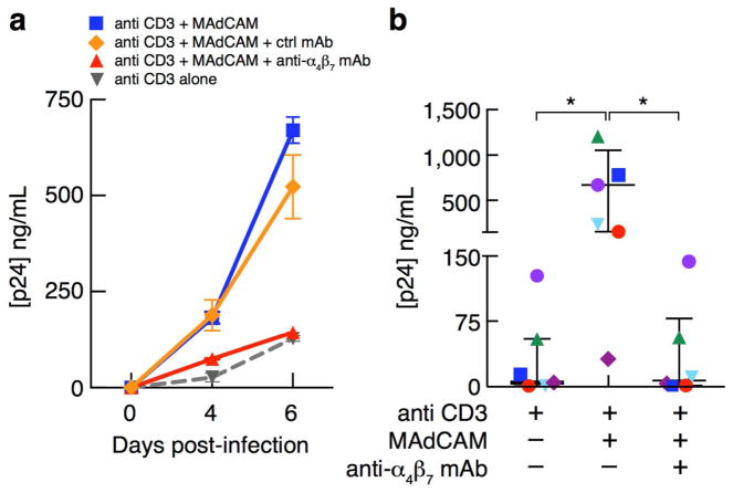
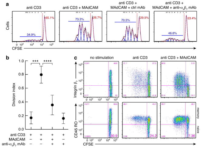
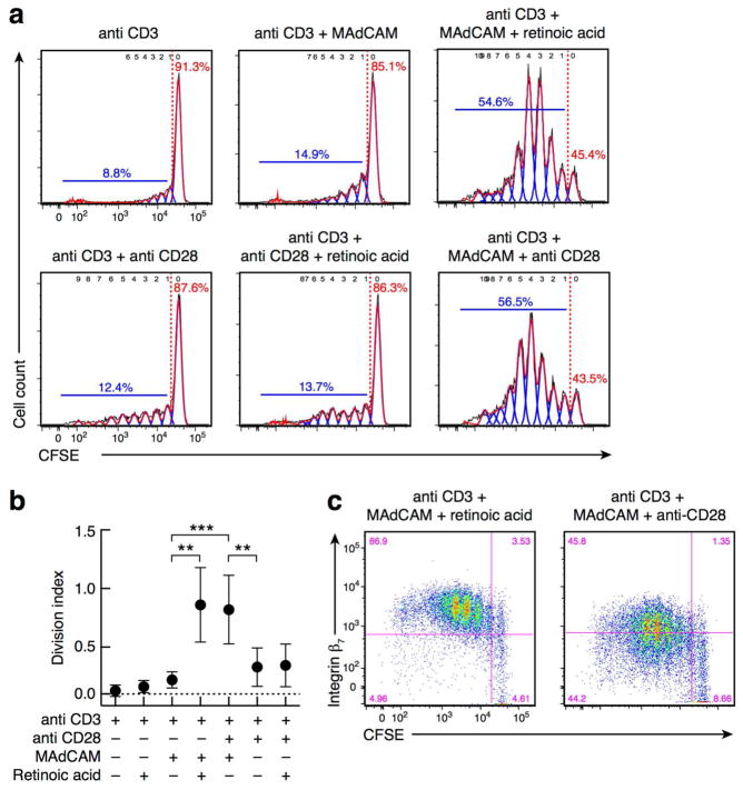
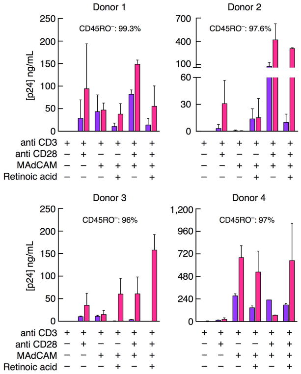
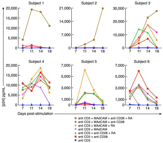
Human gut-associated lymphoid tissues (GALT) play a key role in the acute phase of HIV infection. The propensity of HIV to replicate in these tissues, however, is not fully understood. Access and migration of naive and memory CD4+ T cells to these sites is mediated by interactions between integrin α4β7, expressed on CD4+ T cells, and MAdCAM, expressed on high endothelial venules. We report here that MAdCAM delivers a potent costimulatory signal to naive and memory CD4+ T cells following ligation with α4β7. Such costimulation promotes high levels of HIV replication. An anti-α4β7 mAb that prevents mucosal transmission of SIV blocks MAdCAM signaling through α4β7 and MAdCAM-dependent viral replication. MAdCAM costimulation of memory CD4+ T cells is sufficient to drive cellular proliferation and the upregulation of CCR5, while naive CD4+ T cells require both MAdCAM and retinoic acid to achieve the same response. The pairing of MAdCAM and retinoic acid is unique to the GALT, leading us to propose that HIV replication in these sites is facilitated by MAdCAM-α4β7 interactions. Moreover, complete inhibition of MAdCAM signaling by an anti-α4β7 mAb, an analog of the clinically approved therapeutic vedolizumab, highlights the potential of such agents to control acute HIV infection.
Conflict of interest statement
The authors have no conflict of interest to declare.
The authors have no conflicts of interest to declare.
Figures

Similar articles
-
The V2 loop of HIV gp120 delivers costimulatory signals to CD4+ T cells through Integrin α4β7 and promotes cellular activation and infection.Proc Natl Acad Sci U S A. 2020 Dec 22;117(51):32566-32573. doi: 10.1073/pnas.2011501117. Epub 2020 Dec 7. Proc Natl Acad Sci U S A. 2020. PMID: 33288704 Free PMC article.
-
A Small Molecule, Which Competes with MAdCAM-1, Activates Integrin α4β7 and Fails to Prevent Mucosal Transmission of SHIV-SF162P3.PLoS Pathog. 2016 Jun 27;12(6):e1005720. doi: 10.1371/journal.ppat.1005720. eCollection 2016 Jun. PLoS Pathog. 2016. PMID: 27348748 Free PMC article.
-
The Role of Integrin α4β7 Signaling in Human Immunodeficiency Virus-1 Pathogenesis and Viral Entry in Primary CD4+ T Cells As Revealed by Comparative Phosphoproteomic Signatures.OMICS. 2020 Jul;24(7):437-450. doi: 10.1089/omi.2019.0196. Epub 2020 Jun 9. OMICS. 2020. PMID: 32522079
-
Role of T-cell trafficking in the pathogenesis of HIV disease.Curr Opin HIV AIDS. 2019 Mar;14(2):115-120. doi: 10.1097/COH.0000000000000529. Curr Opin HIV AIDS. 2019. PMID: 30601238 Review.
-
Integrin α4β7 in HIV-1 infection: A critical review.J Leukoc Biol. 2020 Aug;108(2):627-632. doi: 10.1002/JLB.4MR0120-208R. Epub 2020 Apr 9. J Leukoc Biol. 2020. PMID: 32272507 Review.
Cited by
-
Vedolizumab: Beyond Inflammatory Bowel Disease.Med Princ Pract. 2025 Jun 19:1-13. doi: 10.1159/000547015. Online ahead of print. Med Princ Pract. 2025. PMID: 40545809 Free PMC article. Review.
-
Delayed vaginal SHIV infection in VRC01 and anti-α4β7 treated rhesus macaques.PLoS Pathog. 2019 May 13;15(5):e1007776. doi: 10.1371/journal.ppat.1007776. eCollection 2019 May. PLoS Pathog. 2019. PMID: 31083697 Free PMC article.
-
Intestinal endothelial cells increase HIV infection and latency in resting and activated CD4 + T cells, particularly affecting CCR6 + CD4 + T cells.Retrovirology. 2023 May 18;20(1):7. doi: 10.1186/s12977-023-00621-y. Retrovirology. 2023. PMID: 37202790 Free PMC article.
-
Understanding the molecular mechanisms of anti-trafficking therapies and their clinical relevance in inflammatory bowel disease.Mucosal Immunol. 2023 Dec;16(6):859-870. doi: 10.1016/j.mucimm.2023.08.001. Epub 2023 Sep 3. Mucosal Immunol. 2023. PMID: 37574127 Free PMC article. Review.
-
Endothelial Cells Promote Productive HIV Infection of Resting CD4+ T Cells by an Integrin-Mediated Cell Adhesion-Dependent Mechanism.AIDS Res Hum Retroviruses. 2022 Feb;38(2):111-126. doi: 10.1089/AID.2021.0034. Epub 2021 Oct 11. AIDS Res Hum Retroviruses. 2022. PMID: 34465136 Free PMC article.
References
Publication types
MeSH terms
Substances
Grants and funding
LinkOut - more resources
Full Text Sources
Other Literature Sources
Medical
Research Materials
