. 2019 Feb;21(2):319-330.
doi: 10.1038/s41436-018-0048-y. Epub 2018 Jun 6.

Aminoacyl-tRNA synthetase deficiencies in search of common themes

Affiliations

Aminoacyl-tRNA synthetase deficiencies in search of common themes

Sabine A Fuchs et al. Genet Med. 2019 Feb.

Erratum in

  • Correction: Aminoacyl-tRNA synthetase deficiencies in search of common themes.
    Fuchs SA, Schene IF, Kok G, Jansen JM, Nikkels PGJ, van Gassen KLI, Terheggen-Lagro SWJ, van der Crabben SN, Hoeks SE, Niers LEM, Wolf NI, de Vries MC, Koolen DA, Houwen RHJ, Mulder MF, van Hasselt PM. Fuchs SA, et al. Genet Med. 2021 Oct;23(10):2024. doi: 10.1038/s41436-020-00966-1. Genet Med. 2021. PMID: 32934367 Free PMC article. No abstract available.

Abstract

Purpose: Pathogenic variations in genes encoding aminoacyl-tRNA synthetases (ARSs) are increasingly associated with human disease. Clinical features of autosomal recessive ARS deficiencies appear very diverse and without apparent logic. We searched for common clinical patterns to improve disease recognition, insight into pathophysiology, and clinical care.

Methods: Symptoms were analyzed in all patients with recessive ARS deficiencies reported in literature, supplemented with unreported patients evaluated in our hospital.

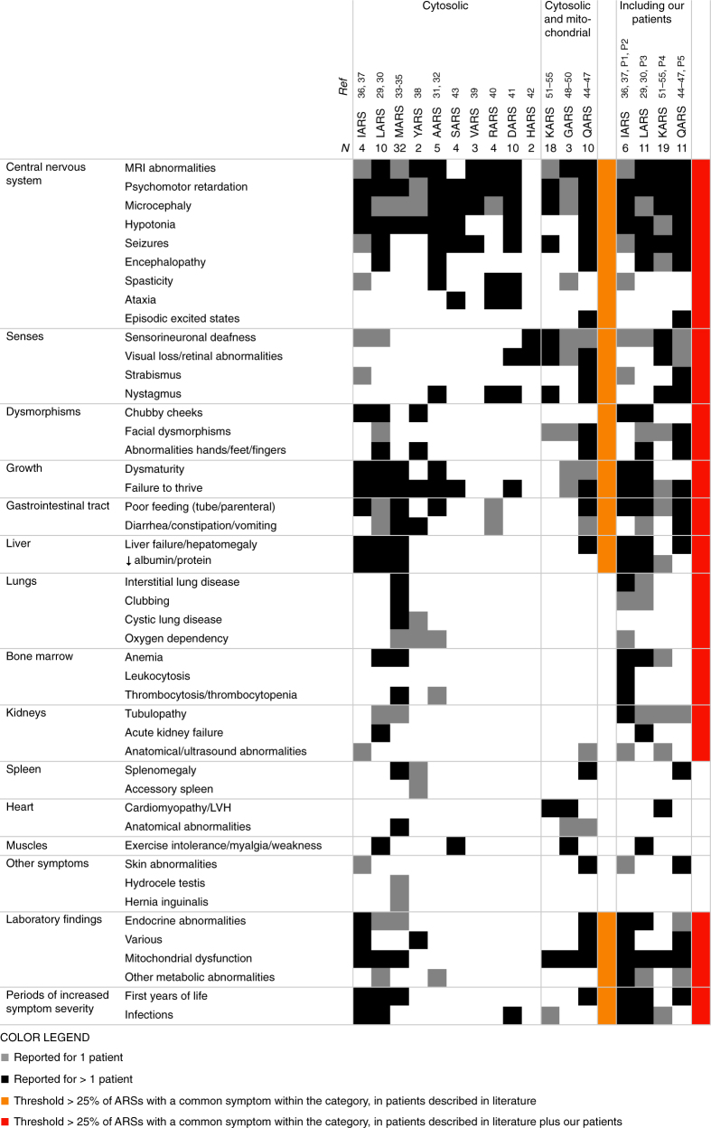
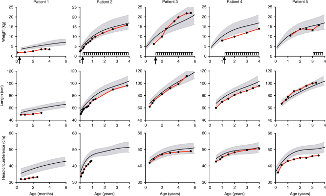
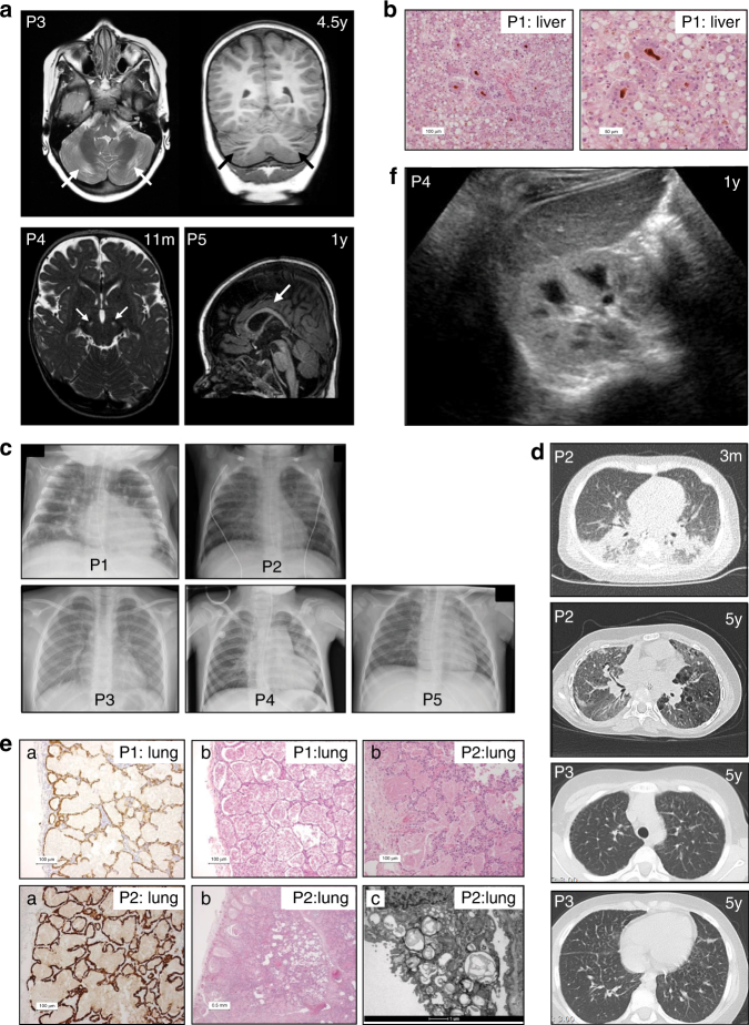
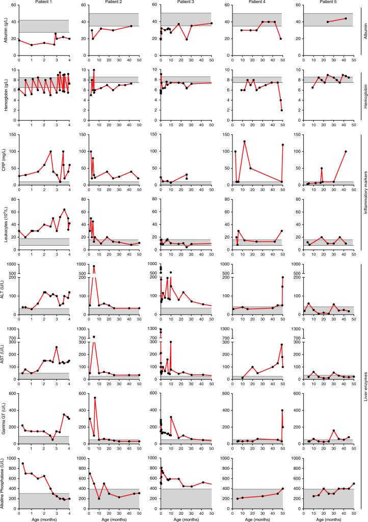
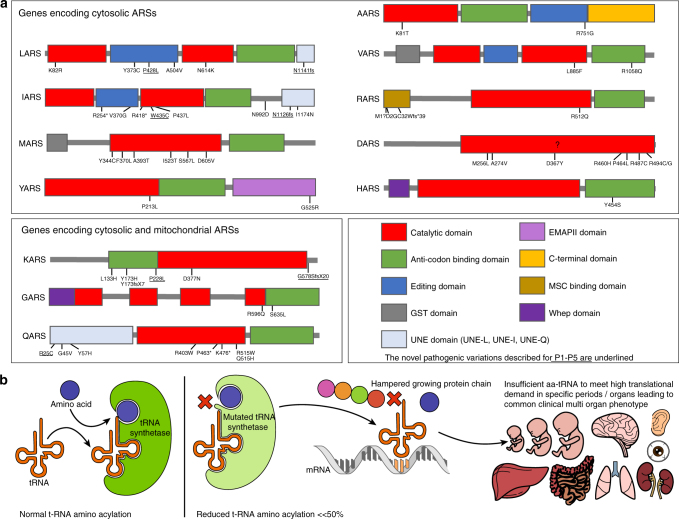
Results: In literature, we identified 107 patients with AARS, DARS, GARS, HARS, IARS, KARS, LARS, MARS, RARS, SARS, VARS, YARS, and QARS deficiencies. Common symptoms (defined as present in ≥4/13 ARS deficiencies) included abnormalities of the central nervous system and/or senses (13/13), failure to thrive, gastrointestinal symptoms, dysmaturity, liver disease, and facial dysmorphisms. Deep phenotyping of 5 additional patients with unreported compound heterozygous pathogenic variations in IARS, LARS, KARS, and QARS extended the common phenotype with lung disease, hypoalbuminemia, anemia, and renal tubulopathy.

Conclusion: We propose a common clinical phenotype for recessive ARS deficiencies, resulting from insufficient aminoacylation activity to meet translational demand in specific organs or periods of life. Assuming residual ARS activity, adequate protein/amino acid supply seems essential instead of the traditional replacement of protein by glucose in patients with metabolic diseases.

Keywords: Aminoacyl-tRNA synthetase deficiency; Clinical phenotype; Cytosolic translation.


Conflict of interest statement

The authors declare no conflicts of interest.

Figures

Fig. 1
Fig. 1. Clinical symptoms of patients with autosomal recessive ARS deficiencies reported in literature (left part) and supplemented with P1–5 (right part).
Gray squares represent symptoms reported for 1 patient, black squares symptoms reported for >1 patient. Categories of symptoms occurring in >30% of the individual ARS deficiencies are marked in orange for patients reported in literature and in red after addition of P1–5. MRI magnetic resonance imaging, LVH left ventricular hypertrophy
Fig. 2
Fig. 2. Growth curves of weight, length, and head circumference of P1–5.
Arrows indicate hospitalization due to feeding problems requiring nasogastric tube feeding (P1–4). Striped bars represent periods of feeding by percutaneous endoscopic gastrostomy (P2, P3, P5) or nasogastric tube (P4). Notice that the ranges of the x-axes are not synchronized between patients due to age differences
Fig. 3
Fig. 3. Radiologic and histologic findings.
(a) Brain magnetic resonance image (MRI) scans of P3, P4, and P5. T2-weighted axial and T1-weighted coronal MRI in P3 at age 4.5 years show focal atrophy of the cerebellar cortex. T2-weighted MRI in P4 at age 11 months shows abnormalities in the substantia nigra. T1-weighted MRI in P5 at age 1 year shows delayed myelination with a relatively thin corpus callosum. (b) Histology shows severe cholestasis and steatosis in liver tissue (P1) in H&E staining (pseudorosette formation around bile plugs (brown). (c) Chest X-rays of P1–5. Interstitial abnormalities are visible in P1–P4. (d) Thoracic computed tomography (CT) scan in P2 at age 3 months shows extensive bilateral peribronchial consolidations, bronchus dilation, and subpleural ground glass consolidation with a remarkable dorso-basal distribution; at age 5 years it shows diffuse ground glass abnormalities, cystic lesions in a paraseptal–subpleural–bronchovascular distribution, and some thickening of interlobular septae. Thoracic CT scan in P3 at age 5 years shows thickening of interlobular septae in the upper and lower thorax. (e) Histology shows severe pulmonary alveolar proteinosis in lung tissue (P1 and P2) by staining for pankeratin marker CKAE1/3: (a: highlights the lining of the alveoli with reactive type 2 pneumocytes, b: H&E staining; b: P1 and P2 alveoli are filled with a dense, eosinophilic, amorphous, protein-lipid precipitate; P2 shows granular material in multivesicular bodies and absent formation of lamellar bodies in type 2 pneumocytes, and c: electron microscopy; c: alveoli contain laminated annular structures [lamellar bodies]). (f) Ultrasound of the kidneys in P4 shows a hyperechogenic cortex of the kidneys. The global intensity of the kidney cortex versus medulla and liver is too intense
Fig. 4
Fig. 4. Laboratory findings of P1–5.
Notice that the ranges of the x-axes are not synchronized between patients due to age differences. ALT alanine transaminase, AST aspartate transaminase, CRP C-reactive protein, gamma-GT, gamma-glutamyltransferase
Fig. 5
Fig. 5. Putative disease mechanism for autosomal recessive ARS deficiencies.
(a) Overview of the pathogenic variations found in autosomal recessive ARS deficiencies, concentrated in the domains associated with the canonical function in protein translation. The novel pathogenic variations described for P1–P5 are underlined. (b) Schematic representation of the putative disease mechanism: insufficient aminoacylation to meet translational demand in specific organs or periods. tRNA transfer RNA

Comment in

References

    1. Antonellis A, Green ED. The role of aminoacyl-tRNA synthetases in genetic diseases. Annu Rev Genom Hum Genet. 2008;9:87–107. doi: 10.1146/annurev.genom.9.081307.164204. - DOI - PubMed
    1. Yao P, Fox PL. Aminoacyl-tRNA synthetases in medicine and disease. EMBO Mol Med. 2013;5:332–43. doi: 10.1002/emmm.201100626. - DOI - PMC - PubMed
    1. Storkebaum E. Peripheral neuropathy via mutant tRNA synthetases: Inhibition of protein translation provides a possible explanation. BioEssays. 2016;38:818–29. doi: 10.1002/bies.201600052. - DOI - PMC - PubMed
    1. Scheper GC, van der Knaap MS, Proud CG. Translation matters: protein synthesis defects in inherited disease. Nat Rev Genet. 2007;8:711–23. doi: 10.1038/nrg2142. - DOI - PubMed
    1. Antonellis A, Ellsworth RE, Sambuughin N, et al. Glycyl tRNA synthetase mutations in Charcot-Marie-Tooth disease type 2D and distal spinal muscular atrophy type V. Am J Hum Genet. 2003;72:1293–9. doi: 10.1086/375039. - DOI - PMC - PubMed

Publication types

MeSH terms

Substances