. 2018 May 29:(761):1-177.
doi: 10.3897/zookeys.761.24214. eCollection 2018.

A revision of dragon millipedes I: genus Desmoxytes Chamberlin, 1923, with the description of eight new species (Diplopoda, Polydesmida, Paradoxosomatidae)

Affiliations

A revision of dragon millipedes I: genus Desmoxytes Chamberlin, 1923, with the description of eight new species (Diplopoda, Polydesmida, Paradoxosomatidae)

Ruttapon Srisonchai et al. Zookeys. .

Abstract

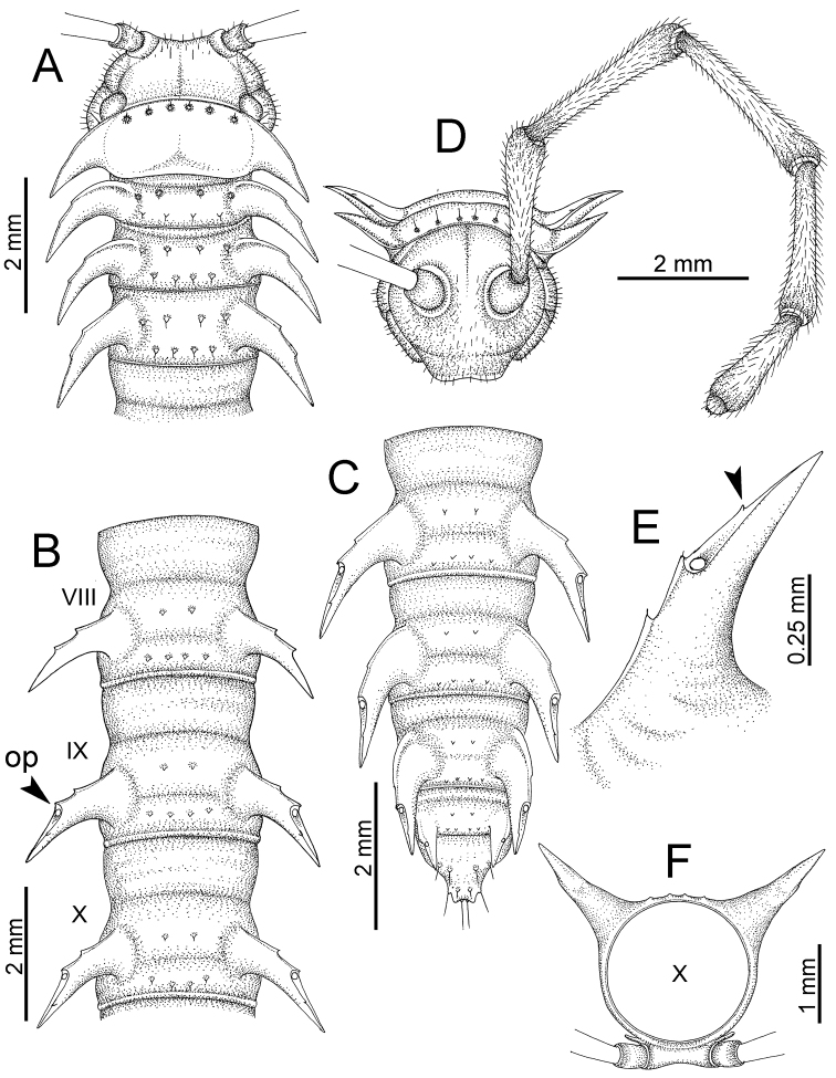
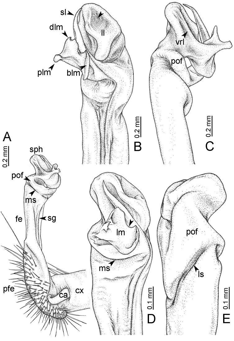
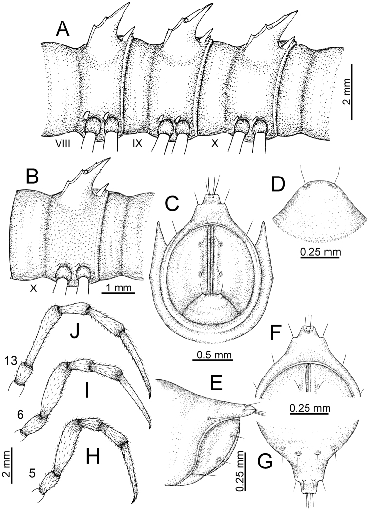
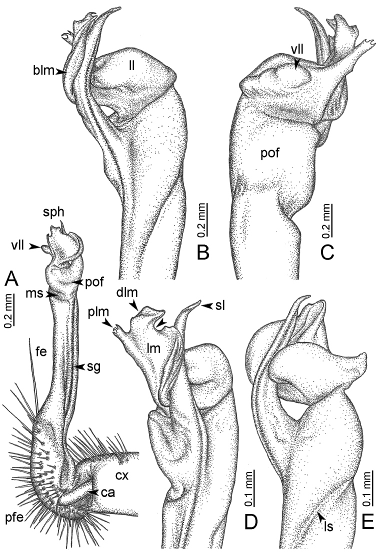
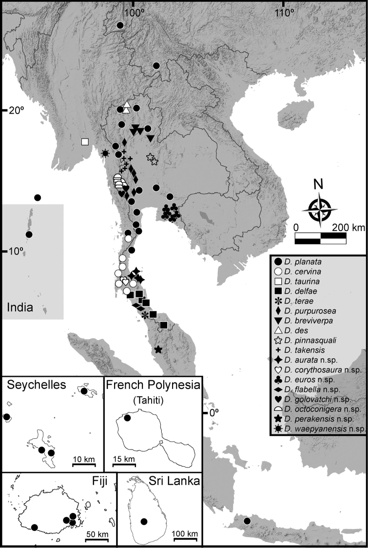
The dragon millipede genus Desmoxytes s.l. is split into five genera, based on morphological characters and preliminary molecular phylogenetic analyses. The present article includes a review of Desmoxytes s.s., while future articles will deal with Hylomus Cook and Loomis, 1924 and three new genera which preliminarily are referred to as the 'acantherpestes', 'gigas', and 'spiny' groups. Diagnostic morphological characters of each group are discussed. Hylomus is resurrected as a valid genus and the following 33 species are assigned to it: H. asper (Attems, 1937), comb. n., H. cattienensis (Nguyen, Golovatch & Anichkin, 2005), comb. n., H. cervarius (Attems, 1953), comb. n., H. cornutus (Zhang & Li, 1982), comb. n., H. draco Cook & Loomis, 1924, stat. rev., H. enghoffi (Nguyen, Golovatch & Anichkin, 2005), comb. n., H. eupterygotus (Golovatch, Li, Liu & Geoffroy, 2012), comb. n., H. getuhensis (Liu, Golovatch & Tian, 2014), comb. n., H. grandis (Golovatch, VandenSpiegel & Semenyuk, 2016), comb. n., H. hostilis (Golovatch & Enghoff, 1994), comb. n., H. jeekeli (Golovatch & Enghoff, 1994), comb. n., H. lingulatus (Liu, Golovatch & Tian, 2014), comb. n., H. laticollis (Liu, Golovatch & Tian, 2016), comb. n., H. longispinus (Loksa, 1960), comb. n., H. lui (Golovatch, Li, Liu & Geoffroy, 2012), comb. n., H. minutuberculus (Zhang, 1986), comb. n., H. nodulosus (Liu, Golovatch & Tian, 2014), comb. n., H. parvulus (Liu, Golovatch & Tian, 2014), comb. n., H. phasmoides (Liu, Golovatch & Tian, 2016), comb. n., H. pilosus (Attems, 1937), comb. n., H. proximus (Nguyen, Golovatch & Anichkin, 2005), comb. n., H. rhinoceros (Likhitrakarn, Golovatch & Panha, 2015), comb. n., H. rhinoparvus (Likhitrakarn, Golovatch & Panha, 2015), comb. n., H. scolopendroides (Golovatch, Geoffroy & Mauriès, 2010), comb. n., H. scutigeroides (Golovatch, Geoffroy & Mauriès, 2010), comb. n., H. similis (Liu, Golovatch & Tian, 2016), comb. n., H. simplex (Golovatch, VandenSpiegel & Semenyuk, 2016), comb. n., H. simplipodus (Liu, Golovatch & Tian, 2016), comb. n., H. specialis (Nguyen, Golovatch & Anichkin, 2005), comb. n., H. spectabilis (Attems, 1937), comb. n., H. spinitergus (Liu, Golovatch & Tian, 2016), comb. n., H. spinissimus (Golovatch, Li, Liu & Geoffroy, 2012), comb. n. and H. variabilis (Liu, Golovatch & Tian, 2016), comb. n.Desmoxytes s.s. includes the following species: D. breviverpa Srisonchai, Enghoff & Panha, 2016; D. cervina (Pocock,1895); D. delfae (Jeekel, 1964); D. des Srisonchai, Enghoff & Panha, 2016; D. pinnasquali Srisonchai, Enghoff & Panha, 2016; D. planata (Pocock, 1895); D. purpurosea Enghoff, Sutcharit & Panha, 2007; D. takensis Srisonchai, Enghoff & Panha, 2016; D. taurina (Pocock, 1895); D. terae (Jeekel, 1964), all of which are re-described based mainly on type material. Two new synonyms are proposed: Desmoxytes pterygota Golovatch & Enghoff, 1994, syn. n. (= Desmoxytes cervina (Pocock, 1895)), Desmoxytes rubra Golovatch & Enghoff, 1994, syn. n. (= Desmoxytes delfae (Jeekel, 1964)). Six new species are described from Thailand: D. aurata Srisonchai, Enghoff & Panha, sp. n., D. corythosaurus Srisonchai, Enghoff & Panha, sp. n., D. euros Srisonchai, Enghoff & Panha, sp. n., D. flabella Srisonchai, Enghoff & Panha, sp. n., D. golovatchi Srisonchai, Enghoff & Panha, sp. n., D. octoconigera Srisonchai, Enghoff & Panha, sp. n., as well as one from Malaysia: D. perakensis Srisonchai, Enghoff & Panha, sp. n., and one from Myanmar: D. waepyanensis Srisonchai, Enghoff & Panha, sp. n. The species can mostly be easily distinguished by gonopod structure in combination with other external characters; some cases of particularly similar congeners are discussed. All species of Desmoxytes s.s. seem to be endemic to continental Southeast Asia (except the 'tramp' species D. planata). Some biological observations (relationship with mites, moulting) are recorded for the first time. Complete illustrations of external morphological characters, an identification key, and distribution maps of all species are provided.

Keywords: Southeast Asia; aposematic; dragon millipede; new species; taxonomy.


Figures

Figure 1.
Figure 1.
Some original and additional labels of some lectotypes and paratypes. A Desmoxytes planata (Pocock, 1895) (NHMUK) B D. cervina (Pocock, 1895) (NHMUK) C D. taurina (Pocock, 1895) (NHMUK) D D. taurina (Pocock, 1895) (ZMUC) D Hylomus draco Cook & Loomis, 1924 (USNM).
Figure 2.
Figure 2.
Shape of gonopod of Desmoxytes s.l. A Desmoxytes s.s. (D. planata (Pocock, 1895)) – specimen from Wat Puang Malai B the ‘acantherpestes’ group (specimen from Kanchanaburi, Thailand) C the ‘gigas’ group (specimen from Krabi, Thailand) D the ‘spiny’ group (specimen from Krabi, Thailand) E, F Hylomus Cook & Loomis, 1924 (E = H. draco Cook & Loomis, 1924 stat. rev. (paratype) F = Hylomus sp. (specimen from Laos)).
Figure 3.
Figure 3.
Type of paranota (paraterga) of Desmoxytes s.l. A–D Desmoxytes s.s. A D. delfae (Jeekel, 1964), specimen from Tham Khan Ti Phol B D. planata (Pocock, 1895), specimen from Suan Sai Thong Restaurant C D. purpurosea Enghoff et al., 2007, specimen from Hup Pa Tard D D. cervina (Pocock, 1895), specimen from Wat Satit Khirirom E the ‘acantherpestes’ group F the ‘gigas’ group G the ‘spiny’ group H–P Hylomus Cook & Loomis, 1924 H H. draco Cook & Loomis, 1924, stat. rev., paratype I H. cervarius (Attems, 1953), comb. n., ZMUM specimen J H. rhinoceros (Likhitrakarn et al., 2015), comb. n., paratype K H. scolopendroides (Golovatch et al., 2010), comb. n., paratype L H. nodulosus (Liu et al., 2014), comb. n., paratype M H. eupterygotus (Golovatch et al., 2012), comb. n., paratype N H. scutigeroides (Golovatch et al., 2010), comb. n., paratype O H. simplex (Golovatch et al., 2016), comb. n., paratype P H. laticollis (Liu et al., 2016), comb. n., paratype.
Figure 4.
Figure 4.
Common habitats for Desmoxytes Chamberlin, 1923. A, B limestone mountain C rock wall and plants D rock wall and small cave.
Figure 5.
Figure 5.
General body characters of DesmoxytesD. planata (Pocock, 1895), male specimen from Wat Puang Malai. A whole body B anterior body part C head region D body ring E body rings 5–7, showing sternal lobe between coxae 4 and gonopods on ring 7 F male femora 5 or 6.
Figure 6.
Figure 6.
General body characters of Desmoxytes (D. planata (Pocock, 1895), male specimen from Wat Puang Malai) – SEM images. A, B anterior body part C collum D, E body rings 9–10 F last ring and telson G posteriormost rings H body ring 10 I mouth parts J leg 13 (right) K sternal lobe between male coxae 4 L gonopods on ring 7 M tip of tarsus and claw of leg 13 N tip of epiproct.
Figure 7.
Figure 7.
Diagrammatic drawings of right gonopod of DesmoxytesD. planata (Pocock, 1895), specimen from Wat Puang Malai. A mesal view B dorsal view C ventral view.
Figure 8.
Figure 8.
SEM images of right gonopod of Desmoxytes planata (Pocock, 1895), specimen from Wat Puang Malai. A lateral view B mesal view C submesal view D ventral view.
Figure 9.
Figure 9.
Photographs of live Desmoxytes aurata sp. n. and habitat. A, B male paratype C female paratype D mating couple E habitat.
Figure 10.
Figure 10.
Desmoxytes aurata sp. n. (male paratype). A anterior body part B body rings 8–10 (op = ozopore) C posteriormost body rings and telson D head and antenna E paraterga of ring 10 F body ring 10.
Figure 11.
Figure 11.
Desmoxytes aurata sp. n. (paratypes). A body rings 8–10 B sculpture of ring 10 C, E last ring and telson D hypoproct F, G epiproct H male leg 5 (right) I male leg 6 (right) J male leg 13 (right).
Figure 12.
Figure 12.
Desmoxytes aurata sp. n. (male holotype, male paratypes) – sternal lobe between male coxae 4. A, B lateral view C ventral view D–F caudal view.
Figure 13.
Figure 13.
Desmoxytes aurata sp. n. (paratype) – right gonopod. A mesal view B dorsal view C ventral view D submesal view (arrow = indentation) E lateral view (arrow = furrow).
Figure 14.
Figure 14.
Desmoxytes aurata sp. n. (paratype) – right gonopod. A lateral view B mesal view C ventral view D subdorsal view (arrow = indentation) E dorsal view F subdorsal view (arrow = furrow).
Figure 15.
Figure 15.
Desmoxytes breviverpa Srisonchai et al., 2016 (paratype) – right gonopod (modified from Srisonchai et al. 2016). A mesal view B dorsal view C ventral view D submesal view (arrow = indentation) E lateral view.
Figure 16.
Figure 16.
Photographs of live Desmoxytes cervina (Pocock, 1895) – males. A specimen from Wat Satit Khiri Rom B specimen from Phrayathtan Cave (Myanmar) C specimen from Ban Song Phi Nong D specimen from Wat Suwan Khuha.
Figure 17.
Figure 17.
Photographs of live Desmoxytes cervina (Pocock, 1895) and habitat. A–E specimens from Ratchaprapa Dam A female B juvenile C, D male with parasitic mites E mating couple F habitat.
Figure 18.
Figure 18.
Desmoxytes cervina (Pocock, 1895) – specimen from Wat Satit Khirirom. A anterior body part B body rings 8–10 (op = ozopore) C posteriormost body rings and telson D head and antenna E paraterga of ring 10 (arrow = tiny denticle) F body ring 10.
Figure 19.
Figure 19.
Desmoxytes cervina (Pocock, 1895) – specimen from Wat Satit Khirirom. A body rings 8–10 B sculpture of ring 10 C, E last ring and telson D hypoproct F, G epiproct H male leg 5 (right) I male leg 6 (right) J male leg 13 (right).
Figure 20.
Figure 20.
Desmoxytes cervina (Pocock, 1895), specimens from Wat Satit Khirirom – sternal lobe between male coxae 4. A, B lateral view C, D ventral view E, F caudal view.
Figure 21.
Figure 21.
Desmoxytes cervina (Pocock, 1895), specimen from Wat Satit Khirirom – right gonopod. A mesal view B dorsal view (arrow = furrow) C ventral view D submesal view (arrow = indentation) E lateral view.
Figure 22.
Figure 22.
Desmoxytes cervina (Pocock, 1895), specimen from Wat Satit Khirirom – right gonopod. A lateral view B mesal view C ventral view D subdorsal view (arrow = indentation) E dorsal view F subdorsal view (arrow = furrow).
Figure 23.
Figure 23.
Photographs of live Desmoxytes corythosaurus sp. n. and habitat A, B male paratypes C female paratype D juvenile E habitat.
Figure 24.
Figure 24.
Desmoxytes corythosaurus sp. n. (male paratype). A anterior body part B body rings 8–10 (op = ozopore) C posteriormost body rings and telson D head and antenna E paraterga of ring 10 F body ring 10.
Figure 25.
Figure 25.
Desmoxytes corythosaurus sp. n. (male paratypes). A body rings 8–10 B sculpture of ring 10 C, E last ring and telson D hypoproct F, G epiproct H male leg 5 (right) I male leg 6 (right) J male leg 13 (right).
Figure 26.
Figure 26.
Desmoxytes corythosaurus sp. n. – sternal lobe between male coxae 4. A lateral view (male holotype) B ventral view (male holotype) C caudal view (male holotype) D caudal view (male paratype).
Figure 27.
Figure 27.
Desmoxytes corythosaurus sp. n. (paratype) – right gonopod. A mesal view B dorsal view C ventral view D submesal view E lateral view (arrow = furrow).
Figure 28.
Figure 28.
Desmoxytes corythosaurus sp. n. (paratype) – right gonopod. A lateral view B mesal view C ventral view D subdorsal view E dorsal view F subdorsal view (arrow = furrow).
Figure 29.
Figure 29.
Photographs of live Desmoxytes delfae (Jeekel, 1964) – males. A specimen from Khao Chi Chan Bureau of Monks B specimen from Tham Tone Din C specimen from Wat Tham Sue (Tiger Cave) D early adult from La-ngu Subdistrict.
Figure 30.
Figure 30.
Photographs of live Desmoxytes delfae (Jeekel, 1964) and habitat. A–C specimens from Tham Khan Ti Phol A female B old female C newly moulted adult female D habitat.
Figure 31.
Figure 31.
Desmoxytes delfae (Jeekel, 1964), specimen from Tham Khan Ti Phol. A anterior body part B body rings 8–10 (op = ozopore) C posteriormost body rings and telson D head and antenna E paraterga of ring 10 (arrow = tiny denticle) F body ring 10.
Figure 32.
Figure 32.
Desmoxytes delfae (Jeekel, 1964), specimens from Tham Khan Ti Phol. A body rings 8–10 B sculpture of ring 10 C, E last ring and telson D hypoproct F, G epiproct H male leg 5 (right) I male leg 6 (right) J male leg 13 (right).
Figure 33.
Figure 33.
Desmoxytes delfae (Jeekel, 1964), specimens from Tham Khan Ti Phol – sternal lobe between male coxae 4. A lateral view B ventral view C, D caudal view E some specimens from Tham Khao Ting – caudal view.
Figure 34.
Figure 34.
Desmoxytes delfae (Jeekel, 1964), specimen from Khao Chi Chan Bureau of Monks – right gonopod. A mesal view B dorsal view C ventral view D submesal view (arrow = indentation) E lateral view (arrow = furrow).
Figure 35.
Figure 35.
Desmoxytes delfae (Jeekel, 1964), specimen from Khao Chi Chan Bureau of Monks – right gonopod. A lateral view B mesal view C ventral view D subdorsal view (arrow = indentation) E dorsal view F subdorsal view (arrow = furrow).
Figure 36.
Figure 36.
Desmoxytes des Srisonchai et al. 2016 (paratype) – right gonopod (modified from Srisonchai et al. 2016). A mesal view B submesal view C process (plm) of lamina medialis and distal lobe (dlm) of lamina medialis D lateral view E dorsal view (arrow = indentation) F ventral view.
Figure 37.
Figure 37.
Photographs of live Desmoxytes euros sp. n. and habitat. A, B male paratypes C female paratype D juvenile E egg cluster F cluster of stadium 1 juveniles G habitat.
Figure 38.
Figure 38.
Desmoxytes euros sp. n. (male paratype). A anterior body part B body rings 8–10 (op = ozopore) C posteriormost body rings and telson D head and antenna E paraterga of ring 10 (arrow = tiny denticle) F body ring 10.
Figure 39.
Figure 39.
Desmoxytes euros sp. n. (male paratype). A body rings 8–10 B sculpture of ring 10 C, E last ring and telson D hypoproct F, G epiproct H male leg 5 (right) I male leg 6 (right) J male leg 13 (right).
Figure 40.
Figure 40.
Desmoxytes euros sp. n. (male paratype) – sternal lobe between male coxae 4. A lateral view B ventral view C caudal view.
Figure 41.
Figure 41.
Desmoxytes euros sp. n. (paratype) – right gonopod. A mesal view B dorsal view C ventral view D submesal view (arrow = indentation) E lateral view.
Figure 42.
Figure 42.
Desmoxytes euros sp. n. (paratype) – right gonopod. A lateral view B mesal view C ventral view D subdorsal view (arrow = indentation) E dorsal view F subdorsal view.
Figure 43.
Figure 43.
Photographs of live Desmoxytes flabella sp. n. and habitat. A male paratype B female paratype C mating couple D habitat.
Figure 44.
Figure 44.
Desmoxytes flabella sp. n. (male paratype). A anterior body part B body rings 8–10 (op = ozopore) C posteriormost body rings and telson D head and antenna E paraterga of ring 10 (arrow = tiny denticle) F body ring 10.
Figure 45.
Figure 45.
Desmoxytes flabella sp. n. (male paratype) A body rings 8–10 B sculpture of ring 10 C, E last ring and telson D hypoproct F, G epiproct H male leg 5 (right) I male leg 6 (right) J male leg 13 (right).
Figure 46.
Figure 46.
Desmoxytes flabella sp. n. (male paratype) – sternal lobe between male coxae 4. A lateral view B ventral view C caudal view.
Figure 47.
Figure 47.
Desmoxytes flabella sp. n. (paratype) – right gonopod. A mesal view B dorsal view (arrows = furrows) C ventral view D submesal view (arrow = indentation) E lateral view.
Figure 48.
Figure 48.
Desmoxytes flabella sp. n. (paratype) – right gonopod. A lateral view B mesal view C ventral view D subdorsal view (arrows = indentations) E dorsal view (arrows = furrows) F subdorsal view.
Figure 49.
Figure 49.
Photographs of live Desmoxytes golovatchi sp. n. and habitat. A male paratype B female paratype C mating couple D habitat.
Figure 50.
Figure 50.
Desmoxytes golovatchi sp. n. (male paratype). A anterior body part B body rings 8–10 (op = ozopore) C posteriormost body rings and telson D head and antenna E paraterga of ring 10 (arrow = tiny denticle) F body ring 10.
Figure 51.
Figure 51.
Desmoxytes golovatchi sp. n. (male paratype). A body rings 8–10 B sculpture of ring 10 C, E last ring and telson D hypoproct F, G epiproct H male leg 5 (right) I male leg 6 (right) J male leg 13 (right).
Figure 52.
Figure 52.
Desmoxytes golovatchi sp. n. (male paratype) – sternal lobe between male coxae 4. A lateral view B ventral view C, D caudal view.
Figure 53.
Figure 53.
Desmoxytes golovatchi sp. n. (paratype) – right gonopod. A mesal view B dorsal view C ventral view D submesal view (arrow = indentation) E lateral view.
Figure 54.
Figure 54.
Desmoxytes golovatchi sp. n. (paratype) – right gonopod. A lateral view B mesal view C ventral view D subdorsal view (arrow = indentation) E dorsal view F subdorsal view.
Figure 55.
Figure 55.
Photographs of live Desmoxytes octoconigera sp. n. and habitat. A male paratype B female paratype C mating couple D habitat.
Figure 56.
Figure 56.
Desmoxytes octoconigera sp. n. (male paratype). A anterior body part B body rings 8–10 (op = ozopore) C posteriormost body rings and telson D head and antenna E paraterga of ring 10 (arrow = tiny denticle) F body ring 10.
Figure 57.
Figure 57.
Desmoxytes octoconigera sp. n. (male paratype). A body rings 8–10 B sculpture of ring 10 C, E last ring and telson D hypoproct F, G epiproct H male leg 5 (right) I male leg 6 (right) J male leg 13 (right).
Figure 58.
Figure 58.
Desmoxytes octoconigera sp. n. (male paratype) – sternal lobe between male coxae 4. A lateral view B ventral view C caudal view.
Figure 59.
Figure 59.
Desmoxytes octoconigera sp. n. (paratype) – right gonopod. A mesal view B dorsal view C ventral view D submesal view (arrow = indentation) E lateral view.
Figure 60.
Figure 60.
Desmoxytes octoconigera sp. n. (paratype) – right gonopod. A lateral view B mesal view C ventral view D subdorsal view (arrow = indentation) E dorsal view F subdorsal view.
Figure 61.
Figure 61.
Desmoxytes perakensis sp. n. (male paratype). A anterior body part B body rings 8–10 (op = ozopore) C posteriormost body rings and telson D head and antenna E paraterga of ring 10 (arrow = tiny denticle) F body ring 10.
Figure 62.
Figure 62.
Desmoxytes perakensis sp. n. (male paratype). A body rings 8–10 B sculpture of ring 10 C, E last ring and telson D hypoproct F, G epiproct H male leg 5 (right) I male leg 6 (right) J male leg 13 (right).
Figure 63.
Figure 63.
Desmoxytes perakensis sp. n. (male paratype) – sternal lobe between male coxae 4. A lateral view B ventral view C caudal view.
Figure 64.
Figure 64.
Desmoxytes perakensis sp. n. (paratype) – right gonopod. A mesal view B submesal view (arrow = indentation) C ventral view D dorsal view E lateral view (arrows = furrows).
Figure 65.
Figure 65.
Desmoxytes perakensis sp. n. (paratype) – right gonopod. A lateral view B mesal view C ventral view D subdorsal view (arrow = indentation) E dorsal view F subdorsal view (arrows = furrows).
Figure 66.
Figure 66.
Desmoxytes pinnasquali Srisonchai et al., 2016 (paratype) – right gonopod (modified from Srisonchai et al. 2016). A mesal view B dorsal view C ventral view D subdorsal view (arrow = indentation) E lateral view.
Figure 67.
Figure 67.
Photographs of live Desmoxytes planata (Pocock, 1895) – males. A specimen from Xishuangbanna, Mengla (China) B specimen from Suan Sai Thong Restaurant, Phrae (Thailand) C specimen from Lampane Village, Nguwun Chuang River (Myanmar) D specimen from Wat Khao Tham Ma Rong, Prachuap Khiri Khan (Thailand).
Figure 68.
Figure 68.
Photographs of live Desmoxytes planata (Pocock, 1895) and habitats. A female B mating couple C–E synanthropic areas F, G limestone area near synanthropic area.
Figure 69.
Figure 69.
Desmoxytes planata (Pocock, 1895), specimen from Suan Sai Thong Restaurant. A anterior body part B body rings 8–10 (op = ozopore) C posteriormost body rings and telson D head and antenna E paraterga of ring 10 (arrow = tiny denticle) F body ring 10.
Figure 70.
Figure 70.
Desmoxytes planata (Pocock, 1895), specimen from Suan Sai Thong Restaurant. A body rings 8–10 B sculpture of ring 10 C, E last ring and telson D hypoproct F, G epiproct H male leg 5 (right) I male leg 6 (right) J male leg 13 (right).
Figure 71.
Figure 71.
Desmoxytes planata (Pocock, 1895), population from Suan Sai Thong Restaurant – sternal lobe between male coxae 4. A lateral view B ventral view C caudal view (specimen from Fiji only) D–F caudal view.
Figure 72.
Figure 72.
Desmoxytes planata (Pocock, 1895), specimen from Wat Puang Malai – right gonopod. A mesal view B dorsal view C ventral view D submesal view (arrow = indentation) E lateral view.
Figure 73.
Figure 73.
Desmoxytes planata (Pocock, 1895), specimen from Wat Puang Malai – right gonopod. A lateral view B mesal view C ventral view D subdorsal view E dorsal view (unlabeled arrow = indentation) F subdorsal view.
Figure 74.
Figure 74.
Desmoxytes planata (Pocock, 1895), specimen from Fiji, Viti Levu – right gonopod. A lateral view B mesal view C ventral view D subdorsal view (arrow = indentation) E dorsal view F subdorsal view.
Figure 75.
Figure 75.
Photographs of live Desmoxytes purpurosea Enghoff et al., 2007 (specimens from Hup Pa Tard) and habitat. A male B female C early adult male D mating couple E habitat.
Figure 76.
Figure 76.
Desmoxytes purpurosea Enghoff et al., 2007 (male paratype). A anterior body part B body rings 8–10 (op = ozopore) C posteriormost body rings and telson D head and antenna E paraterga of ring 10 (arrow = tiny denticle) F body ring 10.
Figure 77.
Figure 77.
Desmoxytes purpurosea Enghoff et al., 2007 (male paratype). A body rings 8–10 B sculpture of ring 10 C, E last ring and telson D hypoproct F, G epiproct H male leg 5 (right) I male leg 6 (right) J male leg 13 (right).
Figure 78.
Figure 78.
Desmoxytes purpurosea Enghoff et al., 2007 (male paratypes) – sternal lobe between male coxae 4. A, D ventral view B caudal view C lateral view.
Figure 79.
Figure 79.
Desmoxytes purpurosea Enghoff et al., 2007 (specimen from Hup Pa Tard) – right gonopod. A mesal view B submesal view (arrow = indentation) C lateral view D dorsal view (Kanchababuri A and B populations) E subdorsal view F ventral view.
Figure 80.
Figure 80.
Desmoxytes purpurosea Enghoff et al., 2007 – right gonopod. A mesal view (specimen from Hup Pa Tard) B, C process (plm) of lamina medialis (Lamphun and Uthai Thani populations) D dorsal view (specimen from Tham Than Lod cave) E, F process (plm) of lamina medialis (Kanchababuri A and B populations).
Figure 81.
Figure 81.
Desmoxytes purpurosea Enghoff et al., 2007 (specimen from Hup Pa Tard) – right gonopod. A lateral view B mesal view C ventral view D subdorsal view (arrow = indentation) E dorsal view F subdorsal view.
Figure 82.
Figure 82.
Desmoxytes takensis Srisonchai et al., 2016 (specimen from Chao Por Phawo Shrine) and habitat. A male B female C mating couple D early adult E moulting chamber F habitat.
Figure 83.
Figure 83.
Desmoxytes takensis Srisonchai et al., 2016 (paratype) – right gonopod (modified from Srisonchai et al. 2016). A mesal view B dorsal view C ventral view D submesal view (arrow = indentation) E lateral view.
Figure 84.
Figure 84.
Desmoxytes taurina (Pocock, 1895), lectotype. A anterior body part B body rings 8–10 (op = ozopore) C posteriormost body rings and telson D head and antenna E paraterga of ring 10 (arrow = tinydenticle) F body ring 10.
Figure 85.
Figure 85.
Desmoxytes taurina (Pocock, 1895), lectotype. A body rings 8–10 B sculpture of ring 10 C, E last ring and telson D hypoproct F, G epiproct H male leg 5 (right) I male leg 6 (right) J male leg 13 (right).
Figure 86.
Figure 86.
Desmoxytes taurina (Pocock, 1895), lectotype – sternal lobe between male coxae 4. A lateral view B ventral view C caudal view.
Figure 87.
Figure 87.
Desmoxytes taurina (Pocock, 1895), lectotype – right gonopod. A mesal view B mesal view C submesal view (arrow = indentation) D lateral view E dorsal view F ventral view.
Figure 88.
Figure 88.
Photographs of live Desmoxytes terae (Jeekel, 1964) (specimens from Tham Tone Din) and habitat. A male B female C habitat.
Figure 89.
Figure 89.
Desmoxytes terae (Jeekel, 1964), specimen from Tham Tone Din. A anterior body part B body rings 8–10 C posteriormost body rings and telson D head and antenna E paraterga of ring 10 F body ring 10.
Figure 90.
Figure 90.
Desmoxytes terae (Jeekel, 1964), specimen from Tham Tone Din. A body rings 8–10 B sculpture of ring 10 C, E last ring and telson D hypoproct F, G epiproct H male leg 5 (right) I male leg 6 (right) J male leg 13 (right).
Figure 91.
Figure 91.
Desmoxytes terae (Jeekel, 1964), specimen from Tham Tone Din – sternal lobe between male coxae 4. A lateral view B mesal view C–E caudal view.
Figure 92.
Figure 92.
Desmoxytes terae (Jeekel, 1964), holotype – right gonopod. A mesal view B dorsal view C ventral view D submesal view (arrow = indentation) E lateral view.
Figure 93.
Figure 93.
Photographs of live Desmoxytes waepyanensis sp. n. and habitat. A male paratype B female paratype C juvenile D mating couple E habitat.
Figure 94.
Figure 94.
Desmoxytes waepyanensis sp. n. (male paratype). A anterior body part B body rings 8–10 (op = ozopore) C posteriormost body rings and telson D head and antenna E paraterga of ring 10 (arrow = tiny denticle) F body ring 10.
Figure 95.
Figure 95.
Desmoxytes waepyanensis sp. n. (male paratype). A body rings 8–10 B sculpture of ring 10 C, E last ring and telson D hypoproct F, G epiproct H male leg 5 (right) I male leg 6 (right) J male leg 13 (right).
Figure 96.
Figure 96.
Desmoxytes waepyanensis sp. n. (male paratype) – sternal lobe between male coxae 4. A lateral view B ventral view C caudal view.
Figure 97.
Figure 97.
Desmoxytes waepyanensis sp. n. (paratype) – right gonopod. A mesal view B dorsal view C ventral view D subdorsal view (arrow = indentation) E lateral view.
Figure 98.
Figure 98.
Desmoxytes waepyanensis sp. n. (paratype) – right gonopod. A lateral view B mesal view C ventral view D subdorsal view (arrow = indentation) E dorsal view F subdorsal view.
Figure 99.
Figure 99.
Type localities of all Desmoxytes species. white circle = described species (1 = D. breviverpa, 2 = D. cervina, 3 = D. delfae, 4 = D. des, 5 = D. pinnasquali, 6 = D. planata, 7 = D. purpurosea, 8 = D. takensis, 9 = D. taurina, 10 = D. terae). black circle = new species described in this study (1 = D. aurata, sp. n. 2 = D. corythosaurus sp. n., 3 = D. euros sp. n., 4 = D. flabella sp. n., 5 = D. golovatchi sp. n., 6 = D. octoconigera sp. n., 7 = D. perakensis sp. n., 8 = D. waepyanensis sp. n.).
Figure 100.
Figure 100.
Known distribution of all Desmoxytes species based on all recorded data (D. planata, D. cervina and D. delfae are shown in the representative localities).

References

    1. Adis J, Hansen B, Wilck L, Adis I, Hirschel K. (2000) A spinning apparatus documented in Polydesmida for the first time. Fragmenta Faunistica Supplement 43: 139–148.
    1. Attems C. (1914) Die indo-australischen Myriopoden. Archiv für Naturgeschichte, Berlin 80A: 1–398. https://biodiversitylibrary.org/page/46057676
    1. Attems C. (1936) Diplopoda of India. Memoirs of the Indian Museum 11: 133–323.
    1. Attems C. (1937) Myriapoda 3. Polydemoidea I, Fam. Strongylosomidae. Das Tierreich 68: 1–300.
    1. Attems C. (1953) Myriapoden von Indochina. Expedition von C. Dawydoff (1938–1939).

LinkOut - more resources