. 2018 May 23:12:139.
doi: 10.3389/fncel.2018.00139. eCollection 2018.

Meclofenamic Acid Reduces Reactive Oxygen Species Accumulation and Apoptosis, Inhibits Excessive Autophagy, and Protects Hair Cell-Like HEI-OC1 Cells From Cisplatin-Induced Damage

Affiliations

Meclofenamic Acid Reduces Reactive Oxygen Species Accumulation and Apoptosis, Inhibits Excessive Autophagy, and Protects Hair Cell-Like HEI-OC1 Cells From Cisplatin-Induced Damage

He Li et al. Front Cell Neurosci. .

Abstract

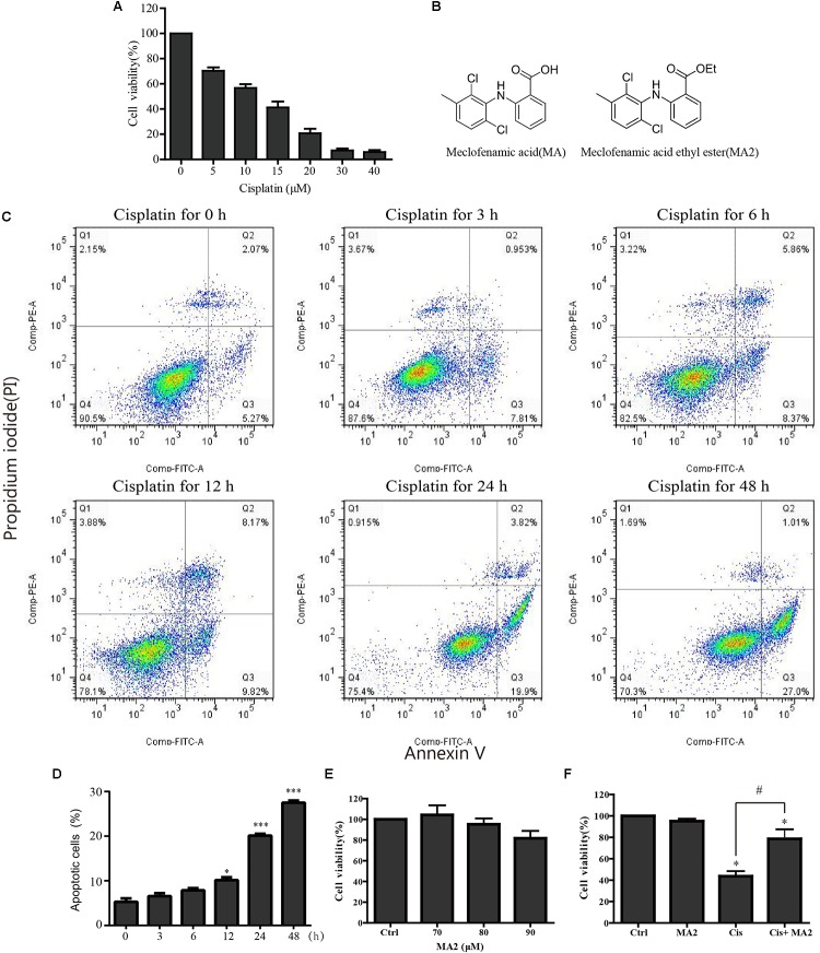
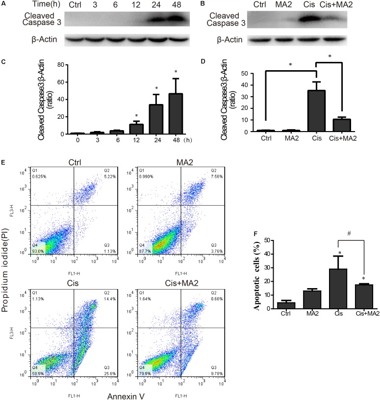
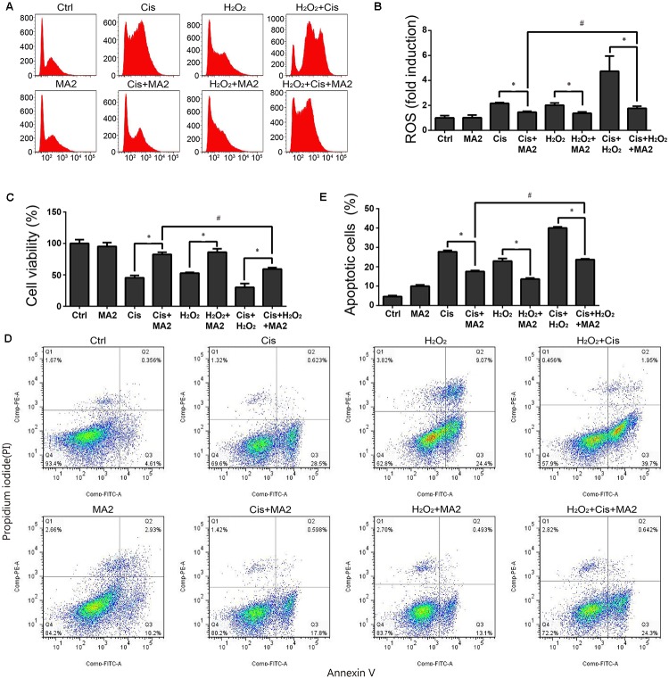
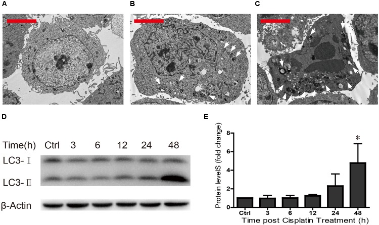
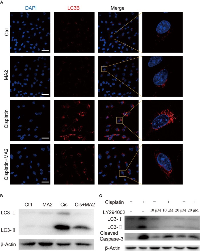
Hearing loss is the most common sensory disorder in humans, and a significant number of cases is due to the ototoxicity of drugs such as cisplatin that cause hair cell (HC) damage. Thus, there is great interest in finding agents and mechanisms that protect HCs from ototoxic drug damage. It has been proposed that epigenetic modifications are related to inner ear development and play a significant role in HC protection and HC regeneration; however, whether the m6A modification and the ethyl ester form of meclofenamic acid (MA2), which is a highly selective inhibitor of FTO (fatmass and obesity-associated enzyme, one of the primary human demethylases), can affect the process of HC apoptosis induced by ototoxic drugs remains largely unexplored. In this study, we took advantage of the HEI-OC1 cell line, which is a cochlear HC-like cell line, to investigate the role of epigenetic modifications in cisplatin-induced cell death. We found that cisplatin injury caused reactive oxygen species accumulation and increased apoptosis in HEI-OC1 cells, and the cisplatin injury was reduced by co-treatment with MA2 compared to the cisplatin-only group. Further investigation showed that MA2 attenuated cisplatin-induced oxidative stress and apoptosis in HEI-OC1 cells. We next found that the cisplatin-induced upregulation of autophagy was significantly inhibited after MA2 treatment, indicating that MA2 inhibited the cisplatin-induced excessive autophagy. Our findings show that MA2 has a protective effect and improves the viability of HEI-OC1 cells after cisplatin treatment, and they provide new insights into potential therapeutic targets for the amelioration of cisplatin-induced ototoxicity.

Keywords: HEI-OC1 cells; autophagy; cisplatin; meclofenamic acid; reactive oxygen species.


Figures

FIGURE 1
FIGURE 1
(A) HEI-OC1 cells were incubated with different concentrations of cisplatin (from 0 to 40 μM) for 48 h, and cell viability was measured with the CCK-8 kit. (B) The chemical structure of MA and MA2. (C) Flow cytometry was used to measure apoptosis after 15 μM cisplatin treatments for different times. (D) Quantification of the data in (C). (E) After treatment with different concentrations of MA2 (0, 70, 80, and 90 μM) for 48 h, the cell viability was measured with the CCK-8 kit. (F) The cells were incubated with 15 μM cisplatin in the presence or absence of 80 μM MA2 for 48 h, and the cell viability was measured with the CCK-8 kit. p < 0.05 compared to the DMSO-only group. #p < 0.05 comparing the cisplatin-only group and the cisplatin + MA2 group, determined using one-way ANOVA.
FIGURE 2
FIGURE 2
(A) HEI-OC1 cells were incubated with 15 μM cisplatin for 0, 3, 6, 12, 24, or 48 h, and then western blots were performed with anti-cleaved-caspase3 antibody. (B) HEI-OC1 cells were incubated with 15 μM cisplatin for 48 h with or without 80 μM MA2, and then western blots were performed with anti-cleaved-caspase3 antibody. (C) Quantification of the western blot in (A). (D) Quantification of the western blot in (B). (E) Flow cytometry was used to measure the rate of apoptosis after different treatments. HEI-OC1 cells were incubated with 15 μM cisplatin for 48 h with or without 80 μM MA2 and then stained with Annexin V-FITC/PI. Cells were analyzed by flow cytometry for a cell count of 20,000, and each experiment was repeated at least three times. In the flow cytometry plots, the Annexin V-negative and PI-negative quadrant (lower left quadrant, Q4) defines viable cells, the Annexin V-positive and PI-negative quadrant (lower right quadrant, Q3) defines early apoptotic cells, and the Annexin V-positive and PI-positive quadrant (upper right quadrant, Q2) defines late apoptotic cells and necrotic cells. (F) Quantification of the data in (E). The proportion of apoptotic cells (Q2 + Q3) increased significantly after cisplatin treatment. Data are shown as the mean ± SEM of three replicates, p < 0.05 compared with the control. #p < 0.05 comparing the cisplatin-only group to the cisplatin + MA2 group using one-way ANOVA.
FIGURE 3
FIGURE 3
(A) HEI-OC1 cells were incubated with 15 μM cisplatin and H2O2 for 48 h with or without 80 μM MA2 and then stained with CellROX® Deep Red. Cells were analyzed by flow cytometry for a cell count of 20,000, and all experiments were repeated at least three times. The levels of intracellular ROS were increased significantly after cisplatin treatment, and MA2 significantly inhibited the production of intracellular ROS. (B) Quantification of the data in (A). (C) The cells were incubated with different treatments, and the cell viability was measured with the CCK-8 kit. (D) Flow cytometry was used to measure apoptosis after different treatments. (E) Quantification of the data in (D). Data are presented as the means ± SD of triplicate determinations from three independent experiments. p < 0.05 compared with the control. #p < 0.05 comparing the cisplatin + MA2 group to the cisplatin + H2O2+MA2 group using one-way ANOVA.
FIGURE 4
FIGURE 4
TEM was used to determine cell morphology in (A) the undamaged HEI-OC1 cells, (B) after cisplatin treatment for 24 h, and (C) after cisplatin treatment for 48 h (arrows in the pictures indicate the autophagic lysosomes). (D) Western blot results showing LC3-II expression changes after different exposure times (0, 3, 6, 12, 24, and 48 h). β-actin served as the sample loading control. (E) Quantification of the western blot in d. p < 0.05 compared with the control. n = 3. Scale bars = 2 μm.
FIGURE 5
FIGURE 5
(A) Immunofluorescence staining with LC3B (red) in HEI-OC1 cells after 15 μM cisplatin treatment for 48 h with or without 80 μM MA2. (B) Western blotting showed changes of LC3-II expression after cisplatin and MA2 treatment. GAPDH served as the sample loading control. (C) Western blotting showed changes of LC3 and cleaved caspase-3 expression after 15 μM cisplatin treatment for 48 h with or without 10 or 20 μM LY294002. Scale bars = 50 μm.
FIGURE 6
FIGURE 6
(A) The quality of mRNA analyzed by an Agilent 2100 Bioanalyzer. (B) The m6A/A ratio in the four groups of samples normalized to the DMSO control group. There were no significant differences in the m6A/A ratio in any of the groups.

References

    1. Baehrecke E. H. (2005). Autophagy: dual roles in life and death? 6 505–510. 10.1038/nrm1666 - DOI - PubMed
    1. Bangs J. D., Crain P. F., Hashizume T., Mccloskey J. A., Boothroyd J. C. (1992). Mass spectrometry of mRNA cap 4 from trypanosomatids reveals two novel nucleosides. 267 9805–9815. - PubMed
    1. Berg A. L., Spitzer J. B., Garvin J. H., Jr. (1999). Ototoxic impact of cisplatin in pediatric oncology patients. 109 1806–1814. 10.1097/00005537-199911000-00016 - DOI - PubMed
    1. Cecconi F., Levine B. (2008). The role of autophagy in mammalian development: cell makeover rather than cell death. 15 344–357. 10.1016/j.devcel.2008.08.012 - DOI - PMC - PubMed
    1. Chai R., Kuo B., Wang T., Liaw E. J., Xia A., Jan T. A., et al. (2012). Wnt signaling induces proliferation of sensory precursors in the postnatal mouse cochlea. 109 8167–8172. 10.1073/pnas.1202774109 - DOI - PMC - PubMed

LinkOut - more resources