Review
. 2018 May 23:9:935.
doi: 10.3389/fimmu.2018.00935. eCollection 2018.

Autophagy and Its Interaction With Intracellular Bacterial Pathogens

Affiliations
Review

Autophagy and Its Interaction With Intracellular Bacterial Pathogens

Mariana da Silva Siqueira et al. Front Immunol. .

Abstract

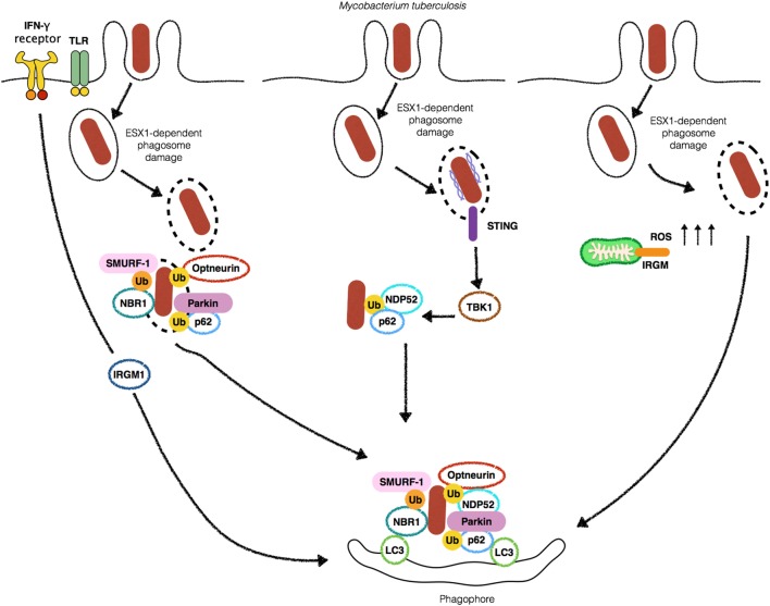
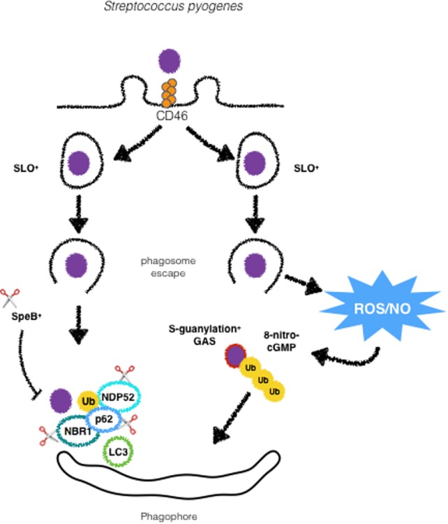
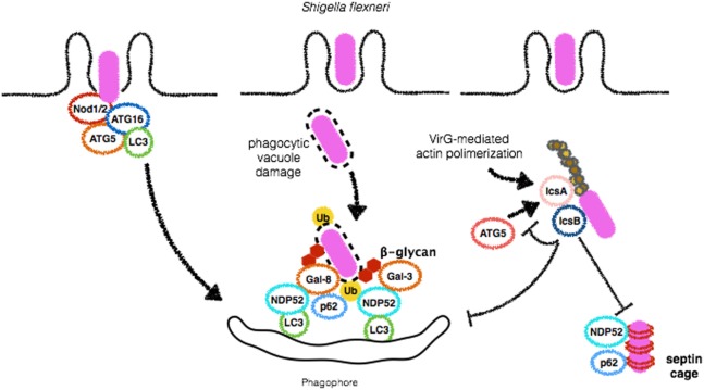
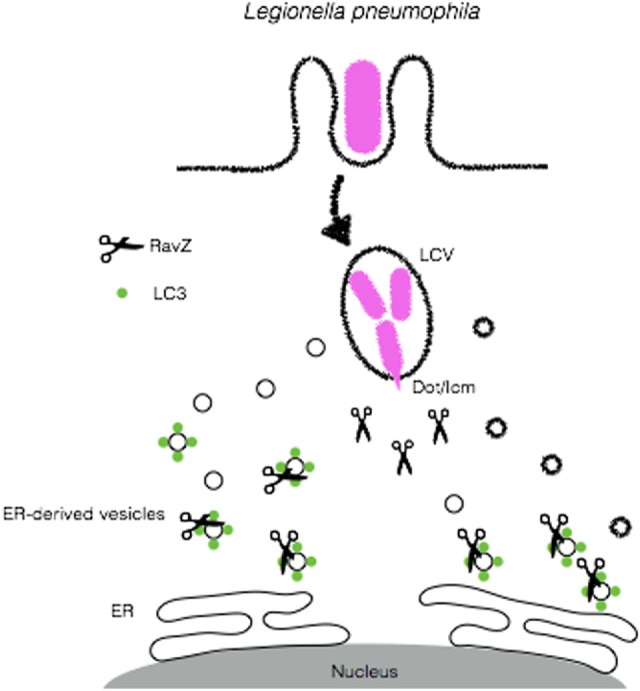
Cellular responses to stress can be defined by the overwhelming number of changes that cells go through upon contact with and stressful conditions such as infection and modifications in nutritional status. One of the main cellular responses to stress is autophagy. Much progress has been made in the understanding of the mechanisms involved in the induction of autophagy during infection by intracellular bacteria. This review aims to discuss recent findings on the role of autophagy as a cellular response to intracellular bacterial pathogens such as, Streptococcus pyogenes, Mycobacterium tuberculosis, Shigella flexneri, Salmonella typhimurium, Listeria monocytogenes, and Legionella pneumophila, how the autophagic machinery senses these bacteria directly or indirectly (through the detection of bacteria-induced nutritional stress), and how some of these bacterial pathogens manage to escape from autophagy.

Keywords: Legionella pneumophila; Listeria monocytogenes; Mycobacterium tuberculosis; Salmonella typhimurium; Shigella flexneri; Streptococcus pyogenes; autophagy; infection.


Figures

Figure 1
Figure 1
Autophagy targets Mycobacterium tuberculosis (Mtb) by different mechanisms. Stimulation with IFN-γ or Toll-like receptors (TLRs) ligands leads to an increase in the localization of (Mtb) into autophagosomes (left). 6-kDa early secretory antigenic target (ESAT-6) secretion system 1 (ESX-1)-induced phagosomal damage induces the exposure of Mtb to cytosolic autophagy adaptors such as Optneurin, p62, NDP52 and NBR1 which bind to ubiquitin associated with Mtb as a consequence of the E3-ligases SMURF-1 and Parkin, culminating with targeting of the bacteria for autophagic degradation (center). Extracellular bacterial DNA from Mtb is detected by STING to activate TBK1 and lead to Mtb ubiquitination and recruitment of p62 and NDP52 (middle-left). IRGM-induced increase in ROS provokes autophagic targeting of Mtb (right).
Figure 2
Figure 2
GAS is targeted by xenophagy by different mechanisms. Following activation of CD46, GAS is directed to autophagosomes. Streptolysin O promotes escape from phagosomes and ubiquitination and recognition by autophagic adaptors p62, NDP52 and NBR1. SpeB producing strains are able to degrade such adaptors to escape from xenophagy. GAS can also undergo modifications by ROS/NO-induced 8-nitro-cGMP via S-guanylation of its surface proteins followed by ubiquitination and targeting to autophagosomes.
Figure 3
Figure 3
Shigella flexneri employs different mechanisms to escape from autophagy. During bacterial entry into host cells, Nod1 and Nod2 recruit ATG16L1 to initiate autophagosome formation in order to restrict S. flexneri replication (left). Vacuole damage leads to β-glycan exposure and recognition by Galectins 3 and 8 and recruitment of NDP52, followed by bacterial ubiquitination and binding to p62 and NDP52, culminating to S. flexneri targeting for autophagic degradation (center). IcsB plays a central role in disguising autophagic machinery. This protein competes with IcsA/VirG for binding to ATG5, preventing p62, and NDP52 binding, septin caging and autophagosome formation (right). Septin caging and further recruitment of autophagic adaptors are blocked by IcsB expression.
Figure 4
Figure 4
Mechanisms of autophagy induction by Salmonella Typhimurium. Upon entry in epithelial cells, S. Typhimurium resides in a specialized compartment, the Salmonella-containing vacuole (SCV). A fraction of the bacterial population damages and/or escapes from SCV and initiates to replicate in the cytosol. Either still within the SCV or free in the cytosolic compartment, S. Typhimurium triggers autophagy by several means. β-glycan present in vacuole remnants is recognized by Galectin-8 (Gal-8) and targets bacteria to autophagosomes. S. Typhimurium can also be ubiquitinated by the E3-ligases LRSAM or LUBAC, allowing its recognition by autophagic adaptors Optneurin, p62 or NDP52. RNF166, another E3-ligase, ubiquitinates p62 to increase the ability of this protein to bind bacteria-associated ubiquitin. Diacylglycerol (DAG) recognition and autophagy induction upon S. Typhimurium infection are not depicted here.
Figure 5
Figure 5
ActA and phospholipases (plc) A and B are major Listeria monocytogenes virulence factors involved in the escape from autophagy. Upon Listeriolysin O-dependent escape from phagosome wild-type L. monocytogenes escapes from autophagic targeting due to the expression of ActA, that blocks the recruitment of p62 and NDP52 to the bacterial surface. The expression of plcA/B stalls the formation of autophagosomal membranes. Galectin recognition of damaged vacuoles and autophagy induction are not shown here.
Figure 6
Figure 6
Legionella pneumophila disrupts autophagy to create a replicative niche. L. pneumophila secretes RavZ through its Dot/Icm apparatus in order to deconjugate LC3 from ER-derived vesicles and block autophagy in order to escape from autophagic degradation.
Figure 7
Figure 7
Bacteria-induced amino acid starvation stress triggers xenophagy. Shigella flexneri induces a persistent amino acid starvation that leads to GCN2 activation and inhibition of mTORC1 to allow the formation of bacteria targeted autophagosomes (left). In contrast, Salmonella Typhimurium (center) and Listeria monocytogenes (right) trigger only a transient amino acid starvation and inhibition of mTORC1, allowing its reactivation at later time-points to block the formation of autophagosomes.

Similar articles

Cited by

References

    1. Hussey S, Travassos LH, Jones NL. Autophagy as an emerging dimension to adaptive and innate immunity. Semin Immunol (2009) 21:233–41.10.1016/j.smim.2009.05.004 - DOI - PMC - PubMed
    1. Rikihisa Y. Glycogen autophagosomes in polymorphonuclear leukocytes induced by rickettsiae. Anat Rec (1984) 208:319–27.10.1002/ar.1092080302 - DOI - PubMed
    1. Gutierrez MG, Master SS, Singh SB, Taylor GA, Colombo MI, Deretic V. Autophagy is a defense mechanism inhibiting BCG and Mycobacterium tuberculosis survival in infected macrophages. Cell (2004) 119:753–66.10.1016/j.cell.2004.11.038 - DOI - PubMed
    1. Nakagawa I, Amano A, Mizushima N, Yamamoto A, Yamaguchi H, Kamimoto T, et al. Autophagy defends cells against invading group A Streptococcus. Science (2004) 306:1037–40.10.1126/science.1103966 - DOI - PubMed
    1. Amano A, Nakagawa I, Yoshimori T. Autophagy in innate immunity against intracellular bacteria. J Biochem (2006) 140:161–6.10.1093/jb/mvj162 - DOI - PubMed

Publication types

MeSH terms