Case Reports
. 2018 Apr 30;12(4):1-5.
doi: 10.3941/jrcr.v12i4.3103. eCollection 2018 Apr.

Pneumorrhachis after Recreational Drug Use

Affiliations
Case Reports

Pneumorrhachis after Recreational Drug Use

Bilal A Sethi et al. J Radiol Case Rep. .

Abstract

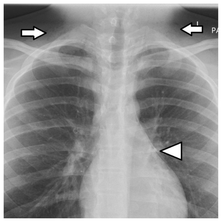
Pneumorrhachis, the presence of air in the spinal canal, is an unusual and alarming radiographic finding. The etiology is most commonly traumatic or iatrogenic but it can occur as a spontaneous phenomenon in association with pneumomediastinum. We report the case of a 16 year old male who presented with throat discomfort and a feeling of altered voice after recreational drug use. Examination confirmed widespread subcutaneous emphysema above the clavicles and plain radiograph and computed tomography imaging confirmed the presence of extensive pneumomediastinum and pneumorrhachis. The patient was managed conservatively and made a full recovery. The clinical and imaging features of spontaneous pneumorrhachis are presented as well as a review of the literature with regard to pathogenesis, management and outcome. Knowledge and understanding of this unusual phenomenon is important to properly direct patient care.

Keywords: Pneumorrhachis; computed tomography; diagnosis; imaging; intraspinal air; spinal pneumatosis.


Figures

Figure 1
Figure 1
16 year old male with subcutaneous emphysema and pneumomediastinum. Technique: Plain posterioranterior (PA) chest radiograph. Findings: Free gas in the soft tissues of the neck (white arrows). Sharp lucency adjacent to the left heart border (white arrowhead). Clear lungs with no visible pneumothorax.
Figure 2
Figure 2
16 year old male with subcutaneous emphysema and pneumorrhachis. Technique: Contrast enhanced CT. Axial view through the upper thorax in lung windows. Findings: Extensive free gas in the soft tissues (black arrows) as noted on the chest radiograph and free gas in the spinal canal (white arrow).
Figure 3
Figure 3
16 year old male with pneumomediastinum, pneumorrhachis and pneumothorax. Technique: Contrast enhanced CT. Sagittal multiplanar reformatted view of the thorax in lung windows. Findings: Free gas tracking into the posterior mediastinum (black arrowhead) and free gas in the anterior mediastinum (white arrow). Free gas in the spinal canal (black arrow). Tiny free gas in the pleura (white arrowhead).
Figure 4
Figure 4
16 year old male with pneumomediastinum Technique: Contrast enhanced CT. Axial view through the mid thorax in lung windows. Findings: Free gas in the mediastinum (white arrow). Intrapleural gas along the lung fissures (black arrows).
Figure 5
Figure 5
16 year old male with subcutaneous emphysema and pneumorrhachis. Technique: Contrast enhanced CT. Axial view through the mid thorax in lung windows. Findings: Free gas is seen tracking up to involve the fascial spaces of the suprahyoid neck (black arrows). Free gas in the cervical spinal canal (white arrow).
Figure 6
Figure 6
16 year old male with pneumomediastinum and subcutaneous emphysema. Technique: Plain posterioranterior (PA) chest radiograph (4 days after initial presentation). Findings: Reduction in the amount of free gas adjacent to the left heart border (white arrowhead) and in soft tissues of the neck (white arrows) compared to previous chest radiograph.

References

    1. Gordon IJ, Hardman DR. The traumatic pneumomyelogram. A previously undescribed entity. Neuroradiology. 1977;13:107–108. - PubMed
    1. Newbold RG, Wiener MD, Vogler JB, 3rd, et al. Traumatic pneumorrhachis. AJR Am J Roentgenol. 1987;148:615–616. - PubMed
    1. Belotti EA, Rizzi M, Rodoni-Cassis P, Ragazzi M, Zanolari-Caledrerari M, Bianchetti MG. Air within the spinal canal in spontaneous mediastinum. Chest. 2010;137(5):1197–1200. doi: 10.1378/chest.09-0514. - DOI - PubMed
    1. Cacares M, Ali SZ, Braud R, Weiman D, Garrett HE., Jnr Spontaneous pneumomediastinum: a comparative study and review of the literature. Ann Thorac Surg. 2008;86(3):962–6. doi: 10.1016/j.athoracsur.2008.04.067. - DOI - PubMed
    1. Macklin CC. Histological indications of the sites of air leakage from the lung alveoli into the vascular sheaths during local overinflation of the living cat’s lung. Can Med Assoc J. 1938 Apr;38(4):401–2. - PMC - PubMed

Publication types