Case Reports
. 2018 Apr 30;12(4):6-14.
doi: 10.3941/jrcr.v12i4.3360. eCollection 2018 Apr.

Torsion of a Myomatous, Non-gravid Uterus: CT Findings

Affiliations
Case Reports

Torsion of a Myomatous, Non-gravid Uterus: CT Findings

Elizabeth Hui Ting Cheong et al. J Radiol Case Rep. .

Abstract

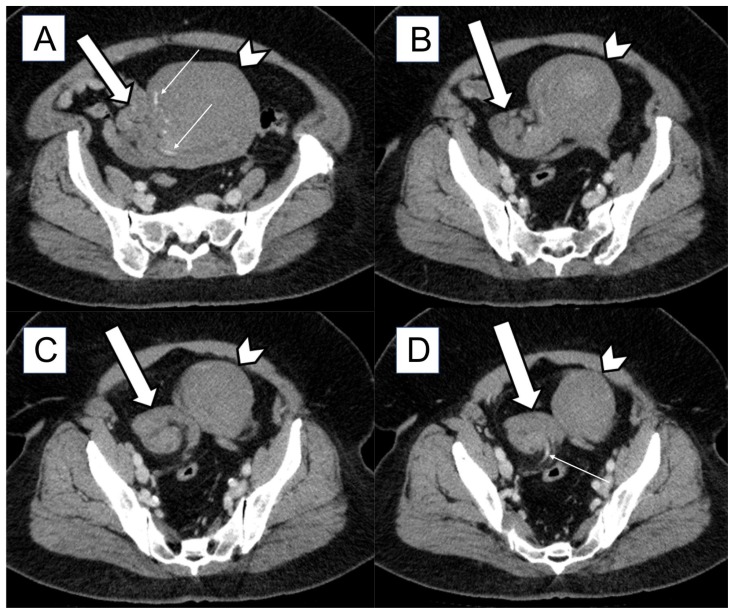
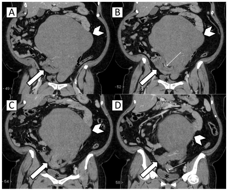
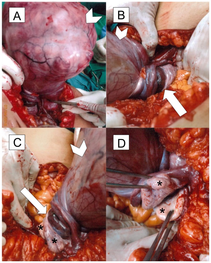
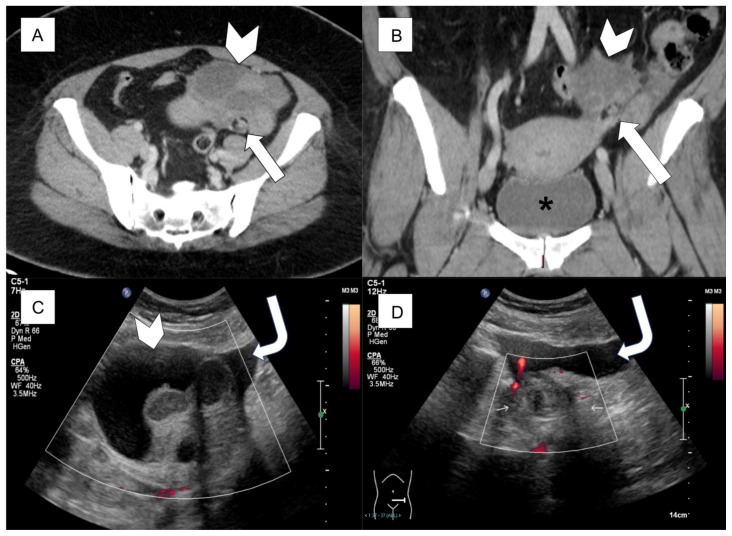
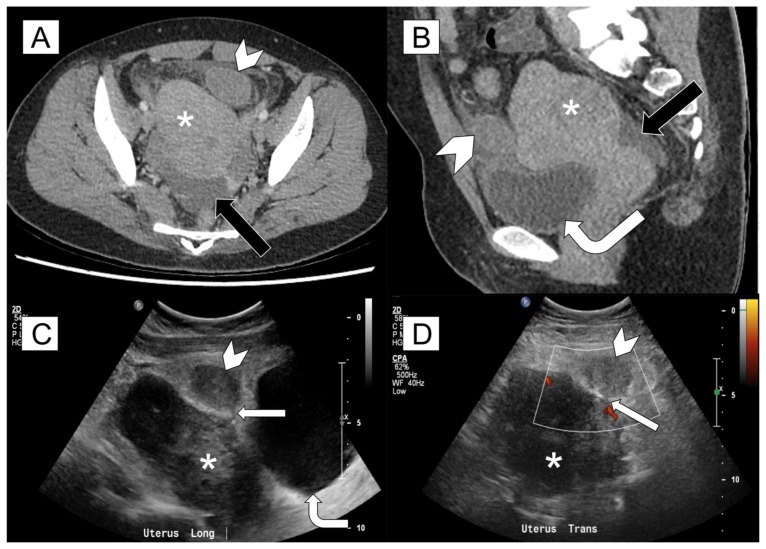
Uterine torsion is a rare condition which is part of a spectrum of gynecological emergencies. If diagnosis is delayed or the condition is left untreated, severe consequences such as infertility and life-threatening complications can arise. As presenting features are often non-specific and can be similar to commonly encountered gastrointestinal and urinary conditions, computed tomography is usually the first imaging modality utilized in an emergency setting. It is therefore important that the radiologist is familiar with recognizing computed tomography features of uterine torsion. We share our findings in a patient with uterine torsion in a non-gravid uterus, diagnosed on contrast-enhanced computed tomography with multiplanar reconstruction.

Keywords: Uterine torsion; adnexal torsion; computed tomography; genitourinary; ovarian torsion; ultrasonography.


Figures

Figure 1
Figure 1
52-year-old nulliparous, menopausal lady with torsion of the uterus and adnexa FINDINGS: Axial contrast-enhanced CT of abdomen and pelvis in the portal venous phase from superior to inferior (A to D) demonstrates the uterus (white arrowheads) twisted along its long axis, with a whorled structure (white block arrows) in the right hemipelvis representing a twisted lower uterine segment. Also noted is swirling of enhancing vessels (thin white arrows), indicating that both adnexa are incorporated into the whorled structure. The uterus is massive and contains multiple fibroids. TECHNIQUE: CT (Toshiba Aquilion ONE) portal venous phase, 579 mA, 120 kV, 3.00 mm slice thickness, 80 ml Omnipaque 350
Figure 2
Figure 2
52-year-old nulliparous, menopausal lady with torsion of the uterus and adnexa FINDINGS: Coronal contrast-enhanced CT of abdomen and pelvis in the portal venous phase from anterior to posterior (A to D) demonstrates the uterus (white arrowheads) twisted along its long axis, with a whorled structure (white block arrows) in the right hemipelvis representing a twisted lower uterine segment. Also noted is swirling of enhancing vessels (thin white arrows), indicating that both adnexa are incorporated into the whorled structure. The uterus is massive and contains multiple fibroids. The urinary bladder (*) is located caudal to the above-mentioned changes. TECHNIQUE: CT (Toshiba Aquilion ONE) portal venous phase, 579 mA, 120 kV, 3.00 mm slice thickness, 80 ml Omnipaque 350
Figure 3
Figure 3
52-year-old nulliparous, menopausal lady with torsion of the uterus and adnexa FINDINGS: Intra-operative images show a grossly enlarged uterus (white arrowheads) which is twisted around its cervix (white arrows), along with the adnexa and ovaries (*).
Figure 4
Figure 4
45-year-old lady with torsion of the adnexa FINDINGS: A, B: Axial and coronal contrast-enhanced CT of abdomen and pelvis in the portal venous phase show a predominantly cystic mass (white arrowheads) containing internal septation and peripheral solid-appearing components in the left adnexa. Postero-inferior to the mass, there is a whorled structure representing a thickened and twisted pedicle (white block arrows). The urinary bladder (*) is seen caudal to the uterus. C: Ultrasound image confirms a complex cystic mass (white arrowhead) in the left adnexa with no internal vascularity. Free fluid is seen around this mass (curved white arrow). D: Ultrasound image shows a thick, avascular whorled structure (thin white arrows) in the left adnexa, corresponding to the thickened, twisted pedicle seen on CT. Free fluid is seen around this whorled structure (curved white arrow). TECHNIQUE: A, B: CT (Toshiba Aquilion ONE) portal venous phase, 750 mA, 120 kV, 3.00 mm slice thickness, 80 ml Omnipaque 350 C, D: Transabdominal ultrasonography on Philips iU22 using 5-1 MHz curved transducer
Figure 5
Figure 5
33-year-old lady with torsion of a pedunculated uterine leiomyoma FINDINGS: A, B: Axial and MPR sagittal contrast-enhanced CT of abdomen and pelvis in the portal venous phase show a circumscribed soft tissue mass (white arrowheads) at the left antero-lateral aspect of a bulky uterus containing fibroids (*). There is surrounding fat stranding and a small amount of free fluid in the pelvis (black arrows). The urinary bladder (curved white arrow) is adjacent to the uterus. C, D: Ultrasound images confirm the presence of a soft tissue mass (white arrowheads) connected to the uterus (*) via a thin stalk (straight white arrows). No internal vascularity is seen within this mass. The urinary bladder (curved white arrow) is adjacent to the uterus. TECHNIQUE: A, B: CT (Toshiba Aquilion ONE) portal venous phase, 663 mA, 120 kV, 3.00 mm slice thickness, 70 ml Omnipaque 350. C, D: Transabdominal ultrasonography on Philips iU22 using 5-1 MHz curved transducer

Similar articles

Cited by

References

    1. Luk SY, Leung JLY, Cheung ML, So S, Fung SH, Cheng SC. Torsion of a Nongravid Myomatous Uterus: Radiological Features and Literature Review. Hong Kong Med J. 2010 Aug;16(4):304–6. - PubMed
    1. Grover S, Sharma Y, Mittal S. Uterine Torsion: A Missed Diagnosis in Young Girls? Journal of Pediatric and Adolescent Gynecology. 2009 Feb;22(1):5–8. - PubMed
    1. Jeong YY, Kang HK, Park JG, Choi HS. CT Features of Uterine Torsion. European Radiology. 2003 Dec;13:249–50. - PubMed
    1. Iraha Y, Okada M, Iraha R, Azama K, Yamashiro T, Tsubakimoto M, et al. CT and MR Imaging of Gynecologic Emergencies. RadioGraphics. 2017 Jul;37(5):1569–86. - PubMed
    1. Yitta S, Hecht EM, Slywotzky CM, Bennett GL. Added value of multiplanar reformation in the multidetector CT evaluation of the female pelvis: a pictorial review. RadioGraphics. 2009 Nov;29(7):1987–2003. - PubMed

Publication types