

Resting-state connectivity in neurodegenerative disorders: Is there potential for an imaging biomarker?
- PMID: 29876270
- PMCID: PMC5988031
- DOI: 10.1016/j.nicl.2018.03.013
Resting-state connectivity in neurodegenerative disorders: Is there potential for an imaging biomarker?
Abstract
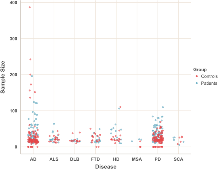
Biomarkers in whichever modality are tremendously important in diagnosing of disease, tracking disease progression and clinical trials. This applies in particular for disorders with a long disease course including pre-symptomatic stages, in which only subtle signs of clinical progression can be observed. Magnetic resonance imaging (MRI) biomarkers hold particular promise due to their relative ease of use, cost-effectiveness and non-invasivity. Studies measuring resting-state functional MR connectivity have become increasingly common during recent years and are well established in neuroscience and related fields. Its increasing application does of course also include clinical settings and therein neurodegenerative diseases. In the present review, we critically summarise the state of the literature on resting-state functional connectivity as measured with functional MRI in neurodegenerative disorders. In addition to an overview of the results, we briefly outline the methods applied to the concept of resting-state functional connectivity. While there are many different neurodegenerative disorders cumulatively affecting a substantial number of patients, for most of them studies on resting-state fMRI are lacking. Plentiful amounts of papers are available for Alzheimer's disease (AD) and Parkinson's disease (PD), but only few works being available for the less common neurodegenerative diseases. This allows some conclusions on the potential of resting-state fMRI acting as a biomarker for the aforementioned two diseases, but only tentative statements for the others. For AD, the literature contains a relatively strong consensus regarding an impairment of the connectivity of the default mode network compared to healthy individuals. However, for AD there is no considerable documentation on how that alteration develops longitudinally with the progression of the disease. For PD, the available research points towards alterations of connectivity mainly in limbic and motor related regions and networks, but drawing conclusions for PD has to be done with caution due to a relative heterogeneity of the disease. For rare neurodegenerative diseases, no clear conclusions can be drawn due to the few published results. Nevertheless, summarising available data points towards characteristic connectivity alterations in Huntington's disease, frontotemporal dementia, dementia with Lewy bodies, multiple systems atrophy and the spinocerebellar ataxias. Overall at this point in time, the data on AD are most promising towards the eventual use of resting-state fMRI as an imaging biomarker, although there remain issues such as reproducibility of results and a lack of data demonstrating longitudinal changes. Improved methods providing more precise classifications as well as resting-state network changes that are sensitive to disease progression or therapeutic intervention are highly desirable, before routine clinical use could eventually become a reality.
Keywords: Biomarker; Neurodegeneration; Resting-state; Review; fMRI.
Figures


References
-
- Agosta F., Pievani M., Geroldi C., Copetti M., Frisoni G.B., Filippi M. Resting state fMRI in Alzheimer's disease: beyond the default mode network. Neurobiol. Aging. 2012;33(8):1564–1578. - PubMed
-
- Agosta F., Canu E., Valsasina P., Riva N., Prelle A., Comi G., Filippi M. Divergent brain network connectivity in amyotrophic lateral sclerosis. Neurobiol. Aging. 2013;34(2):419–427. - PubMed
-
- Agosta F., Sala S., Valsasina P., Meani A., Canu E., Magnani G.…Filippi M. Brain network connectivity assessed using graph theory in frontotemporal dementia. Neurology. 2013;81(2):134–143. - PubMed
-
- Agosta F., Caso F., Stankovic I., Inuggi A., Petrovic I., Svetel M.…Filippi M. Cortico-striatal-thalamic network functional connectivity in hemiparkinsonism. Neurobiol. Aging. 2014;35(11):2592–2602. - PubMed
Publication types
MeSH terms
Substances
LinkOut - more resources
Full Text Sources
Other Literature Sources
Medical
