. 2018 Mar 5:18:972-985.
doi: 10.1016/j.nicl.2018.02.021. eCollection 2018.

Repairing the brain with physical exercise: Cortical thickness and brain volume increases in long-term pediatric brain tumor survivors in response to a structured exercise intervention

Affiliations

Repairing the brain with physical exercise: Cortical thickness and brain volume increases in long-term pediatric brain tumor survivors in response to a structured exercise intervention

Kamila U Szulc-Lerch et al. Neuroimage Clin. .

Abstract

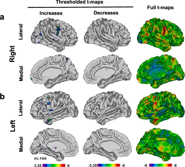
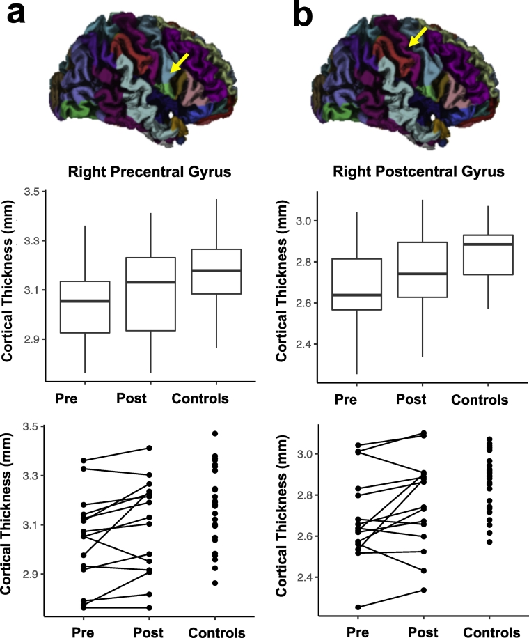
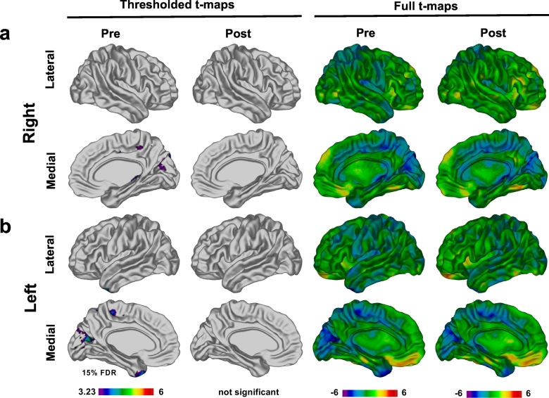
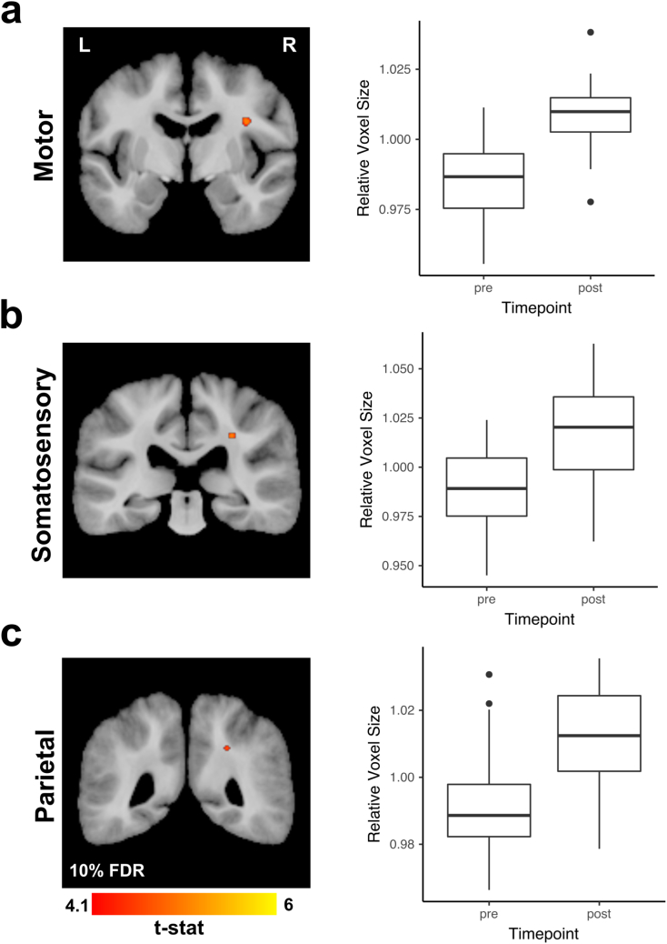
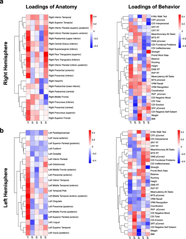
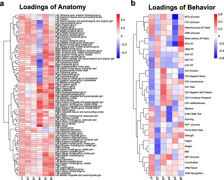
There is growing evidence that exercise induced experience dependent plasticity may foster structural and functional recovery following brain injury. We examined the efficacy of exercise training for neural and cognitive recovery in long-term pediatric brain tumor survivors treated with radiation. We conducted a controlled clinical trial with crossover of exercise training (vs. no training) in a volunteer sample of 28 children treated with cranial radiation for brain tumors (mean age = 11.5 yrs.; mean time since diagnosis = 5.7 yrs). The endpoints were anatomical T1 MRI data and multiple behavioral outcomes presenting a broader analysis of structural MRI data across the entire brain. This included an analysis of changes in cortical thickness and brain volume using automated, user unbiased approaches. A series of general linear mixed effects models evaluating the effects of exercise training on cortical thickness were performed in a voxel and vertex-wise manner, as well as for specific regions of interest. In exploratory analyses, we evaluated the relationship between changes in cortical thickness after exercise with multiple behavioral outcomes, as well as the relation of these measures at baseline. Exercise was associated with increases in cortical thickness within the right pre and postcentral gyri. Other notable areas of increased thickness related to training were present in the left pre and postcentral gyri, left temporal pole, left superior temporal gyrus, and left parahippocampal gyrus. Further, we observed that compared to a separate cohort of healthy children, participants displayed multiple areas with a significantly thinner cortex prior to training and fewer differences following training, indicating amelioration of anatomical deficits. Partial least squares analysis (PLS) revealed specific patterns of relations between cortical thickness and various behavioral outcomes both after training and at baseline. Overall, our results indicate that exercise training in pediatric brain tumor patients treated with radiation has a beneficial impact on brain structure. We argue that exercise training should be incorporated into the development of neuro-rehabilitative treatments for long-term pediatric brain tumor survivors and other populations with acquired brain injury. (ClinicalTrials.gov, NCT01944761).

Keywords: Brain recovery; Cortical thickness; Cranial radiation; Exercise; Neuroplasticity; Pediatric brain tumor.


Figures

Fig. 1
Fig. 1
Study design. Participants were quasi-randomly assigned to start either: (a) No Training, or (b) Exercise Training first. All participants were evaluated immediately prior to starting Exercise Training (Pre) and 12-weeks later after its completion (Post). About half of the participants were also evaluated 12-weeks prior to starting the Exercise Training (Baseline) and about half were evaluated 12-weeks after completing the program (Follow-up). Training occurred in either a Group or Combined (group/home) setting.
Fig. 2
Fig. 2
Vertex-wise analysis. T-maps illustrating statistically significant (FDR 5%) regions of cortical thickness increases and decreases observed in patients in the Group setting immediately after completion of the Exercise Training in right (a) and left (b) hemispheres. The areas of cortical thickness increase were broader and more pronounced than areas of decrease. The strongest areas of increase were observed in right and left precentral gyri, right postcentral gyrus and left temporal lobe. Smaller areas of increase were observed in occipital, parietal and frontal lobes in both hemispheres. Areas of cortical thinning were also observed (full t-maps, blue areas) although only very small, isolated foci survived FDR correction.
Fig. 3
Fig. 3
Results of region of interest (ROI) based cortical thickness analysis in participants in a Group setting. Comparison of cortical thickness in right precentral (a) and postcentral gyrus (b) before (Pre) and after (Post) Exercise Training. These results are contrasted with cortical thickness estimates in healthy age- and gender-matched controls. Normalization of cortical thickness Post Exercise Training to thickness of controls was observed in these areas. These results are illustrated using box-plots and line plots connecting data points of individual participants.
Fig. 4
Fig. 4
Comparison of cortical thickness in patients to controls. Results of vertex-wise analysis comparing cortical thickness in participants in Group setting to age- and gender-matched controls. In this analysis, we co-varied for scanner type because of imbalance between all controls and Group setting participants. Before exercise areas of thinner cortex were observed in participants (FDR 15%) in the occipital and parietal lobes in right (a) and left (b) hemispheres. These differences were no longer significant after completion of exercise program.
Fig. 5
Fig. 5
Deformation based morphometry (DBM) results. DBM analysis revealed several areas of statistically significant (FDR 10%) volume increases in all participants regardless of assignment to the training setting (Group or Combined). These areas included WM underlying the motor cortex (a), somatosensory cortex (b) and parietal lobe (c). Magnitude of these observed volume increases are illustrated in the box-plots from a single voxel within a cluster with the highest t-value.
Fig. 6
Fig. 6
Partial least squares (PLS) analysis of relationships between changes in cortical thickness and behavior after Exercise Training for participants in the Group setting. Each column of loadings of anatomy maps within a heat-map onto the same column of the loadings of behavior. The first column always explains the greatest amount of orthogonal variance between variables. Definitions of all the abbreviations have been provided in Supplementary Fig. 1.
Fig. 7
Fig. 7
Partial least squares (PLS) analysis of cortical thickness and behavioral data at Baseline for all participants. Please refer to Supplementary Fig. 1 for definitions of all the abbreviations.

References

    1. Akers K.G., Martinez-Canabal A., Restivo L., Yiu A.P., De Cristofaro A., Hsiang H.-L.L., Wheeler A.L., Guskjolen A., Niibori Y., Shoji H., Ohira K., Richards B.A., Miyakawa T., Josselyn S.A., Frankland P.W. Hippocampal neurogenesis regulates forgetting during adulthood and infancy. Science. 2014;344:598–602. - PubMed
    1. Anderson B.J., Eckburg P.B., Relucio K.I. Alterations in the thickness of motor cortical subregions after motor-skill learning and exercise. Learn. Mem. 2002;9:1–9. - PMC - PubMed
    1. Ang E., Gomez-Pinilla F. Potential therapeutic effects of exercise to the brain. Curr. Med. Chem. 2007;14:2564–2571. - PubMed
    1. Avants B., Gee J.C. Geodesic estimation for large deformation anatomical shape averaging and interpolation. NeuroImage. 2004;23:S139–S150. - PubMed
    1. Avants B.B., Tustison N.J., Song G., Cook P.A., Klein A., Gee J.C. A reproducible evaluation of ANTs similarity metric performance in brain image registration. NeuroImage. 2011;54:2033–2044. - PMC - PubMed

Publication types

Associated data