. 2018 May 23:5:70.
doi: 10.3389/fmed.2018.00070. eCollection 2018.

Detection of Central Nervous System Infiltration by Myeloid and Lymphoid Hematologic Neoplasms Using Flow Cytometry Analysis: Diagnostic Accuracy Study

Affiliations

Detection of Central Nervous System Infiltration by Myeloid and Lymphoid Hematologic Neoplasms Using Flow Cytometry Analysis: Diagnostic Accuracy Study

Laiz Cameirão Bento et al. Front Med (Lausanne). .

Abstract

Introduction: Infiltration of the central nervous system (CNS) by hematologic or lymphoid malignant cells can cause extensive neurological damage, be progressive and fatal. However, usually, the cerebrospinal fluid (CSF) has low cellularity and rapid cell degeneration, which can impair cytometry analysis. Storage and transport measures, sample preparation, and staining protocols can interfere with diagnostic accuracy.

Objective: To calculate the diagnostic performance of flow cytometry (FC) using a cell stabilizer for sample preservation compared to cytomorphology in the detection of CNS infiltration by lymphoid and hematologic neoplasms.

Methods: Cell samples from all consecutive patients with suspected infiltration by hematological malignancies evaluated between January 2014 and December 2016 were included. Cases were analyzed by FC using a cell preservation medium and cytomorphology. Sensitivity and specificity were calculated.

Results: From 414 CSF samples, 72 had a phenotype compatible with characteristics of infiltration by hematological disease, whereas cytology was positive for 35 cases. FC showed higher sensitivity and specificity when compared to cytomorphology, particularly in cases with cellularity under 5 leukocytes/mm3.

Conclusion: We demonstrated that collecting CSF in a medium that preserves the stability of the sample improves accuracy when compared to cytomorphology, particularly in low-volume and low-cellularity samples.

Keywords: central nervous system; cerebrospinal fluid; cytology; flow cytometry; neoplasms.


Figures

Figure 1
Figure 1
Negative flow cytometry result for suspected central nervous fluid (CSF) infiltration by hematological malignancies. Presumptive diagnosis: central nervous system disorder. (A,B) Normal forward-scattered light (FSC size) and side-scattered light (complexity—SSC); (C–E) normal antigenic expression of CD3 in T-lymphocytes, CD4 and CD8 expression in CD3+CD4+ and CD3+CD8+ lymphocyte subtypes, CD14 expression in monocytes and CD38 expression and CD56+CD3 natural killer (NK) cells; (F,G) polyclonal CD19+CD20+ B-lymphoid population for kappa (73%) and lambda (27%) light chains. Sample with 3 leukocytes/mm3 and 1 red blood cell/mm3, acquisition of 8,425 events in the FACS Canto II (Becton Dickinson), and analysis performed with the software Infinicyt (Cytognos). The frequency of populations in the viable cell region and CD45+ (3,385 events) was 68.6% CD3 T-lymphocytes—in that, 52.6% CD4 T-lymphocytes and 13.9% CD8 T-lymphocytes; 0.9% NK cells; 0.7% mature and polyclonal B-lymphocytes (kappa/lambda ratio: 2.38); and 29.8% monocyte lineage cells.
Figure 2
Figure 2
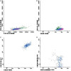
Flow cytometry results of suspected central nervous system infiltration by B-cell non-Hodgkin lymphoma. Presumptive diagnosis: B-cell non-Hodgkin lymphoma. (A,B) Normal forward-scattered light (FSC size) and side-scattered light (complexity—SSC); (C) mature CD19+ and CD20+ B-lymphoid population; (D) mature monoclonal lambda-light-chain-expressing B-lymphoid population. Sample collected in Transfix®/ethylenediaminetetracetic acid with an approximate volume of 5 ml and 2 leukocytes/mm3 and 0 red blood cells/mm3. Samples with 8,575 events acquired in Canto II (Becton Dickinson) and analyzed using the Infinicyt software (Cytognos). Frequency of populations in the viable cell region and CD45+ cell (1,000 events).
Figure 3
Figure 3
Flow cytometry results of suspected central nervous system infiltration by B-cell acute lymphoblastic leukemia. Presumptive diagnosis: suspected B-cell lymphoblastic leukemia. (A) Forward-scattered light (FSC size) and side-scattered light (complexity—SSC); (B) low-complexity blast population weakly expressing CD45 (light blue); (C) population of CD19+ and CD10+ B-lymphoid blasts; (D) strong expression of CD58 and moderate expression of CD38 in blast population; (E) CD10+ and CD34+ expression; (F) strong expression of CD10 in blast population. Sample collected in Transfix®/ethylenediaminetetracetic acid with an approximate volume of 5 ml and 3 leukocytes/mm3 and 0 red blood cells/mm3. Samples with 20,904 events acquired in Canto II (Becton Dickinson) and analyzed using the Infinicyt software (Cytognos). The frequency of populations in the viable cell region and CD45+ was 14,210 events.
Figure 4
Figure 4
Flow cytometry results of suspected central nervous system infiltration by T-cell acute lymphoblastic leukemia. Presumptive diagnosis: T-cell lymphoblastic leukemia. (A) Forward-scattered light (FSC size) and side-scattered light (complexity—SSC); (B) low-complexity blast population (light blue) moderately/strongly expressing CD45; (C) CD4/CD8 double positive blast population; (D) intracytoplasmic CD3-positive and surface CD3-negative blasts. Sample collected in Transfix/ethylenediaminetetracetic acid with an approximate volume of 5 ml and 3 leukocytes/mm3 and 10 red blood cells/mm3. Samples with 11,575 events acquired in Canto II (Becton Dickinson) and analyzed using the Infinicyt software (Cytognos). The frequency of populations in the viable cell region and CD45+ cell was 7,913 events.
Figure 5
Figure 5
Flow cytometry results of suspected central nervous system infiltration by immature myeloid population. Presumptive diagnosis: acute myeloid leukemia. (A) Forward-scattered light (FSC size) and side-scattered light (complexity—SSC); (B) low-complexity blast population (light blue) strongly expressing CD45; (C) blast population with abnormal CD117 expression and absent CD34 expression. Sample collected in Transfix/ethylenediaminetetracetic acid with an approximate volume of 5 ml and 1 leukocyte/mm3 and 2 red blood cells/mm3. Samples with 6,375 events acquired in Canto II (Becton Dickinson) and analyzed using the Infinicyt software (Cytognos). The frequency of populations in the viable cell region and CD45+ was 2,541 events.
Figure 6
Figure 6
Positive results for flow cytometry and results found for cytomorphology.
Figure 7
Figure 7
Samples grouped according to cellularity.

References

    1. Kraan J, Gratama JW, Haioun C, Orfao A, Plonquet A, Porwit A, et al. Flow cytometric immunophenotyping of cerebrospinal fluid. Curr Protoc Cytom (2008) Chapter 6:Unit 6.25.10.1002/0471142956.cy0625s45 - DOI - PMC - PubMed
    1. Bromberg JE, Breems DA, Kraan J, Bikker G, van der Holt B, Smitt PS, et al. CSF flow cytometry greatly improves diagnostic accuracy in CNS hematologic malignancies. Neurology (2007) 68(20):1674–9.10.1212/01.wnl.0000261909.28915.83 - DOI - PubMed
    1. Pittman M, Treese S, Chen L, Frater JL, Nguyen TT, Hassan A, et al. Utility of flow cytometry of cerebrospinal fluid as a screening tool in the diagnosis of central nervous system lymphoma. Arch Pathol Lab Med (2013) 137(11):1610–8.10.5858/arpa.2012-0313-OA - DOI - PubMed
    1. Chamberlain MC. Carcinomatous meningitis. Arch Neurol (1997) 54(1):16–7.10.1001/archneur.1997.00550130008003 - DOI - PubMed
    1. Gonzalez-Vitale JC, Garcia-Bunuel R. Meningeal carcinomatosis. Cancer (1976) 37(6):2906–11.10.1002/1097-0142(197606)37:6<2906::AID-CNCR2820370648>3.0.CO;2-D - DOI - PubMed

LinkOut - more resources