Review
. 2018 Oct;17(5):654-682.
doi: 10.1007/s12311-018-0952-3.

Cerebellar Modules and Their Role as Operational Cerebellar Processing Units: A Consensus paper [corrected]

Affiliations
Review

Cerebellar Modules and Their Role as Operational Cerebellar Processing Units: A Consensus paper [corrected]

Richard Apps et al. Cerebellum. 2018 Oct.

Erratum in

Abstract

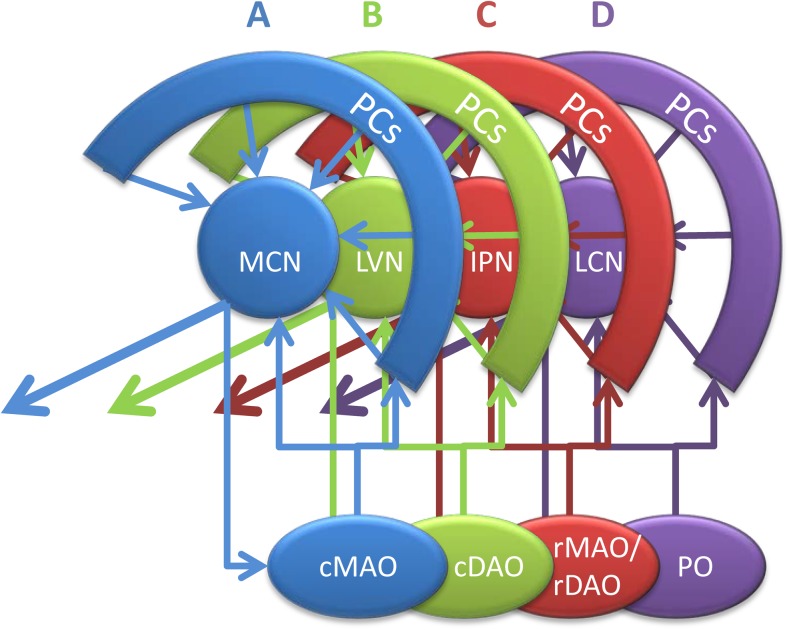
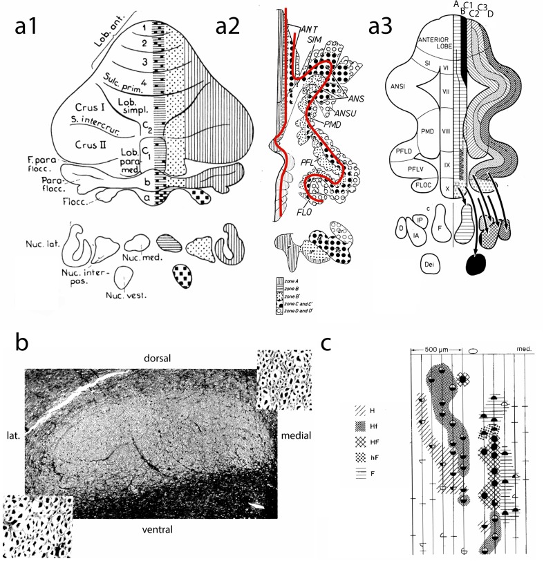
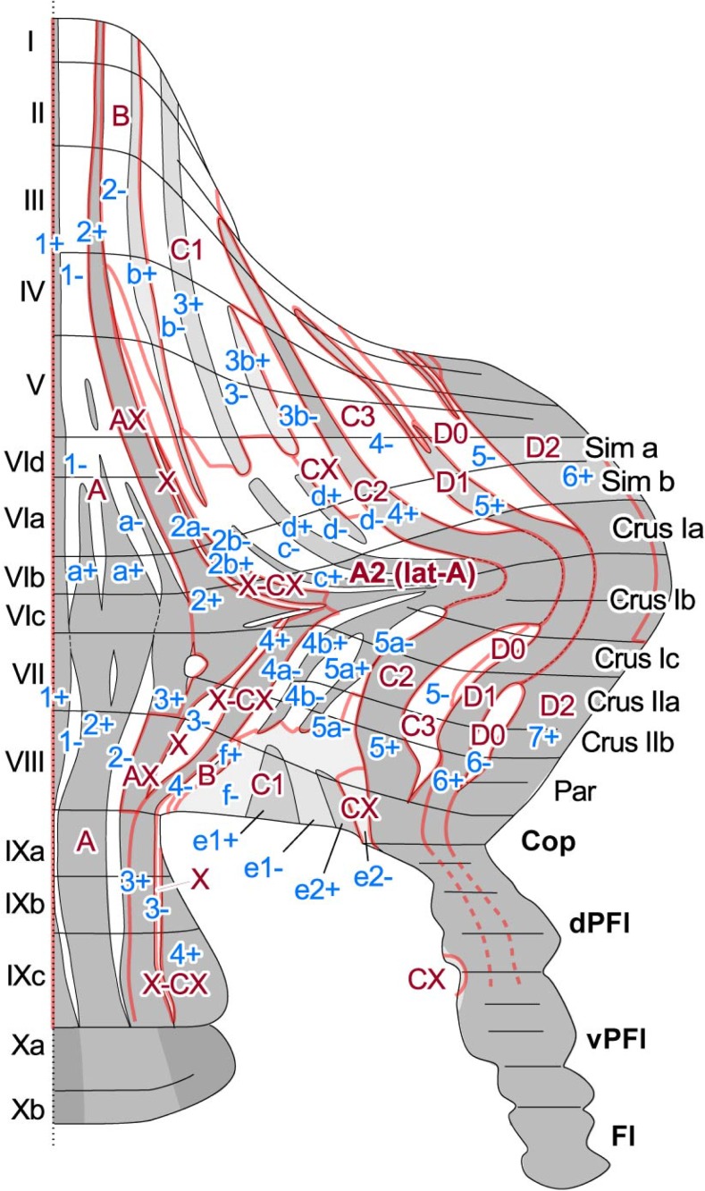
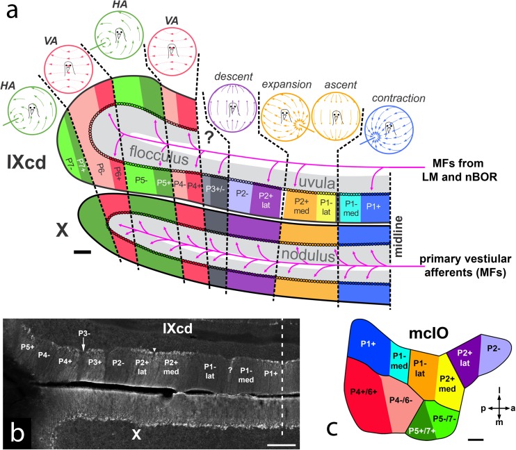
The compartmentalization of the cerebellum into modules is often used to discuss its function. What, exactly, can be considered a module, how do they operate, can they be subdivided and do they act individually or in concert are only some of the key questions discussed in this consensus paper. Experts studying cerebellar compartmentalization give their insights on the structure and function of cerebellar modules, with the aim of providing an up-to-date review of the extensive literature on this subject. Starting with an historical perspective indicating that the basis of the modular organization is formed by matching olivocorticonuclear connectivity, this is followed by consideration of anatomical and chemical modular boundaries, revealing a relation between anatomical, chemical, and physiological borders. In addition, the question is asked what the smallest operational unit of the cerebellum might be. Furthermore, it has become clear that chemical diversity of Purkinje cells also results in diversity of information processing between cerebellar modules. An additional important consideration is the relation between modular compartmentalization and the organization of the mossy fiber system, resulting in the concept of modular plasticity. Finally, examination of cerebellar output patterns suggesting cooperation between modules and recent work on modular aspects of emotional behavior are discussed. Despite the general consensus that the cerebellum has a modular organization, many questions remain. The authors hope that this joint review will inspire future cerebellar research so that we are better able to understand how this brain structure makes its vital contribution to behavior in its most general form.

Keywords: Aldolase C; Cerebellum; Climbing fibers; Compartments; Functional organization; Longitudinal stripes; Microzones; Mossy fibers; Purkinje cells; Zebrin.


Conflict of interest statement

The authors declare that they have no conflict of interest.

Figures

Fig. 1
Fig. 1
Simplified diagram illustrating the four main modules of the right cerebellum seen from medial. The elementary modular connections are based on the projection of longitudinally arranged strips of Purkinje cells (PCs) to four main target nuclei and their olivocerebellar input from selective inferior olivary subnuclei. As such two vermal Purkinje cell zones (A and B) are recognized, together with their respective targets, the medial cerebellar nucleus (MCN) and lateral vestibular nucleus (LVN) and their sources of climbing fibers, caudal parts of the medial accessory (cMAO) and dorsal accessory (cDAO) olives, respectively. The C zones of the paravermis targets the interposed nuclei (IPN) and receives climbing fibers from the rostral (r) MAO and rDAO, while the D zones targets the lateral cerebellar nucleus (LCN) and receive from the principal olive (PO). Note that olivary subnuclei are also reciprocally connected according the same scheme. The interconnected olivocorticonuclear entity is referred to as module and each have a specific output. All modules (apart from the B module) have been further subdivided. Note that the modules of the vestibulocerebellum are not indicated in this diagram. Modified after Ruigrok [6]
Fig. 2
Fig. 2
a1 Diagram of the corticonuclear projection of the cerebellum, showing the vermal, intermediate, and lateral zones of Jansen and Brodal [24]. Nomenclature of the lobules according to Bolk [25]. a2 Diagram of the flattened cerebellar cortex of the cat showing the corticonuclear projection (after Voogd [26]). The red lines indicate the direction of the folial chains of vermis and hemisphere. a3 Corticonuclear projection shown in diagrams of the flattened cerebellar cortex of the cat from Groenewegen et al. [19]. b Superior cerebellar peduncle of the cat, Häggqvist stain. Note small myelinated fibers in the medial third and coarse fibers in lateral two-thirds [after 24]. c Microzones with different climbing fiber inputs in the B zone of the cerebellum of the cat. Stimulation of the ipsilateral and contralateral ulnar and sciatic nerves results in Purkinje cells with similar responses in microzones as indicated by different hatching and stippling: H (hindlimb), Hf (mainly hindlimb), HF (hind- and forelimb), hF (mainly forelimb), F (forelimb), after Andersson and Oscarsson [27]. ANS, ANSI ansiform lobule; ANSU ansula; D dentate nucleus; Dei Deiters nucleus; F fastigial nucleus; F. parafloc parafloccular fissure; FLO, FLOC flocculus; IA anterior interposed nucleus; IP posterior interposed nucleus; Lc. Lateral nucleus pars convexa; Lob. Paramed paramedian lobule; Lob.ant, ANT anterior lobe; Lob.simpl simple lobule; Lr, lateral nucleus pars rotunda; Nuc.interpos interposed nucleus; Nuc.lat lateral nucleus; Nuc.med. medial nucleus; Nuc.vest. vestibular nucleus; Parafloc paraflocculus; PFL(D,V) paraflocculus (dorsalis, ventralis); PMD paramedian lobule; S.intercrur intercrural sulcus; SIM, SI primary fissure simplex lobul; Sulc.prim
Fig. 3
Fig. 3
Schematic of positional correlation between zebrin II (aldolase C) striped pattern and the cerebellar module mapped on the unfolded rat cerebellar cortex in the rat. Based on Sugihara and Shinoda [10]
Fig. 4
Fig. 4
a Diagram of the optic flow modules in the pigeon vestibulocerebellum (VbC; folia IXcd and X) (based on data from [–69]. The lateral half of the VbC is the flocculus, the medial half is the uvula (IXcd)/nodulus (X). Each module is represented by a depiction of the optic flowfield that maximally excites the complex spike activity (CSA) of the Purkinje cells (PCs). The ZII+ and ZII− stripes in IXcd are also indicated. (All PCs in X are uniformly ZII+). There are seven optic flow modules, each spanning a ZII+/− stripe pair (see text for details). P3+/− PCs do not respond to optic flow. The magenta arrows indicate the primary vestibular afferents, which project as mossy fibers (MFs) to X. Magenta arrows also show the optic flow MF inputs from the nucleus of the basal optic root (nBOR) and pretectal nucleus lentiformis mesencephali (LM) to the ZII+ stripes in IXcd. b Coronal section through ventral IXcd and dorsal X, showing the ZII expression. The inverted triangle indicates the “notch” where PCs are absent, and bisects the P2+ stripe in to medial and lateral halves (P2+med, P2+lat). The “?” indicates a ZII+ stripe, 1 to 3 PCs in width, which similarly divides the P1−stripe (P1−med, P1−lat). The vertical dashed line indicates the midline. c Dorsal view of the medial column of the inferior olive (mcIO) and is color-coded to match the ZII stripes in (a), to indicate the topography of the climbing fiber projections (based on data from [32, 33]). a anterior, p posterior, m medial, l lateral. Scale bars: 200 μm in (a), 300 μm in (b), 100 μm in (c)
Fig. 5
Fig. 5
Physiological difference between zebrin-identified cerebellar modules. a Schematic drawing of unfolded cerebellar surface, adapted from [–69], depicting post-mortem immunohistochemically determined recording locations of PC, with color-coded simple spike firing rate. Note the higher firing rate in ZII− PCs and the consistent presence of the difference, even in nearby pairs. b Summary of (a) demonstrating the significant difference in average simple spike firing rate between ZII+ and ZII− PCs, recorded in vivo. c Complex spike firing rates show a similar difference, with higher firing rates in ZII- than in ZII+ PCs. d Pharmacological block of TRPC3 with two difference blockers, genestein, and pyr3, selectively affects PC simple spike activity in ZII− PCs, indicating the contribution of TRPC3 to creating this difference
Fig. 6
Fig. 6
An example of a functional Purkinje cell module in the lobule III–IV of the cerebellar cortex. GC clusters belonging to different microzones (identified by the zebrin band pattern in red and gray) communicate with specific groups of PCs (one example in black). In this example, a group of PCs (120 μm width spanning P1− and P1+ zebrin stripes) close to the midline receives GC inputs from ipsilateral and contralateral P2+, ipsilateral and contralateral median P1− and P1+ microzones. This organization is conserved across mice. Each GC cluster receives specific MF inputs from different precerebellar nuclei and modalities (identified by the color in the GC pie chart). MFs projections in the GCL are complex and redundant. The other GCs remain silent or unconnected (shaded pie chart). This functional module does not necessarily fit with the anatomical boundaries given by the CF and MF inputs. ML molecular layer, PCL Purkinje cell layer, GCL granule cell layer, MFs Mossy fibers, Ecu external cuneate, SCL lumbar part of the spinocerebellar tract, SCT thoracic part of the spinocerebellar tract, SCC cervical part of the spinocerebellar tract, BPN basal pontine nuclei, CFs climbing fibers, MFs mossy fibers, PCs Purkinje cells, GCs granule cells. Adapted from [113, 199]
Fig. 7
Fig. 7
Multiple modules collaborate in sensorimotor processing. a Superimposed stack of plots of 10 serial (1 out of 4), 40-μm-thick sections showing RABV−/ZII+ Purkinje cells (gray), RABV+/ZII− (yellow), and RABV+/ZII+ (magenta) Purkinje cells in the rat anterior lobe 120 h after injection of RABV in the gastrocnemius muscle (case 1010). Note that a prominent band of RABV+/ZII− Purkinje cells is seen between the P1+ and P2+ zebrin stripes, which mostly is territory of the B zone [240]. The main zebrin+ stripes (p1+ to p6+) are indicated. b Similar superimposed stack of 12 plotted sections 70 h after injection in M1 (case 1151). Note that three separated clusters of RABV infected Purkinje cells are recognized: a vermal one just lateral of P2+, a large paravermal cluster that encompasses P3+, P3−, P4+, P4− zebrin stripes and a hemispheral cluster lateral within the P6+ strip. c Diagram showing multimodular impact of cerebellar zones on three sensorimotor regions. Line thickness is shown relative to 100% of total number of RABV labeled Purkinje cells in the anterior part of the cerebellum (modified after [241]). Modular identity is inferred from its relation with the zebrin pattern [11, 99]
Fig. 8
Fig. 8
The “A” module of the cerebellar vermis can be separated into several different rostrocaudally arranged regions, in some cases corresponding to specific lobules, that are associated with a variety of cognitive, motor and autonomic functions relating to defensive behaviors

Similar articles

Cited by

References

    1. Glickstein M, Doron K. Cerebellum: connections and functions. Cerebellum. 2008;7:589–594. - PubMed
    1. Ito M. The cerebellum and neural control. Raven Press; 1984.
    1. Cerminara NL, Lang EJ, Sillitoe RV, Apps R. Redefining the cerebellar cortex as an assembly of non-uniform Purkinje cell microcircuits. Nat Rev Neurosci. 2015;16:79–93. - PMC - PubMed
    1. Apps R, Strata P. Neuronal circuits for fear and anxiety—the missing link. Nat Rev Neurosci. 2015;16:642. - PubMed
    1. Koziol LF, Budding D, Andreasen N, D'Arrigo S, Bulgheroni S, Imamizu H, Ito M, Manto M, Marvel C, Parker K, Pezzulo G, Ramnani N, Riva D, Schmahmann J, Vandervert L, Yamazaki T. Consensus paper: the cerebellum's role in movement and cognition. Cerebellum. 2014;13:151–177. - PMC - PubMed

Publication types

LinkOut - more resources