. 2018 Aug 10;80(8):1241-1244.
doi: 10.1292/jvms.18-0157. Epub 2018 Jun 5.

Antigenic heterogeneity among phylogenetic clusters of influenza D viruses

Affiliations

Antigenic heterogeneity among phylogenetic clusters of influenza D viruses

Tomoha Odagiri et al. J Vet Med Sci. .

Abstract

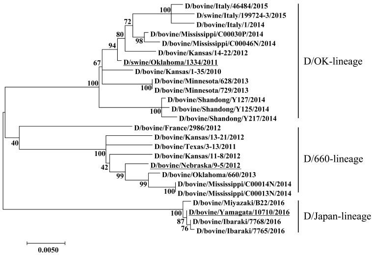
Influenza (flu) D virus, a possible causative agent of bovine respiratory disease, is genetically classified into three clusters: D/OK-, D/660-, and D/Japan-lineages. To evaluate antigenic heterogeneity among these clusters, we compared antibody titers to each lineage virus using bovine sera collected over time following virus infection. Antibody titers to D/Japan-lineage virus rose rapidly in the acute phase of infection, and were 4 times higher than those to the other clustered viruses. In the later phase of infection, titers to D/Japan-lineage virus were equivalent to those to D/OK-lineage virus, and still higher than those to D/660-lineage virus. These results suggest the existence of common and lineage-specific antigenic epitopes in the hemagglutinin-esterase-fusion protein of flu D viruses.

Keywords: antigenicity; cattle; hemagglutinin-esterase-fusion protein; influenza D virus; serology.


Figures

Fig. 1.
Fig. 1.
Phylogenetic tree of the HEF segment of flu D viruses at the nucleotide level. Maximum-likelihood analysis in combination with 500 bootstrap replicates was used to derive tree based on nucleotide sequences of the HEF segment. Bootstrap values are shown above and to the left of the major nodes. Scale bars indicate the number of substitutions per site.Viruses used in this study are underlined.

Similar articles

Cited by

References

    1. Chiapponi C., Faccini S., De Mattia A., Baioni L., Barbieri I., Rosignoli C., Nigrelli A., Foni E.2016. Detection of influenza D virus among swine and cattle, Italy. Emerg. Infect. Dis. 22: 352–354. doi: 10.3201/eid2202.151439 - DOI - PMC - PubMed
    1. Collin E. A., Sheng Z., Lang Y., Ma W., Hause B. M., Li F.2015. Cocirculation of two distinct genetic and antigenic lineages of proposed influenza D virus in cattle. J. Virol. 89: 1036–1042. doi: 10.1128/JVI.02718-14 - DOI - PMC - PubMed
    1. Ducatez M. F., Pelletier C., Meyer G.2015. Influenza D virus in cattle, France, 2011-2014. Emerg. Infect. Dis. 21: 368–371. doi: 10.3201/eid2102.141449 - DOI - PMC - PubMed
    1. Ferguson L., Eckard L., Epperson W. B., Long L. P., Smith D., Huston C., Genova S., Webby R., Wan X. F.2015. Influenza D virus infection in Mississippi beef cattle. Virology 486: 28–34. doi: 10.1016/j.virol.2015.08.030 - DOI - PMC - PubMed
    1. Ferguson L., Olivier A. K., Genova S., Epperson W. B., Smith D. R., Schneider L., Barton K., McCuan K., Webby R. J., Wan X. F.2016. Pathogenesis of influenza D virus in cattle. J. Virol. 90: 5636–5642. doi: 10.1128/JVI.03122-15 - DOI - PMC - PubMed

Substances