

The effects of arousal on apical amplification and conscious state
- PMID: 29877512
- PMCID: PMC5934888
- DOI: 10.1093/nc/niw015
The effects of arousal on apical amplification and conscious state
Abstract
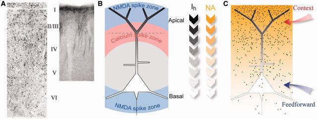
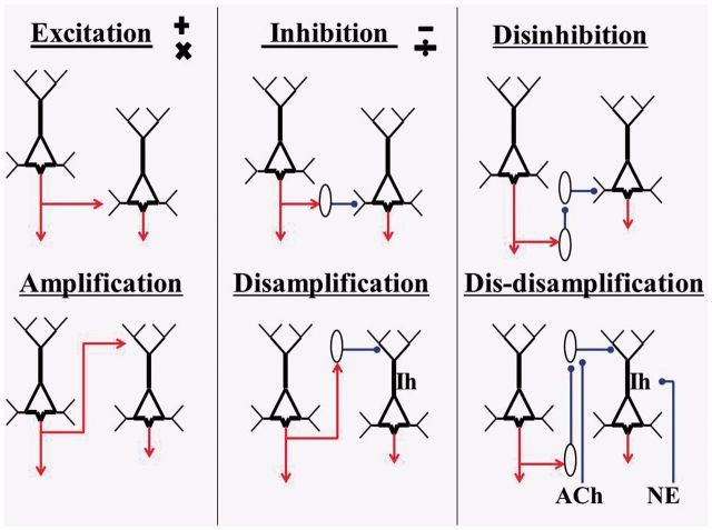
Neocortical pyramidal cells can integrate two classes of input separately and use one to modulate response to the other. Their tuft dendrites are electrotonically separated from basal dendrites and soma by the apical dendrite, and apical hyperpolarization-activated currents (Ih) further isolate subthreshold integration of tuft inputs. When apical depolarization exceeds a threshold, however, it can enhance response to the basal inputs that specify the cell's selective sensitivity. This process is referred to as apical amplification (AA). We review evidence suggesting that, by regulating Ih in the apical compartments, adrenergic arousal controls the coupling between apical and somatic integration zones thus modifying cognitive capabilities closely associated with consciousness. Evidence relating AA to schizophrenia, sleep, and anesthesia is reviewed, and we assess theories that emphasize the relevance of AA to consciousness. Implications for theories of neocortical computation that emphasize context-sensitive modulation are summarized. We conclude that the findings concerning AA and its regulation by arousal offer a new perspective on states of consciousness, the function and evolution of neocortex, and psychopathology. Many issues worthy of closer examination arise.
Keywords: apical amplification; arousal; conscious state; context-sensitive modulation; hyperpolarization-activated currents; schizophrenia.
Figures
Similar articles
-
Cognitive functions of intracellular mechanisms for contextual amplification.Brain Cogn. 2017 Mar;112:39-53. doi: 10.1016/j.bandc.2015.09.005. Epub 2015 Oct 1. Brain Cogn. 2017. PMID: 26428863
-
Modification of current transmitted from apical dendrite to soma by blockade of voltage- and Ca2+-dependent conductances in rat neocortical pyramidal neurons.J Neurophysiol. 1997 Jul;78(1):187-98. doi: 10.1152/jn.1997.78.1.187. J Neurophysiol. 1997. PMID: 9242273
-
Apical tuft input efficacy in layer 5 pyramidal cells from rat visual cortex.J Physiol. 2001 Oct 1;536(Pt 1):167-87. doi: 10.1111/j.1469-7793.2001.00167.x. J Physiol. 2001. PMID: 11579167 Free PMC article.
-
Apical Function in Neocortical Pyramidal Cells: A Common Pathway by Which General Anesthetics Can Affect Mental State.Front Neural Circuits. 2018 Jul 2;12:50. doi: 10.3389/fncir.2018.00050. eCollection 2018. Front Neural Circuits. 2018. PMID: 30013465 Free PMC article. Review.
-
Cellular psychology: relating cognition to context-sensitive pyramidal cells.Trends Cogn Sci. 2025 Jan;29(1):28-40. doi: 10.1016/j.tics.2024.09.002. Epub 2024 Oct 1. Trends Cogn Sci. 2025. PMID: 39353837 Review.
Cited by
-
Computational Modeling of Genetic Contributions to Excitability and Neural Coding in Layer V Pyramidal Cells: Applications to Schizophrenia Pathology.Front Comput Neurosci. 2019 Sep 26;13:66. doi: 10.3389/fncom.2019.00066. eCollection 2019. Front Comput Neurosci. 2019. PMID: 31616272 Free PMC article.
-
Potential of olfactory neuroepithelial cells as a model to study schizophrenia: A focus on GPCRs (Review).Int J Mol Med. 2024 Jan;53(1):7. doi: 10.3892/ijmm.2023.5331. Epub 2023 Dec 1. Int J Mol Med. 2024. PMID: 38038161 Free PMC article. Review.
-
Anesthesia-induced loss of consciousness disrupts auditory responses beyond primary cortex.Proc Natl Acad Sci U S A. 2020 May 26;117(21):11770-11780. doi: 10.1073/pnas.1917251117. Epub 2020 May 12. Proc Natl Acad Sci U S A. 2020. PMID: 32398367 Free PMC article.
-
Perspective on the Multiple Pathways to Changing Brain States.Front Syst Neurosci. 2020 May 8;14:23. doi: 10.3389/fnsys.2020.00023. eCollection 2020. Front Syst Neurosci. 2020. PMID: 32457583 Free PMC article. Review.
-
Somatostatin-Positive Gamma-Aminobutyric Acid Interneuron Deficits in Depression: Cortical Microcircuit and Therapeutic Perspectives.Biol Psychiatry. 2017 Oct 15;82(8):549-559. doi: 10.1016/j.biopsych.2017.05.024. Epub 2017 Jun 8. Biol Psychiatry. 2017. PMID: 28697889 Free PMC article. Review.
References
-
- Amit DJ. (1989). Modeling Brain Function. NY, New York: Cambridge University Press.
-
- Aoki C, Venkatesan C, Go CG. et al. Cellular and subcellular sites for noradrenergic action in the monkey dorsolateral prefrontal cortex as revealed by the immunocytochemical localization of noradrenergic receptors and axons. Cereb Cortex 1998;8:269–77. - PubMed
Publication types
LinkOut - more resources
Full Text Sources
Other Literature Sources