Revealing the Dynamic Modulations That Underpin a Resilient Neural Network for Semantic Cognition: An fMRI Investigation in Patients With Anterior Temporal Lobe Resection
- PMID: 29878076
- PMCID: PMC6041810
- DOI: 10.1093/cercor/bhy116
Revealing the Dynamic Modulations That Underpin a Resilient Neural Network for Semantic Cognition: An fMRI Investigation in Patients With Anterior Temporal Lobe Resection
Abstract
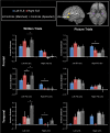
One critical feature of any well-engineered system is its resilience to perturbation and minor damage. The purpose of the current study was to investigate how resilience is achieved in higher cognitive systems, which we explored through the domain of semantic cognition. Convergent evidence implicates the bilateral anterior temporal lobes (ATLs) as a conceptual knowledge hub. While bilateral damage to this region produces profound semantic impairment, unilateral atrophy/resection or transient perturbation has a limited effect. Two neural mechanisms might underpin this resilience to unilateral ATL damage: 1) the undamaged ATL upregulates its activation in order to compensate; and/or 2) prefrontal regions involved in control of semantic retrieval upregulate to compensate for the impoverished semantic representations that follow from ATL damage. To test these possibilities, 34 postsurgical temporal lobe epilepsy patients and 20 age-matched controls were scanned whilst completing semantic tasks. Pictorial tasks, which produced bilateral frontal and temporal activation, showed few activation differences between patients and control participants. Written word tasks, however, produced a left-lateralized activation pattern and greater differences between the groups. Patients with right ATL resection increased activation in left inferior frontal gyrus (IFG). Patients with left ATL resection upregulated both the right ATL and right IFG. Consistent with recent computational models, these results indicate that 1) written word semantic processing in patients with ATL resection is supported by upregulation of semantic knowledge and control regions, principally in the undamaged hemisphere, and 2) pictorial semantic processing is less affected, presumably because it draws on a more bilateral network.
Figures


References
-
- Alpherts WCJ, Vermeulen J, van Rijen PC, da Silva FHL, van Veelen CWM, Dutch Collaborative Epilepsy S. 2006. Verbal memory decline after temporal epilepsy surgery? A 6-year multiple assessments follow-up study. Neurology. 67(4):626–631. - PubMed
-
- Backes WH, Deblaere K, Vonck K, Kessels AG, Boon P, Hofman P, Wilmink JT, Vingerhoets G, Boon PA, Achten R, et al. . 2005. Language activation distributions revealed by fMRI in post-operative epilepsy patients: differences between left- and right-sided resections. Epilepsy Res. 66(1–3):1–12. - PubMed
-
- Badre D, Poldrack RA, Pare-Blagoev EJ, Insler RZ, Wagner AD. 2005. Dissociable controlled retrieval and generalized selection mechanisms in ventrolateral prefrontal cortex. Neuron. 47(6):907–918. - PubMed
-
- Badre D, Wagner AD. 2002. Semantic retrieval, mnemonic control, and prefrontal cortex. Behav Cogn Neurosci Rev. 1:206–218. - PubMed
Publication types
MeSH terms
Substances
Grants and funding
- BB_/Biotechnology and Biological Sciences Research Council/United Kingdom
- MC_UU_00005/18/MRC_/Medical Research Council/United Kingdom
- MR/K026992/1/MRC_/Medical Research Council/United Kingdom
- MR/R023883/1/MRC_/Medical Research Council/United Kingdom
- MR/J004146/1/MRC_/Medical Research Council/United Kingdom
LinkOut - more resources
Full Text Sources
Other Literature Sources
Medical
Molecular Biology Databases

