. 2018 Sep;48(9):1445-1455.
doi: 10.1002/eji.201747400. Epub 2018 Jun 26.

Murine macrophage chemokine receptor CCR2 plays a crucial role in macrophage recruitment and regulated inflammation in wound healing

Affiliations

Murine macrophage chemokine receptor CCR2 plays a crucial role in macrophage recruitment and regulated inflammation in wound healing

Anna E Boniakowski et al. Eur J Immunol. 2018 Sep.

Abstract

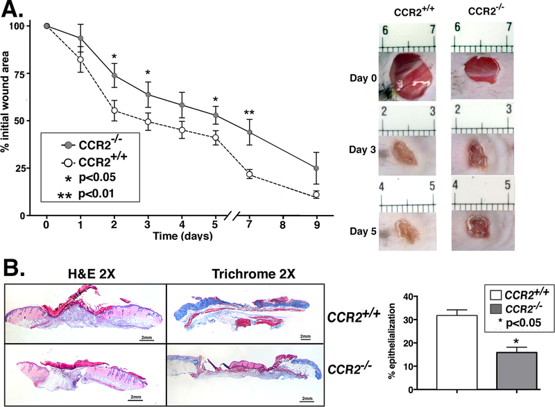
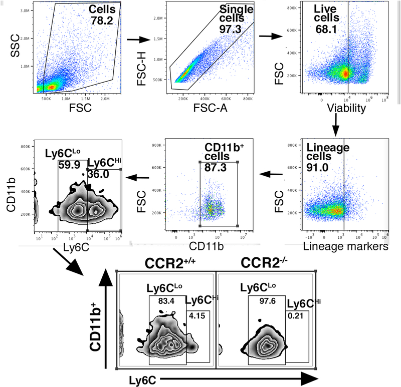
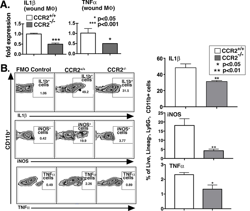
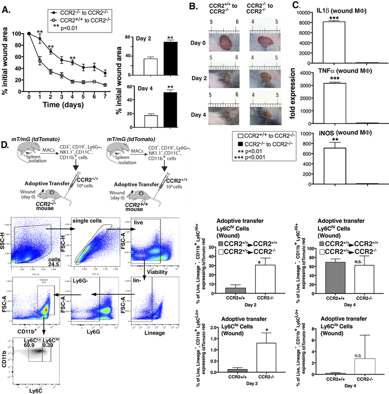
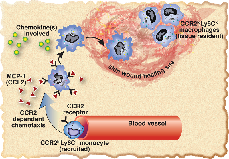
Macrophages play a critical role in the establishment of a regulated inflammatory response following tissue injury. Following injury, CCR2+ monocytes are recruited from peripheral blood to wound tissue, and direct the initiation and resolution of inflammation that is essential for tissue repair. In pathologic states where chronic inflammation prevents healing, macrophages fail to transition to a reparative phenotype. Using a murine model of cutaneous wound healing, we found that CCR2-deficient mice (CCR2-/- ) demonstrate significantly impaired wound healing at all time points postinjury. Flow cytometry analysis of wounds from CCR2-/- and WT mice revealed a significant decrease in inflammatory, Ly6CHi recruited monocyte/macrophages in CCR2-/- wounds. We further show that wound macrophage inflammatory cytokine production is decreased in CCR2-/- wounds. Adoptive transfer of mT/mG monocyte/macrophages into CCR2+/+ and CCR2-/- mice demonstrated that labeled cells on days 2 and 4 traveled to wounds in both CCR2+/+ and CCR2-/- mice. Further, adoptive transfer of monocyte/macrophages from WT mice restored normal healing, likely through a restored inflammatory response in the CCR2-deficient mice. Taken together, these data suggest that CCR2 plays a critical role in the recruitment and inflammatory response following injury, and that wound repair may be therapeutically manipulated through modulation of CCR2.

Keywords: Chemokine receptor; Inflammation; Macrophages; Recruitment; Wound healing.


Conflict of interest statement

CONFLICT OF INTEREST

The authors have no conflicts of interest to disclose.

Figures

Figure 1.
Figure 1.. Wound healing is impaired in CCR2-deficient mice (CCR2−/−).
Wounds were created using 4 mm punch biopsies on the backs of CCR2−/− mice and matched controls (CCR2+/+). A: The change in wound area was recorded daily using ImageJ software (NIH) until complete healing was observed. Representative photographs of the wounds of CCR2−/− mice and controls on days 0, 3 and 5 post-injury are shown (*P < 0.05; **P < 0.01; n=14; data is representative of four independent experiments). B: Wounds were harvested on day 3, paraffin embedded and sectioned. 5 μM sections were stained with hematoxylin and eosin and with Masson’s Trichrome stain. Percent re-epithelialization was calculated by measuring distance traveled by epithelial tongues on both sides of wound divided by total distance for full re-epithelialization. Representative images are shown in 2X magnification (*P < 0.05; n=10; data is representative of two independent experiments). Statistical analysis was performed using a paired Student’s t-test. All data are expressed as mean ± SEM.
Figure 2.
Figure 2.. Loss of CCR2 in Ly6CHi monocytes results in decreased inflammatory Ly6CHi monocyte/macrophages in wound tissue.
A: Wounds were harvested from CCR2−/− and matched controls on day 3 and processed for ex vivo intracellular flow cytometry. The gating strategy used for flow cytometry selected live, lineage- (CD3, CD19, Ter119, NK1.1), non-neutrophil (Ly6G), CD11b+ cells as shown. Flow cytometry quantification of Ly6C revealed two populations of cells in both CCR2−/− and CCR2+/+ wounds, translating to Ly6CHi and Ly6CLo monocyte macrophages (CD3, CD19, Ter119, NK1.1, Ly6G, CD11b+).(n=10; data is representative of two independent experiments).
Figure 3.
Figure 3.. Macrophages isolated from wounds of CCR2−/− mice display decreased inflammatory cytokine gene expression and protein levels.
A: Wound macrophages were isolated from CCR2−/− mice and matched controls on day 3 post-injury by MACS for CD11b+ (CD3,CD19, Ly6G) cells. TNF-α and IL1β gene expression in isolated macrophages was measured by qPCR using 18s for normalization (*P<0.05; **P < 0.01; n=12, data is representative of two independent experiments). B: CCR2−/− and littermate control wounds harvested on day 3 were processed for ex vivo intracellular flow cytometry. Intracellular cytokine quantification by flow cytometry of IL1β, iNOS, and TNF-α in wound macrophages was examined (*P < 0.05, ** P < 0.01; n=10; data is representative of two independent experiments). Statistical analysis was performed using a paired Student’s t-test. All data are expressed as mean ± SEM.
Figure 4.
Figure 4.. Myeloid-specific CCR2 was sufficient to rescue wound healing in CCR2−/− mice.
CD3, CD11c, CD19, Ly6G, NK1.1, CD11b+ single cell suspensions were isolated from CCR2−/− and CCR2+/+ spleens by MACS. 1 × 10⁶ cells were injected intravenously via tail vein into wounded (day 0) CCR2−/− mice and wound closure was measured daily using ImageJ software (**P < 0.01; n=15; data is representative of two independent experiments). B: Representative images are shown from CCR2−/− and CCR2+/+ mice on days 0, 2, and 4. C: Wound macrophages were isolated from adoptively transferred CCR2+/+ to CCR2−/− and CCR2−/− to CCR2−/− mice on day 2 post-injury by MACS and gene expression (IL1β, iNOS, and TNF-α) in isolated macrophages was measured by qPCR using 18s for normalization (**P < 0.01; ***P<0.0001; n=12; data is representative of three independent experiments). D: 1×10⁶ tdTomato-expressing CD3, CD11c, CD19, Ly6G, NK1.1, CD11b+ single cell suspensions were injected via tail vein into wounded (day 0) CCR2−/− and control mice. Wounds were harvested on days 2 and 4 and single cell suspensions were processed for flow cytometry. Percentage of tdTomato+ (CD3, CD19, Ter119, NK1.1, Ly6G, CD11b+) and subsequent stratification by Ly6C designation is shown (*P < 0.05; n=10; data is representative of two independent experiments). Statistical analysis was performed using a paired Student’s t-test. All data are expressed as mean ± SEM.
Figure 5.
Figure 5.. Schematic of CCR2-dependent Ly6CHi cell recruitment to peripheral wounds.
Upon initial tissue injury, CCL2, one of the primary ligands for CCR2, is increased in the wound. This ligand binds the CCR2 receptors that are present on Ly6CHi monocytes, recruiting these cells to the wound, allowing initiation of the macrophage-mediated inflammatory phase of wound healing.

References

    1. Sica A, Erreni M, Allavena P, Porta C. Macrophage polarization in pathology. Cell. Mol. Life Sci 2015; 72:4111–4126. Available at: https://www.scopus.com/inward/record.uri?eid=2-s2.0-84944170532&doi=10.1... DOI: 10.1007/s00018-015-1995-y. - DOI - PMC - PubMed
    1. Martin P Wound Healing--Aiming for Perfect Skin Regeneration. Science (80-. ) 1997; 276:75–81. Available at: http://www.sciencemag.org/cgi/doi/10.1126/science.276.5309.75DOI: 10.1126/science.276.5309.75. - DOI - DOI - PubMed
    1. Okuno Y, Nakamura-Ishizu A, Kishi K, Suda T, Kubota Y. Bone marrow-derived cells serve as proangiogenic macrophages but not endothelial cells in wound healing. Blood 2011; 117:5264–5272.DOI: 10.1182/blood-2011-01-330720. - DOI - PMC - PubMed
    1. Ginhoux F, Greter M, Leboeuf M, Nandi S, See P, Gokhan S, Mehler MF, et al. Fate mapping analysis reveals that adult microglia derive from primitive macrophages. Science (80-. ) 2010; 330:841–5. Available at: http://www.pubmedcentral.nih.gov/articlerender.fcgi?artid=3719181&tool=p...DOI: 10.1126/science.1194637. - DOI - PMC - PubMed
    1. Hoeffel G, Wang Y, Greter M, See P, Teo P, Malleret B, Leboeuf M, et al. Adult Langerhans cells derive predominantly from embryonic fetal liver monocytes with a minor contribution of yolk sac-derived macrophages. J. Exp. Med 2012; 209:1167–81. Available at: http://jem.rupress.org/content/209/6/1167.fullDOI: 10.1084/jem.20120340. - DOI - PMC - PubMed

Publication types